. 2010 Oct 12;107(41):17668-73.
doi: 10.1073/pnas.1004487107. Epub 2010 Sep 27.

Induced pluripotent stem cell models of the genomic imprinting disorders Angelman and Prader-Willi syndromes

Affiliations

Induced pluripotent stem cell models of the genomic imprinting disorders Angelman and Prader-Willi syndromes

Stormy J Chamberlain et al. Proc Natl Acad Sci U S A. .

Abstract

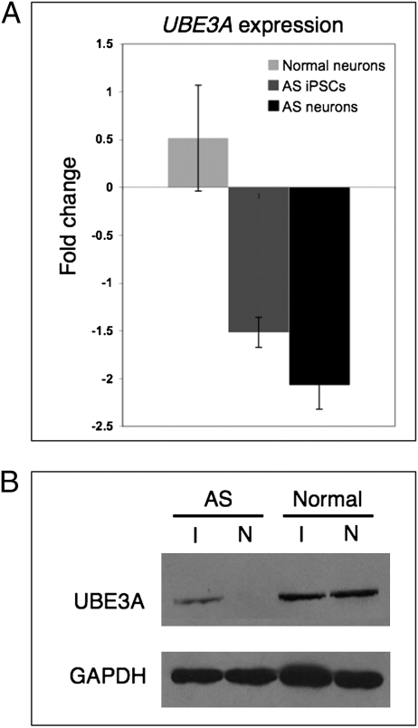
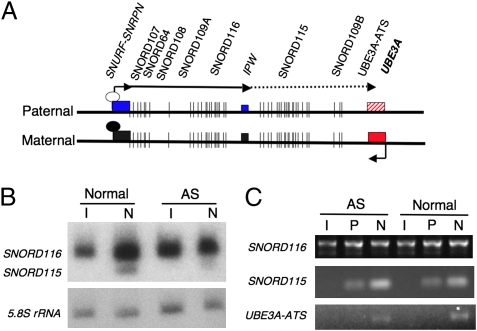
Angelman syndrome (AS) and Prader-Willi syndrome (PWS) are neurodevelopmental disorders of genomic imprinting. AS results from loss of function of the ubiquitin protein ligase E3A (UBE3A) gene, whereas the genetic defect in PWS is unknown. Although induced pluripotent stem cells (iPSCs) provide invaluable models of human disease, nuclear reprogramming could limit the usefulness of iPSCs from patients who have AS and PWS should the genomic imprint marks be disturbed by the epigenetic reprogramming process. Our iPSCs derived from patients with AS and PWS show no evidence of DNA methylation imprint erasure at the cis-acting PSW imprinting center. Importantly, we find that, as in normal brain, imprinting of UBE3A is established during neuronal differentiation of AS iPSCs, with the paternal UBE3A allele repressed concomitant with up-regulation of the UBE3A antisense transcript. These iPSC models of genomic imprinting disorders will facilitate investigation of the AS and PWS disease processes and allow study of the developmental timing and mechanism of UBE3A repression in human neurons.


Conflict of interest statement

The authors declare no conflict of interest.

Figures

Fig. 1.
Fig. 1.
AS iPSCs express markers of pluripotency and have expected karyotypes. (A) Phase-contrast (a–c) images of AS iPSCs show hESC-like morphology. Immunocytochemistry for pluripotency markers on representative iPSC lines shows expression of NANOG (d–f), SSEA4 (g–i), TRA1-60 (j–l), and TRA1-81 (m–p). (B) Karyotype analysis on G-banded chromosomes was performed by Cell Line Genetics. Representative G-banded chromosomes from two AS iPSC lines show normal chromosome counts of 46 XX (ASdel1-0) and 46 XY (ASdel2-0).
Fig. 2.
Fig. 2.
Methylation imprint at the PWS-IC is maintained during reprogramming. (A) Methylation-specific PCR analysis of genomic DNA from normal, AS, and PWS fibroblasts. Primers specific for the methylated allele amplify a band that is 174 bp, whereas primers specific for the unmethylated allele amplify a 100-bp product. Mat, maternal; Pat, paternal. (B) Methylation-specific PCR using genomic DNA from three different AS iPSC lines and two different normal iPSC lines. Neg, negative. (C) Methylation-specific PCR using genomic DNA from three different PWS iPSC lines.
Fig. 3.
Fig. 3.
Functional neurons can be generated from AS iPSCs. (A) Phase-contrast image of 10-wk-old in vitro differentiated neurons generated from AS iPSCs. (B) iPSC-derived neurons stain positively for βIII-TUBULIN (TUJ1, green). (C) S100β-positive astrocytes spontaneously arise in AS iPSC-derived neuron cultures after 6 wk of differentiation. (D) Electrophysiological recordings from a mature AS iPSC-derived neuron after 10 wk of differentiation. (Upper) Train of action potentials evoked by depolarizing current injection (50 pA). (Calibration bars: 20 mV, 0.1 s.) (Lower) Example sweeps of spontaneous EPSCs recorded from the same neuron in the absence or presence of the AMPA receptor antagonist DNQX (10 μM). (Calibration bars: 10 pA, 1 s.) (E) Electrophysiological recordings from an immature AS iPSC-derived neuron after 10 wk of differentiation. (Upper) Response to a 60-pA depolarizing current injection. (Lower) Lack of spontaneous synaptic activity. Calibration bars are the same as in D.
Fig. 4.
Fig. 4.
Paternal UBE3A is repressed in AS iPSC-derived neurons. (A) qRT-PCR analysis of UBE3A expression in AS and normal iPSCs and iPSC-derived neurons. Gene expression is normalized to GAPDH and is presented as the fold change relative to UBE3A expression levels in normal iPSCs. Error bars indicate SD for three independent cultures. (B) Western blot analysis of normal and AS iPSCs (I) and 10-wk-old iPSC-derived neurons (N).
Fig. 5.
Fig. 5.
UBE3A-ATS is only expressed in neuron cultures and correlates with paternal UBE3A repression. (A) Map of the SNURF-SNRPN and UBE3A transcripts. White and black oval-shaped circles indicate the unmethylated and methylated PWS-IC, respectively. The solid line with arrows indicates transcripts expressed in iPSCs, whereas the dotted line indicates neural-specific transcripts. AS iPSCs and their neural derivatives lack the entire maternally inherited allele of this region. (B) Northern blot using total RNA from AS and normal iPSCs (I) and iPSC-derived 10-wk-old neurons (N) shows expression of the snoRNAs SNORD116 and SNORD115 during in vitro neural development. (C) RT-PCR using primers spanning multiple exons of SNORD116, SNORD115, and UBE3A-ATS in iPSCs (I), 3-wk-old iPSC-derived neural precursor cells (P), and 10-wk-old iPSC-derived neuron cultures (N).

Similar articles

Cited by

References

    1. Lossie AC, et al. Distinct phenotypes distinguish the molecular classes of Angelman syndrome. J Med Genet. 2001;38:834–845. - PMC - PubMed
    1. Williams CA, et al. Angelman syndrome 2005: Updated consensus for diagnostic criteria. Am J Med Genet A. 2006;140:413–418. - PubMed
    1. Cassidy SB, Driscoll DJ. Prader-Willi syndrome. Eur J Hum Genet. 2009;17:3–13. - PMC - PubMed
    1. Rougeulle C, Glatt H, Lalande M. The Angelman syndrome candidate gene, UBE3A/E6-AP, is imprinted in brain. Nat Genet. 1997;17:14–15. - PubMed
    1. Vu TH, Hoffman AR. Imprinting of the Angelman syndrome gene, UBE3A, is restricted to brain. Nat Genet. 1997;17:12–13. - PubMed

Publication types