. 2010 Oct 12;107(41):17797-802.
doi: 10.1073/pnas.1001907107. Epub 2010 Sep 27.

O-linked β-N-acetylglucosamine transferase is indispensable in the failing heart

Affiliations

O-linked β-N-acetylglucosamine transferase is indispensable in the failing heart

Lewis J Watson et al. Proc Natl Acad Sci U S A. .

Abstract

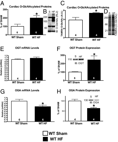
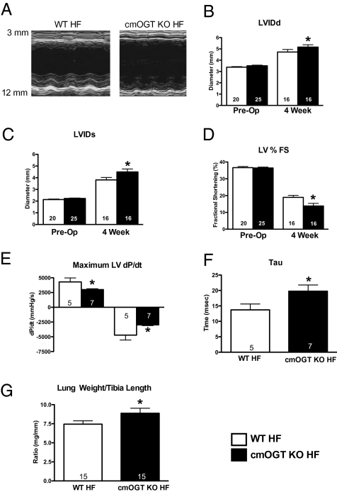
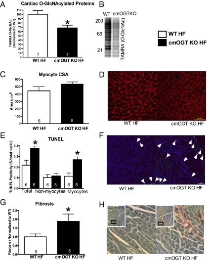
The failing heart is subject to elevated metabolic demands, adverse remodeling, chronic apoptosis, and ventricular dysfunction. The interplay among such pathologic changes is largely unknown. Several laboratories have identified a unique posttranslational modification that may have significant effects on cardiovascular function. The O-linked β-N-acetylglucosamine (O-GlcNAc) posttranslational modification (O-GlcNAcylation) integrates glucose metabolism with intracellular protein activity and localization. Because O-GlcNAc is derived from glucose, we hypothesized that altered O-GlcNAcylation would occur during heart failure and figure prominently in its pathophysiology. After 5 d of coronary ligation in WT mice, cardiac O-GlcNAc transferase (OGT; which adds O-GlcNAc to proteins) and levels of O-GlcNAcylation were significantly (P < 0.05) elevated in the surviving remote myocardium. We used inducible, cardiac myocyte-specific Cre recombinase transgenic mice crossed with loxP-flanked OGT mice to genetically delete cardiomyocyte OGT (cmOGT KO) and ascertain its role in the failing heart. After tamoxifen induction, cardiac O-GlcNAcylation of proteins and OGT levels were significantly reduced compared with WT, but not in other tissues. WT and cardiomyocyte OGT KO mice underwent nonreperfused coronary ligation and were followed for 4 wk. Although OGT deletion caused no functional change in sham-operated mice, OGT deletion in infarcted mice significantly exacerbated cardiac dysfunction compared with WT. These data provide keen insights into the pathophysiology of the failing heart and illuminate a previously unrecognized point of integration between metabolism and cardiac function in the failing heart.


Conflict of interest statement

The authors declare no conflict of interest.

Figures

Fig. 1.
Fig. 1.
Cardiac O-GlcNAcylation changes following 5 d of coronary artery ligation. Hearts from the same animals as in Figs. S1 and S2 were used to determine the changes in cardiac O-GlcNAcylation. (A) Coronary ligation for 5 d produced significantly elevated O-GlcNAcylation according to immunoblot (representative image shown in B). (C) Secondarily, a nonantibody method (Click-iT method) also revealed a similar significant increase (representative image shown in D). (E) OGT mRNA levels were unchanged in the failing heart. (F) OGT protein was significantly elevated in the failing heart (representative image shown in Inset). (G) OGA mRNA levels were significantly decreased in the failing heart. (H) OGA protein levels were reduced (representative image shown in Inset); *P < 0.05 versus WT sham.
Fig. 2.
Fig. 2.
Cardiac-specific deletion of OGT. To determine the efficacy of targeted OGT deletion, MCM Tg mice were bred onto a homozygous OGT floxed mouse line. All mice in Figs. 2–4 are homozygous OGT floxed mice. The cmOGT KO or WT designations refer only to the presence or absence of the MCM transgene. Following induction of the transgene via tamoxifen injections, OGT mRNA levels (A), OGT protein levels (B), and levels of O-GlcNAcylated proteins (C) were significantly reduced in the cmOGT KO hearts. To confirm tissue specificity of OGT ablation, skeletal muscle and lungs were removed and subjected to immunoblot for OGT; no change was observed (B). Cardiac myocyte specific ablation of OGT was confirmed via immunofluorescence. Using antibodies specific for OGT and cardiac troponin, a mosaic loss of green (OGT) signal occurs in the cmOGT KO myocardium compared with the WT, in which the signal remains largely uniform throughout the myocardium (D). In addition, cmOGT KO hearts were stained for O-GlcNAcylated proteins and a similar mosaic pattern emerged (E); *P < 0.05 versus WT.
Fig. 3.
Fig. 3.
OGT ablation exacerbates heart failure. Following cardiac ablation of OGT, mice were subjected to permanent coronary ligation and followed for 4 wk via echocardiography, with representative M-modes shown (A). cmOGT KO mice subjected to coronary ligation showed a significant increase in diastolic (B) and systolic (C) diameters and a significant reduction in fractional shortening by 4 wk (D). Left ventricular dP/dt, an index of cardiac function, was also reduced in the cmOGT KO HF group (E), whereas an elevation in tau provides evidence of impaired relaxation (F). Finally, an increased lung weight-to-tibia length ratio (G) suggests pulmonary edema caused by a slightly elevated LVEDP; *P < 0.05 versus WT HF.
Fig. 4.
Fig. 4.
OGT ablation increases pathological remodeling of the left ventricle. Following 4 wk of coronary ligation, hearts were removed and subjected to biochemical and histological assessments. (A) Reduction in O-GlcNAcylation persisted in the cmOGT KO HF group as determined by Click-it; a representative image is shown in B. Ablation of OGT had no effect on cardiomyocyte size (C) in the failing heart; representative image shown in D. There was an increase in the total number of TUNEL-positive nuclei, and more specifically the number of TUNEL-positive myocytes, in the cmOGT KO group compared with WT (E). A representative image is shown (F) with arrowheads denoting positive nuclei. Following this elevated rate of apoptosis, total fibrotic area was also exacerbated in the cmOGT KO failing hearts (G); representative image is also shown (H). Both TUNEL and fibrotic measurements were made in a section remote from the infarct; *P < 0.05 versus WT HF.

References

    1. Razeghi P, et al. Metabolic gene expression in fetal and failing human heart. Circulation. 2001;104:2923–2931. - PubMed
    1. Garnier A, et al. Depressed mitochondrial transcription factors and oxidative capacity in rat failing cardiac and skeletal muscles. J Physiol. 2003;551:491–501. - PMC - PubMed
    1. Arany Z, et al. Transcriptional coactivator PGC-1 alpha controls the energy state and contractile function of cardiac muscle. Cell Metab. 2005;1:259–271. - PubMed
    1. Cha SH, Rodgers JT, Puigserver P, Chohnan S, Lane MD. Hypothalamic malonyl-CoA triggers mitochondrial biogenesis and oxidative gene expression in skeletal muscle: Role of PGC-1alpha. Proc Natl Acad Sci USA. 2006;103:15410–15415. - PMC - PubMed
    1. Arany Z, et al. Transverse aortic constriction leads to accelerated heart failure in mice lacking PPAR-gamma coactivator 1alpha. Proc Natl Acad Sci USA. 2006;103:10086–10091. - PMC - PubMed

Publication types

Substances