Review
. 2010 Oct 5;107(40):17087-94.
doi: 10.1073/pnas.1011782107. Epub 2010 Sep 27.

Parallel information processing channels created in the retina

Affiliations
Review

Parallel information processing channels created in the retina

Peter H Schiller. Proc Natl Acad Sci U S A. .

Abstract

In the retina, several parallel channels originate that extract different attributes from the visual scene. This review describes how these channels arise and what their functions are. Following the introduction four sections deal with these channels. The first discusses the "ON" and "OFF" channels that have arisen for the purpose of rapidly processing images in the visual scene that become visible by virtue of either light increment or light decrement; the ON channel processes images that become visible by virtue of light increment and the OFF channel processes images that become visible by virtue of light decrement. The second section examines the midget and parasol channels. The midget channel processes fine detail, wavelength information, and stereoscopic depth cues; the parasol channel plays a central role in processing motion and flicker as well as motion parallax cues for depth perception. Both these channels have ON and OFF subdivisions. The third section describes the accessory optic system that receives input from the retinal ganglion cells of Dogiel; these cells play a central role, in concert with the vestibular system, in stabilizing images on the retina to prevent the blurring of images that would otherwise occur when an organism is in motion. The last section provides a brief overview of several additional channels that originate in the retina.


Conflict of interest statement

The author declares no conflict of interest.

Figures

Fig. 1.
Fig. 1.
(A) The basic connections a cone makes with ON and OFF ganglion cells via ON and OFF bipolar cells. The ON bipolar cells have sign inverting synapses as described in the text. (B) The basic connections rods, cones, bipolar cells, horizontal cells, and amacrine cells make in the retina. [Reprinted from Progress in Retinal and Eye Research, Vol 15, Schiller PH, The ON and OFF channels of the mammalian visual system, 173–195, Copyright (1995), with permission from Elsevier]. (C) The responses of an ON and OFF monkey LGN cell before, during, and after the infusion of APB into the eye. APB blocks the ON cell response and has no significant effect on the OFF cell (25). (Reprinted by permission from Macmillan Publishers Ltd: Nature, 297:580–583, copyright 1982.)
Fig. 2.
Fig. 2.
The responses of an ON and an OFF cell in the monkey LGN before and after APB infusion into the eye when the center and the surround of the receptive fields are stimulated with red and green spots of different sizes. The surround activated by the large green spot is turned on briefly during both the on and off cycles of the small red spot. APB application (Lower) silences the ON ganglion cell for both center and surround stimulation. The response of the OFF ganglion cell is unaffected; the cell retains its center-surround antagonism (25). (Reprinted by permission from Macmillan Publishers Ltd: Nature, 297:580–583, copyright 1982.)
Fig. 3.
Fig. 3.
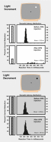
Percent correct performance and eye-movement reaction times made to light and dark stimuli presented in various locations of the visual field to which a trained rhesus monkey had to make saccadic eye movements to be rewarded. Following APB administration, percent correct performance declines and reaction times increase to a bright spot. Percent correct responses and reaction times to a dark spot are unaffected.
Fig. 4.
Fig. 4.
(A) Cross-section of Golgi-stained midget and parasol cells (51). [Reprintedwith permission from Polyak SL (1957) The vertebrate visual system: Its origin, structure, and function and its manifestations in disease with an analysis of its role in the life of animals and in the origin of man, preceded by a historical review of investigations of the eye, and of the visual pathways and centers of the brain (University of Chicago Press, Chicago).] (B) The dendritic arbors of a midget and parasol cell at comparable eccentricities (52). [Reprinted from Watanabe M, Rodieck RW (1989) Parasol and midget ganglion cells of the primate retina. J Comp Neurol 289:434–454 (Copyright 1989 with permission from Wiley).]

Comment in

  • Profile of Peter H. Schiller.
    Nair P. Nair P. Proc Natl Acad Sci U S A. 2011 Mar 22;108(12):4705-7. doi: 10.1073/pnas.1101955108. Epub 2011 Feb 28. Proc Natl Acad Sci U S A. 2011. PMID: 21368197 Free PMC article. No abstract available.

Similar articles

Cited by

References

    1. Schultze M. Zur anatomie und physiologie der retina. Arch Mikrosk Anat. 1866;2:175–286.
    1. Ramon y Cajal S, Craigie EH, Cano J. Recollections of My Life. Philadelphia: American Philosophical Society; 1937.
    1. Hartline H. The responses of single optic nerve fibers of the vertebrate eye to illumination of the retina. Am J Physiol. 1938;121:400–415.
    1. Barlow HB. Summation and inhibition in the frog's retina. J Physiol. 1953;119:69–88. - PMC - PubMed
    1. Kuffler SW. Discharge patterns and functional organization of mammalian retina. J Neurophysiol. 1953;16:37–68. - PubMed

MeSH terms

Substances

LinkOut - more resources