. 2010 Oct 5;107(40):17152-7.
doi: 10.1073/pnas.1007988107. Epub 2010 Sep 27.

Structures of the Escherichia coli ribosome with antibiotics bound near the peptidyl transferase center explain spectra of drug action

Affiliations

Structures of the Escherichia coli ribosome with antibiotics bound near the peptidyl transferase center explain spectra of drug action

Jack A Dunkle et al. Proc Natl Acad Sci U S A. .

Abstract

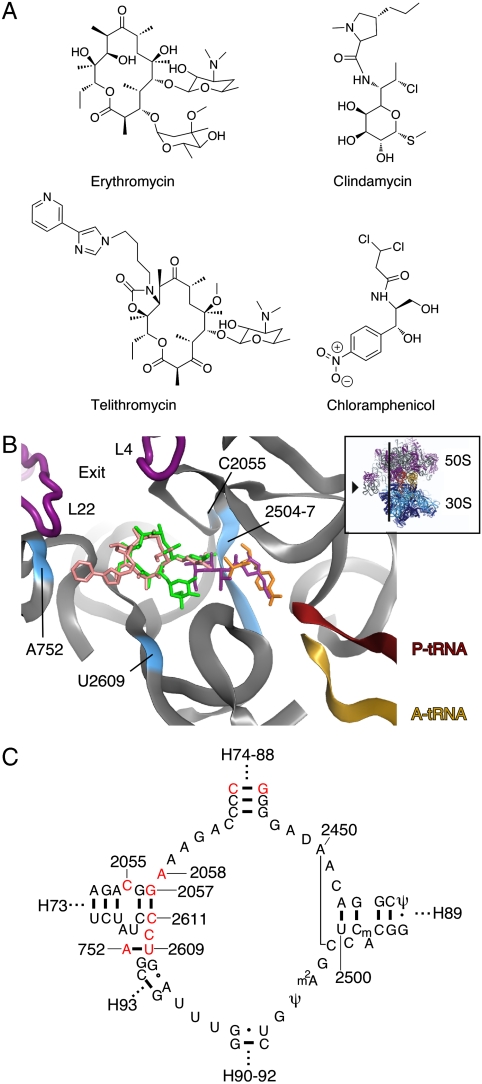
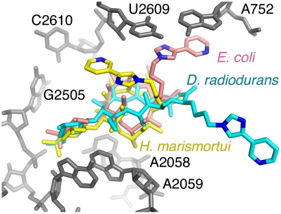
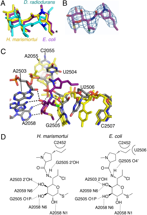
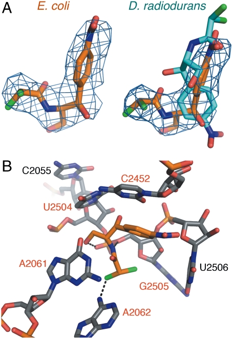
Differences between the structures of bacterial, archaeal, and eukaryotic ribosomes account for the selective action of antibiotics. Even minor variations in the structure of ribosomes of different bacterial species may lead to idiosyncratic, species-specific interactions of the drugs with their targets. Although crystallographic structures of antibiotics bound to the peptidyl transferase center or the exit tunnel of archaeal (Haloarcula marismortui) and bacterial (Deinococcus radiodurans) large ribosomal subunits have been reported, it remains unclear whether the interactions of antibiotics with these ribosomes accurately reflect those with the ribosomes of pathogenic bacteria. Here we report X-ray crystal structures of the Escherichia coli ribosome in complexes with clinically important antibiotics of four major classes, including the macrolide erythromycin, the ketolide telithromycin, the lincosamide clindamycin, and a phenicol, chloramphenicol, at resolutions of ∼3.3 Å-3.4 Å. Binding modes of three of these antibiotics show important variations compared to the previously determined structures. Biochemical and structural evidence also indicates that interactions of telithromycin with the E. coli ribosome more closely resembles drug binding to ribosomes of bacterial pathogens. The present data further argue that the identity of nucleotides 752, 2609, and 2055 of 23S ribosomal RNA explain in part the spectrum and selectivity of antibiotic action.


Conflict of interest statement

The authors declare no conflict of interest.

Figures

Fig. 1.
Fig. 1.
Binding sites for antibiotics in the PTC and peptide exit tunnel. (A) The chemical structures of erythromycin (a macrolide), telithromycin (a ketolide), chloramphenicol (a phenyl propanoside), and clindamycin (a lincosamide) are shown. (B) An overview of the antibiotic binding sites within the 50S subunit. Erythromycin (green), telithromycin (pink), clindamcyin (purple), and chloramphenicol (orange) are shown as stick models. Ribbons denote the sugar phosphate backbone of 23S rRNA (gray) with nucleotides of interest colored light blue, the acceptor ends of A-site tRNA (yellow) and P-site tRNA (red). The location of the peptide exit tunnel is labeled “exit,” and an icon indicating the point of view is shown on the right. (C) The secondary structure of the 3′ region of 23S rRNA showing elements of the PTC and the adjacent peptide exit tunnel. Nucleotides that are divergent between E. coli and H. marismortui are shown in red. D. radiodurans diverges from E. coli at the 2057-2611 base pair and at nucleotides 752 and 2586. Ribosomal RNA helices emanating from this region are marked with dotted lines.
Fig. 2.
Fig. 2.
Telithromycin bound to the E. coli ribosome. A comparison of the conformations reported for telithromcyin bound to the ribosome. 23S rRNA for E. coli is shown in gray. Telithromycin models from H. marismortui (gold), D.radiodurans (cyan), and E. coli (pink) are shown. Nitrogens in the alkyl-aryl arm are also shown for reference.
Fig. 3.
Fig. 3.
Telithromycin and erythromycin footprints on 23S rRNA reveal species-specific binding modes. Interactions of antibiotics erythromycin (E) or telithromycin (T) in solution with ribosomes prepared from archaea (H. halobium) or three different bacterial species (D. radiodurans, E. coli, or S. aureus) as revealed by dimethylsulfate (DMS) probing. (Upper) Protections of A2058 and A2059 by both antibiotics to all the tested ribosomes. (Lower) Protection of A752 from DMS modification by telithromycin in E. coli or S. aureus ribosomes, but not in either H. halobium or D. radiodurans ribosomes. Lanes on the gels are labeled as following: A, an A-specific sequencing reaction; K, unmodified ribosome; 0, ribosome modified with DMS in the absence of antibiotics; E and T, ribosome modified with DMS after preincubation with 50 μM of erythromycin or telithromycin, respectively.
Fig. 4.
Fig. 4.
Clindamycin bound to the E. coli ribosome. (A) The conformations of clindamycin bound to the ribosome. The structural model of clindamycin bound to the D. radiodurans 50S subunit has the pyrrolydinyl propyl group (marked with an asterisk, and showing nitrogen atoms for reference) rotated by 180 degrees. (B) Unbiased difference electron density (Fobs-Fcalc) for clindamycin bound to the E. coli ribosome. The electron density is contoured at 3.5 standard deviations from the mean. (C) E. coli 23S rRNA is superpositioned with H. marismortui 23S rRNA to reveal that some nucleotides (2504–2507) are shifted in space due to the sequence difference at 2055, C in E. coli, A in H. marismortui. E. coli nucleotides are in gray, H. marismortui in yellow. (D) A diagram of the interactions of clindamycin with the H. marismortui (Left) and E. coli (Right) ribosomes. Dashed lines represent hydrogen bonds, and curves represent key van der Waals interactions.
Fig. 5.
Fig. 5.
Chloramphenicol bound to the E. coli ribosome. (A) Unbiased difference electron density (Fobs-Fcalc) contoured at 3.5 standard deviations from the mean for chloramphenicol bound to the E. coli ribosome (Left). At right, the structure of chloramphenicol in the context of E. coli rRNA is shown (orange) compared to the structure of chloramphenicol reported bound to D. radiodurans ribosomal subunits (cyan). (B) Chloramphenicol (orange) bound to the A-site cleft of the E. coli ribosome. Hydrogen bonds are shown as black dashes. Nucleotides that are sites of resistance mutations to chloramphenicol are labeled in red.

Comment in

  • Designer drugs for discerning bugs.
    Douthwaite S. Douthwaite S. Proc Natl Acad Sci U S A. 2010 Oct 5;107(40):17065-6. doi: 10.1073/pnas.1012547107. Epub 2010 Sep 27. Proc Natl Acad Sci U S A. 2010. PMID: 20876111 Free PMC article. No abstract available.

Similar articles

Cited by

References

    1. Cundcliffe E. Antibiotic biosynthesis: Some thoughts on “why” and “how”. In: Garrett RA, Liljas A, Matheson AT, Moore PB, Noller HF, editors. The Ribosome:Structure, Function, Antibiotics and Cellular Interactions. Washington, DC: ASM Press; 2000. pp. 409–417.
    1. Sohmen D, Harms JM, Schluenzen F, Wilson DN. SnapShot: Antibiotic inhibition of protein synthesis II. Cell. 2009;139 212.e211. - PubMed
    1. Polacek N, Mankin AS. The ribosomal peptidyl transferase center: Structure, function, evolution, inhibition. Crit Rev Biochem Mol Biol. 2005;40:285–311. - PubMed
    1. Vester B, Douthwaite S. Macrolide resistance conferred by base substitutions in 23S rRNA. Antimicrob Agents Chemother. 2001;45:1–12. - PMC - PubMed
    1. Mankin AS. Macrolide myths. Curr Opin Microbiol. 2008;11:414–421. - PMC - PubMed

Publication types

MeSH terms

LinkOut - more resources