Comparative Study
. 2010 Nov 15;588(Pt 22):4519-37.
doi: 10.1113/jphysiol.2010.196618. Epub 2010 Sep 27.

Stromatoxin-sensitive, heteromultimeric Kv2.1/Kv9.3 channels contribute to myogenic control of cerebral arterial diameter

Affiliations
Comparative Study

Stromatoxin-sensitive, heteromultimeric Kv2.1/Kv9.3 channels contribute to myogenic control of cerebral arterial diameter

Xi Zoë Zhong et al. J Physiol. .

Abstract

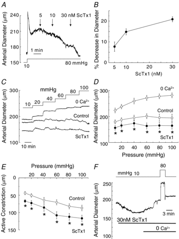
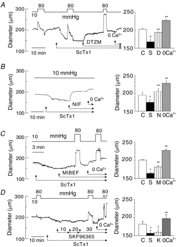
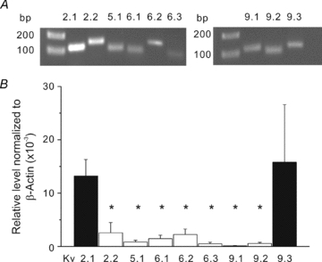
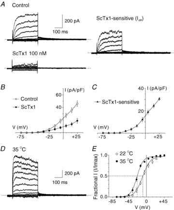
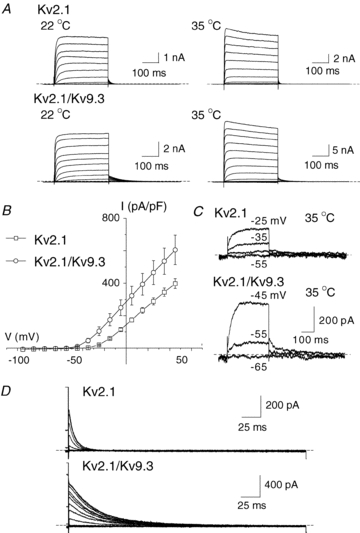
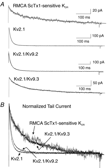
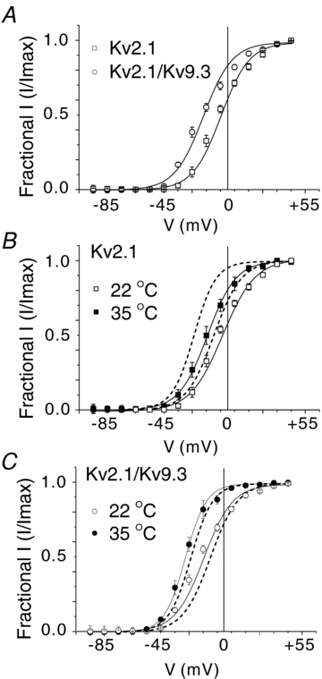
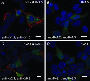
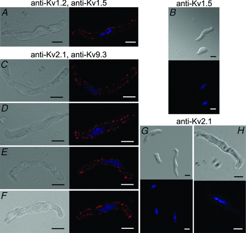
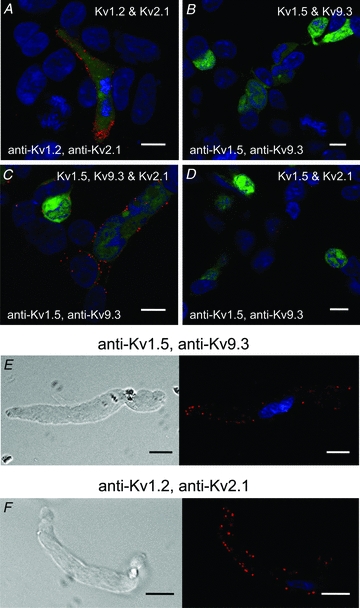
Cerebral vascular smooth muscle contractility plays a crucial role in controlling arterial diameter and, thereby, blood flow regulation in the brain. A number of K(+) channels have been suggested to contribute to the regulation of diameter by controlling smooth muscle membrane potential (E(m)) and Ca(2+) influx. Previous studies indicate that stromatoxin (ScTx1)-sensitive, Kv2-containing channels contribute to the control of cerebral arterial diameter at 80 mmHg, but their precise role and molecular composition were not determined. Here, we tested if Kv2 subunits associate with 'silent' subunits from the Kv5, Kv6, Kv8 or Kv9 subfamilies to form heterotetrameric channels that contribute to control of diameter of rat middle cerebral arteries (RMCAs) over a range of intraluminal pressure from 10 to 100 mmHg. The predominant mRNAs expressed by RMCAs encode Kv2.1 and Kv9.3 subunits. Co-localization of Kv2.1 and Kv9.3 proteins at the plasma membrane of dissociated single RMCA myocytes was detected by proximity ligation assay. ScTx1-sensitive native current of RMCA myocytes and Kv2.1/Kv9.3 currents exhibited functional identity based on the similarity of their deactivation kinetics and voltage dependence of activation that were distinct from those of homomultimeric Kv2.1 channels. ScTx1 treatment enhanced the myogenic response of pressurized RMCAs between 40 and 100 mmHg, but this toxin also caused constriction between 10 and 40 mmHg that was not previously observed following inhibition of large conductance Ca(2+)-activated K(+) (BK(Ca)) and Kv1 channels. Taken together, this study defines the molecular basis of Kv2-containing channels and contributes to our understanding of the functional significance of their expression in cerebral vasculature. Specifically, our findings provide the first evidence of heteromultimeric Kv2.1/Kv9.3 channel expression in RMCA myocytes and their distinct contribution to control of cerebral arterial diameter over a wider range of E(m) and transmural pressure than Kv1 or BK(Ca) channels owing to their negative range of voltage-dependent activation.


Figures

Figure 1
Figure 1. Enhanced RMCA myogenic response by ScTx1
A, representative recording of arterial diameter at 80 mmHg showing concentration-dependent constriction of RMCA treated with 5–30 nm ScTx1. B, mean values ± s.e.m. (n = 4) for the percentage decrease in RMCA diameter in the presence of ScTx1 at 80 mmHg. C and D, representative recordings and mean values ± s.e.m. (n = 7) of RMCA diameter between 10 and 100 mmHg under control conditions, and following 30 nm ScTx1 prior to exposure to zero Ca2+ solution to determine passive diameter at each pressure. E, mean values ± s.e.m. (n = 7) for active constriction in micrometres under control conditions and after treatment with ScTx1 (value for active constriction is the difference between control or ScTx1 diameter and passive diameter). F, representative recording of vasoconstriction evoked by ScTx1 at 10 mmHg and block of constriction at 10 and 80 mmHg in ScTx1 and zero Ca2+ solution. *Significantly different (P < 0.05) from value in control solution.
Figure 2
Figure 2. Inhibition of VGCCs but not NSCCs reversed ScTx1-evoked vasoconstriction
A, representative recording and mean values ± s.e.m. (n = 4) of RMCA diameter at 10 mmHg under control conditions and following sequential treatment with ScTx1 (30 nm), diltiazem (10 μm) and zero Ca2+ solution. Brief steps to 80 mmHg show that the extent of myogenic response was enhanced by ScTx1 and inhibited by diltiazem. B, representative recording and mean values ± s.e.m. (n = 6) of RMCA diameter at 10 mmHg under control conditions and following sequential treatment with ScTx1 (30 nm), nifedipine (1 μm) and zero Ca2+ solution. C, representative recording and mean values ± s.e.m. (n = 4) of RMCA diameter at 10 mmHg under control conditions and following sequential treatment with ScTx1 (30 nm), mibefradil (1 μm) and zero Ca2+ solution. Brief steps to 80 mmHg show that the myogenic response was inhibited by mibefradil to level observed in zero Ca2+. D, representative recording and mean values ± s.e.m. (n = 4) of RMCA diameter at 10 mmHg under control conditions and following sequential treatment with ScTx1 (30 nm), 10, 20 and 30 μm SKF96365(K) and zero Ca2+ solution. SKF96365 did not affect ScTx1-evoked constriction, and brief steps to 80 mmHg indicated that the extent of myogenic response was reduced by SKF96365 confirming effective block of NSCCs was achieved. * and ** are significantly different (P < 0.05) from value in control solution and ScTx1, respectively.
Figure 3
Figure 3. Kv2.1 and Kv9.3 are predominant transcripts expressed in RMCA
A, representative gels indicating generation of amplicons of appropriate sizes using QPCR pimer pairs for Kv2.1, Kv2.2, Kv5.1, Kv6.1–Kv6.3, Kv9.1–9.3 and mRNA extracted from rat brain. Similar results were obtained from 7 additional experiments. B, mean values ± s.e.m. (n = 4–10) for level of Kv2.1, Kv2.2, Kv5.1, Kv6.1–Kv6.3, Kv9.1–9.3 transcript expression relative to β-actin determined by QPCR and mRNA derived from RMCAs from different rats. Relative transcript levels were determined using the 2−ΔΔCt method. Specfic expression of Kv2.1 and Kv9.3 was confirmed using mRNA from isolated RMCA myocytes. *Significantly different (P < 0.05) from value for Kv2.1.
Figure 4
Figure 4. Native ScTx1-sensitive Kv current of RMCA myocytes
A, representative recordings of whole-cell Kv current of an RMCA myocyte in the absence (Control) and presence of 100 nm ScTx1 (left) and the ScTx1-sensitive current (right) determined by digital subtraction of ScTx1 from Control current at 22°C. Voltage steps of 325 ms duration between −95 and +45 mV in increments of 10 mV prior to repolarization to −45 mV were applied from a holding potential of −75 mV. Similar recordings were obtained from 7 additional myocytes from cell isolations of RMCAs of 3 rats. B and C, mean values ± s.e.m. (n = 6) for net whole-cell and ScTx1-sensitive Kv current I–V relations. D, representative recording of RMCA ScTx1-sensitive Kv current at 35°C. E, mean values ± s.e.m. for normalized tail current amplitude versus command step voltage for native current at 22 and 35°C (n = 6 and 3, respectively) that exhibited complete suppression of tail currents following treatment with ScTx1. Continuous lines represent best fits to the data points using a standard Boltzmann function.
Figure 5
Figure 5. Kv2.1 and Kv2.1/Kv9.3 currents in HEK293 cells
A, representative families of whole-cell currents due to expression of Kv2.1 (upper) and Kv2.1/Kv9.3 (lower) channels in HEK 293 cells recorded at 22 and 35°C (left and right, respectively). Voltage steps of 325 ms duration between −95 and +45 mV in increments of 10 mV prior to repolarization to −55 mV were applied from a holding potential of −75 mV. B, mean values ± s.e.m. (n = 6 and 8, respectively) for current density (pA pF−1) versus voltage relation for Kv2.1 and Kv2.1/Kv9.3 channels in HEK 293 cells at 22°C. C, representative Kv2.1 and Kv2.1/Kv9.3 currents recorded in response to command steps to between −55 and −25 mV and between −65 and −45 mV, respectively. Note the activation of current due to heteromultimeric, but not homomultimeric, channels at −55 mV. D, representative families of expanded Kv2.1 and Kv2.1/Kv9.3 tail currents recorded at −50 mV at 22°C illustrating the significantly slower deactivation kinetics of the heteromultimeric channels.
Figure 6
Figure 6. Decay of native ScTx1-sensitive Kv current mimics that of Kv2.1/Kv9.3, but not Kv2.1 or Kv2.1/Kv9.2, channels
A, expanded representative recordings of tail currents of native RMCA ScTx1-sensitive Kv, Kv2.1, Kv2.1/Kv9.2 and Kv2.1/Kv9.3 channels at −45 mV following steps to +25 mV at 22°C. Continuous lines through each recording represent the best fit using a two-exponential function. B, superimposed tail current recordings from panel A with currents for Kv2.1, Kv2.1/Kv9.2 and Kv2.1/Kv9.3 normalized to value of peak tail current of native channels.
Figure 7
Figure 7. Voltage dependence of activation of Kv2.1/Kv9.3, but not Kv2.1, channels mimics that of native Kv current
A, mean values ± s.e.m. for normalized tail current amplitude versus command step voltage for Kv2.1 and Kv2.1/Kv9.3 current at 22°C (n = 6 and 8, respectively). Note the negative shift in voltage dependence of Kv2.1/Kv9.3. B, mean values ± s.e.m. for normalized tail current amplitude versus command step voltage for Kv2.1 current at 22 and 35°C (n = 6 and 3, respectively). Dashed lines indicate the relation for activation of native current from Fig. 4E for comparison; note lack of similarity in voltage dependence of native and recombinant channel current activation at both recording temperatures. C, mean values ± s.e.m. for normalized tail current amplitude versus command step voltage for Kv2.1/Kv9.3 current at 22 and 35°C (n = 8 and 5, respectively). Dashed lines indicate the relation for activation of native current from Fig. 4E for comparison; note the similarity in voltage dependence of native and recombinant channel current activation at both recording temperatures.
Figure 8
Figure 8. PLA detection of plasma membrane expression of recombinant Kv channel protein in HEK 293 cells
A, PLA reaction product indicated by red fluorescent dots was detected at the periphery of GFP-positive HEK 293 cells transfected with cDNAs encoding GFP, Kv1.2 and Kv1.5, but not in non-transfected GFP-negative cells probed with Kv1.2 and Kv1.5 primary antibodies. Here and in subsequent panels, the nuclei of GFP-positive and -negative cells are indicated by the blue Hoechst 33342 stain, the Kv channel cDNAs and primary antibodies used are indicated in the upper right and lower left corners, respectively, the scale bars are 10 μm in length and each image is an optical section of 0.3–0.5 μm thickness at a mid-cell depth. B, lack of PLA reaction product at cell periphery of GFP-positive cells transfected with Kv1.5 only (i.e. no Kv1.2) and probed with Kv1.2 and Kv1.5 primary antibodies. C, PLA signals were detected at the periphery of GFP-positive cells transfected with Kv2.1 and Kv9.3, but not in GFP-negative cells probed with Kv2.1 and Kv9.3 primary antibodies. D, lack of PLA signals at cell periphery of GFP-positive cells transfected with Kv2.1 (i.e. no Kv9.3) and probed with Kv2.1 and Kv9.3 primary antibodies.
Figure 9
Figure 9. PLA detection of plasma membrane expression of Kv channel protein in RMCA myocytes
A, DIC (left) and fluorescence (right) micrographs of an RMCA myocyte probed for co-localization of Kv1.2 and Kv1.5 with PLA signals at cell periphery when probed with Kv1.2 and Kv1.5 primary antibodies. Here and in subsequent panels, the nuclei of GFP-positive and -negative cells are indicated by the blue Hoechst 33342 stain, the primary antibodies used are indicated above the panels, the scale bars are 10 μm in length and each image is an optical section of 0.3–0.5 μm thickness at a mid-cell depth. B, lack of PLA signals in two myocytes probed for Kv1.2 and Kv1.5 co-localization when Kv1.2 primary antibody was omitted. C–F, four representative RMCA myocytes exhibiting PLA signals when probed with Kv2.1 and Kv9.3 primary antibodies. G, lack of PLA signals in three myocytes probed for Kv2.1 and Kv9.3 co-localization when Kv9.3 primary antibody was omitted. H, single RMCA myocyte at high magnification showing lack of PLA signals for Kv2.1 and Kv9.3 co-localization when Kv9.3 primary antibody was omitted.
Figure 10
Figure 10. PLA detection of Kv subunit proteins in adjacent channel complexes in HEK 293 cells and RMCA myocytes
A, PLA signals were detected at the periphery of GFP-positive cells transfected with Kv1.2 and Kv2.1, but not in GFP-negative cells probed with Kv1.2 and Kv2.1 primary antibodies. B, PLA signals were not detected at the periphery of GFP-positive cells transfected with Kv1.5 and Kv9.3 and probed with Kv1.5 and Kv9.3 primary antibodies. C, PLA signals were detected at the periphery of GFP-positive cells transfected with Kv1.5, Kv9.3 and Kv2.1, but not in GFP-negative cells probed with Kv1.5 and Kv9.3 primary antibodies. D, PLA signals were not detected at cell periphery of GFP-positive cells transfected with Kv1.5 and Kv2.1 and probed with Kv1.5 and Kv9.3 primary antibodies. E, representative RMCA myocyte exhibiting PLA signals at cell periphery when probed with Kv1.5 and Kv9.3 primary antibodies. F, representative RMCA myocyte exhibiting PLA signals at cell periphery when probed with Kv1.2 and Kv2.1 primary antibodies.

Similar articles

Cited by

References

    1. Albarwani S, Nemetz LT, Madden JA, Tobin AA, England SK, Pratt PF, Rusch NJ. Voltage-gated K+ channels in rat small cerebral arteries: molecular identity of the functional channels. J Physiol. 2003;551:751–763. - PMC - PubMed
    1. Amberg GC, Rossow CF, Navedo MF, Santana LF. NFATc3 regulates Kv2.1 expression in arterial smooth muscle. J Biol Chem. 2004;279:47326–47334. - PubMed
    1. Amberg GC, Santana LF. Kv2 channels oppose myogenic constriction of rat cerebral arteries. Am J Physiol Cell Physiol. 2006;291:C348–C356. - PubMed
    1. Belevych AE, Beck R, Tammaro P, Poston L, Smirnov SV. Developmental changes in the functional characteristics and expression of voltage-gated K+ channel currents in rat aortic myocytes. Cardiovasc Res. 2002;54:152–161. - PubMed
    1. Bocksteins E, Labro AJ, Mayeur E, Bruyns T, Timmermans JP, Adriaensen D, Snyders DJ. Conserved negative charges in the N-terminal tetramerization domain mediate efficient assembly of Kv2.1 and Kv2.1/Kv6.4 channels. J Biol Chem. 2009;284:31625–31634. - PMC - PubMed

Publication types