Comparative Study
. 2010 Nov 15;588(Pt 22):4579-91.
doi: 10.1113/jphysiol.2010.193243. Epub 2010 Sep 27.

Angio-adaptation in unloaded skeletal muscle: new insights into an early and muscle type-specific dynamic process

Affiliations
Comparative Study

Angio-adaptation in unloaded skeletal muscle: new insights into an early and muscle type-specific dynamic process

Emilie Roudier et al. J Physiol. .

Abstract

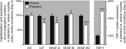
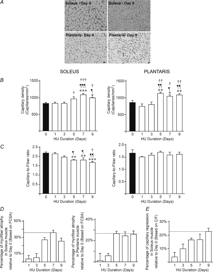
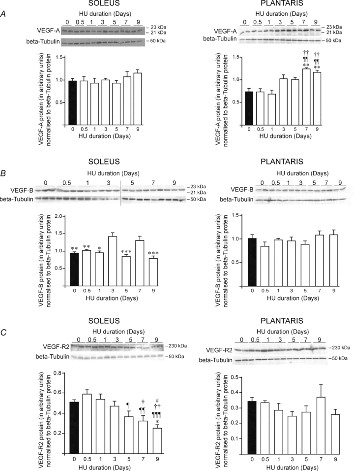
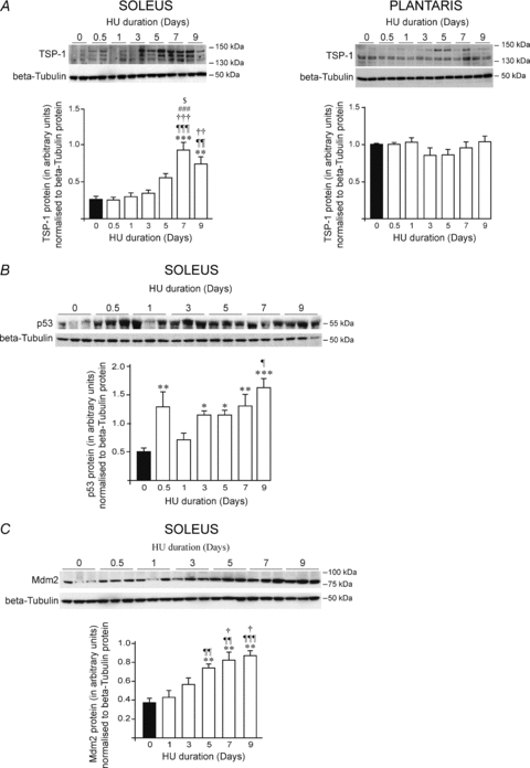
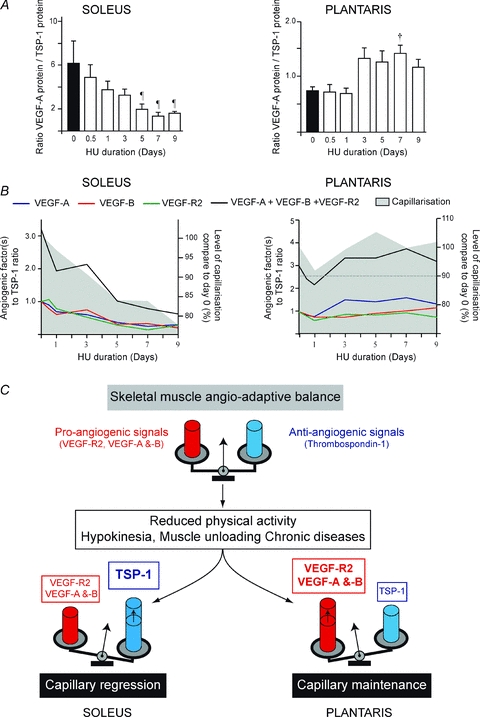
With a remarkable plasticity, skeletal muscle adapts to an altered functional demand. Muscle angio-adaptation can either involve the growth or the regression of capillaries as respectively observed in response to endurance training or muscle unloading. Whereas the molecular mechanisms that regulate exercise-induced muscle angiogenesis have been extensively studied, understanding how muscle unloading can in contrast lead to capillary regression has received very little attention. Here we have investigated the consequences of a 9 day time course hindlimb unloading on both capillarization and expression of angio-adaptive molecules in two different rat skeletal muscles. Both soleus and plantaris muscles were atrophied similarly. In contrast, our results have shown different angio-adaptive patterns between these two muscles. Capillary regression occurred only in the soleus, a slow-twitch and oxidative postural muscle. Conversely, the level of capillarization was preserved in the plantaris, a fast-twitch and glycolytic muscle. We have also measured the time course protein expression of key pro- and anti-angiogenic signals (VEGF-A, VEGF-B, VEGF-R2, TSP-1). Our results have revealed that the angio-adaptive response to unloading was muscle-type specific, and that an integrated balance between pro- and anti-angiogenic signals plays a determinant role in regulating this process. In conclusion, we have brought new evidence that measuring the ratio between pro- and anti-angiogenic signals in order to evaluate muscle angio-adaptation was a more accurate approach than analysing the expression of molecular factors taken individually.


Figures

Figure 1
Figure 1. Muscle-type differences in capillarization and angio-adaptive proteins
Relative levels of capillarization and angio-adaptive proteins in the plantaris muscle compared to the soleus muscle, under control conditions (i.e. day 0). Values have been normalized to soleus levels. Data are expressed as means ± s.e.m. (n = 6 rats/group). Significant difference between soleus and plantaris muscles: *P ≤ 0.05; **P ≤ 0.01; ***P ≤ 0.001.
Figure 2
Figure 2. Hindlimb unloading and skeletal muscle capillarization
A, representative images of capillaries staining in soleus (left) and plantaris (right) muscles from control animals (day 0, top panel) and after 9 days of hindlimb unloading (day 9, bottom panel). Scale bar, 50 μm. B–C, quantitative measurement of capillary density (B) and capillary-to-fibre ratio (C) in soleus (left panel) and plantaris (right panel) muscles following time course hindlimb unloading (HU, from days 0 to 9). Data are expressed as means ± s.e.m. (n = 6 rats/group). Significantly different from day 0: *P ≤ 0.05; **P ≤ 0.01; ***P ≤ 0.001. Significantly different from day 1: ¶P ≤ 0.05; ¶¶P ≤ 0.01; ¶¶¶P ≤ 0.001. Significantly different from day 3: †P ≤ 0.05; ††P ≤ 0.01; †††P ≤ 0.001. Significantly different from day 5: #P ≤ 0.05. Percentage of muscle atrophy (D) and capillary regression (E) based on mean FCSA and C/F values, respectively. Data are expressed as means ± s.e.m. (n = 6 rats/group). FCSA, mean fibre cross-sectional area; C/F, capillary-to-fibre ratio.
Figure 3
Figure 3. VEGF-A, VEGF-B and VEGF receptor-2 protein expression in response to hindlimb unloading
Animals were subjected to a 9-day time course hindlimb unloading. Protein levels were measured by western blotting in soleus and plantaris muscles, left and right panels respectively. β-Tubulin protein was used as a loading control. Top-panels of A, B and C show images of VEGF-A, VEGF-B and VEGF-R2 protein expressions, respectively. Bottom panels of A, B and C show densitometric analyses of VEGF-A, VEGF-B or VEGF-R2 proteins relative to β-tubulin. Densitometric analyses show means ± s.e.m. (n = 6 rats/group). Significant differences: vs. day 0 **P ≤ 0.01; ***P ≤ 0.001; vs. day 0.5: ¶P ≤ 0.05; ¶¶P ≤ 0.01; ¶¶¶P ≤ 0.001; vs. day 1: †P ≤ 0.05; ††P ≤ 0.01; †††P ≤ 0.001; vs. day 3 #P ≤ 0.05; ##P ≤ 0.01; ##P ≤ 0.001; vs. day 7 ‡‡P ≤ 0.01.
Figure 4
Figure 4. TSP-1, p53 and Mdm2 protein expressions in response to hindlimb unloading
Animals were subjected to a 9 day time course hindlimb unloading. Protein levels were measured by Western blotting in soleus and plantaris muscles. β-Tubulin protein was used as a loading control. Top-panels of A, B and C show representative pictures of TSP-1, p53 and Mdm2 protein expressions, respectively. Bottom panels of A, B and C show densitometric analyses of TSP-1, p53 or Mdm2 proteins relative to β-tubulin. Data are means ± s.e.m. (n = 6 rats/group). Significant differences are vs. day 0 *P≤0.05; **P ≤ 0.01; ***P ≤ 0.001; vs. day 0.5 ¶P ≤ 0.05; ¶¶P ≤ 0.01; ¶¶¶P ≤ 0.001; vs. day 1 †P ≤ 0.05; ††P ≤ 0.01; †††P ≤ 0.001; vs. day 3 #P ≤ 0.05; ###P ≤ 0.001; and vs. day 5, $P ≤ 0.05.
Figure 5
Figure 5. Hindlimb unloading and the angioadaptive balance in soleus and plantaris muscles
The ratios between the relative expression of pro-angiogenic proteins (VEGF-A, VEGF-B and VEGF-R2) and the anti-angiogenic TSP-1 proteins were calculated during the time course of hindlimb unloading. A shows VEGF-A to TSP-1 ratio over the time of hindlimb unloading in soleus and plantaris muscles (left and right panels, respectively). Data are means ± s.e.m. (n = 6 rats/group). Significant differences are vs. day 0.5, ¶P ≤ 0.05; and vs. day 1 †P ≤ 0.05. B illustrates the time course of changes in muscle capillarization and in the ratios VEGF-A, VEGF-B, VEGF-R2, or the sum of all pro-angiogenic proteins measured (VEGF-A+VEGF-B+VEGF-R2), relative to TSP-1 in soleus and plantaris (left and right panels, respectively). Ratios at different HU time points were normalized to day 0 (equal to 1.0 arbitrary unit). Muscle capillarization at different HU time points is expressed as a percentage of capillarization at day 0. C, schematic illustration of how the angio-adaptative balance is differently regulated between soleus and plantaris muscles in response to hindlimb unloading.

Similar articles

Cited by

References

    1. Alway SE, Lowe DA, Chen KD. The effects of age and hindlimb supension on the levels of expression of the myogenic regulatory factors MyoD and myogenin in rat fast and slow skeletal muscles. Exp Physiol. 2001;86:509–517. - PubMed
    1. An WG, Kanekal M, Simon MC, Maltepe E, Blagosklonny MV, Neckers LM. Stabilization of wild-type p53 by hypoxia-inducible factor 1α. Nature. 1998;392:405–408. - PubMed
    1. Bellomo D, Headrick JP, Silins GU, Paterson CA, Thomas PS, Gartside M, Mould A, Cahill MM, Tonks ID, Grimmond SM, Townson S, Wells C, Little M, Cummings MC, Hayward NK, Kay GF. Mice lacking the vascular endothelial growth factor-B gene (Vegfb) have smaller hearts, dysfunctional coronary vasculature, and impaired recovery from cardiac ischemia. Circ Res. 2000;86:E29–35. - PubMed
    1. Blagosklonny MV, Giannakakou P, Wojtowicz M, Romanova LY, Ain KB, Bates SE, Fojo T. Effects of p53-expressing adenovirus on the chemosensitivity and differentiation of anaplastic thyroid cancer cells. J Clin Endocrinol Metab. 1998;83:2516–2522. - PubMed
    1. Booth FW, Thomason DB. Molecular and cellular adaptation of muscle in response to exercise: perspectives of various models. Physiol Rev. 1991;71:541–585. - PubMed

Publication types

LinkOut - more resources