. 2010 Dec 1;588(Pt 23):4785-94.
doi: 10.1113/jphysiol.2010.196568. Epub 2010 Sep 27.

Lactate per se improves the excitability of depolarized rat skeletal muscle by reducing the Cl- conductance

Affiliations

Lactate per se improves the excitability of depolarized rat skeletal muscle by reducing the Cl- conductance

Frank Vincenzo de Paoli et al. J Physiol. .

Abstract

Studies on rats have shown that lactic acid can improve excitability and function of depolarized muscles. The effect has been related to the ensuing reduction in intracellular pH causing inhibition of muscle fibre Cl(-) channels. However, since several carboxylic acids with structural similarities to lactate can inhibit muscle Cl(-) channels it is possible that lactate per se can increase muscle excitability by exerting a direct effect on these channels. We therefore examined the effects of lactate on the function of intact muscles and skinned fibres together with effects on pH and Cl(-) conductance (G(cl)). In muscles where extracellular compound action potentials (M-waves) and tetanic force response to excitation were reduced by (mean ± s.e.m.) 82 ± 4% and 83 ± 2%, respectively, by depolarization with 11 mm extracellular K(+), both M-waves and force exhibited an up to 4-fold increase when 20 mm lactate was added. This effect was present already at 5 mm and saturated at 15 mm lactate, and was associated with a 31% reduction in G(Cl). The effects of lactate were completely blocked by Cl(-) channel inhibition or use of Cl(-)-free solutions. Finally, both experiments where effects of lactate on intracellular pH in intact muscles were mimicked by increased CO₂ tension and experiments with skinned fibres showed that the effects of lactate could not be related to reduced intracellular pH. It is concluded that addition of lactate can inhibit ClC-1 Cl(-) channels and increase the excitability and contractile function of depolarized rat muscles via mechanisms not related to a reduction in intracellular pH.


Figures
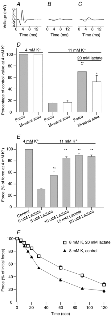
Figure 1
Figure 1. Effect of lactate on M-wave and contractile properties in K+-depressed soleus muscles
A–E, after determination of control tetanic force at 4 mm K+, the intact soleus muscles were incubated for 90 min in control solution with 11 mm K+ and then for 30 min in solution with 11 mm K+ and 20 mm lactate. Tetanic contractions were elicited every 10 min using 60 Hz pulse trains of 2 s duration. Representative M-wave traces from a single soleus muscle recorded during contractions at the end of the incubation period at 4 mm K+ (A), 11 mm K+ (B) and 11 mm K+ with 20 mm lactate (C). D, tetanic force and M-wave area at end of the incubation at 4 mm K+, 11 mm K+, and 11 mm K+ with 20 mm lactate, as indicated by bars (n= 6). E, effect of lactate concentration on tetanic force in soleus muscles at 11 mm K+ (n= 6–29). F, force production in muscles pre-equilibrated at 8 mm K+ for 60 min after which a prolonged contraction was induced by 120 s of 60 Hz continuous stimulation in the presence (open squares) or absence (filled triangles) of 20 mm lactate (n= 4). In all panels the data show means and s.e.m. Significantly different from the corresponding value at 4 mm K+: *P < 0.05, **P < 0.01.
Figure 2
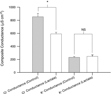
Figure 2. Effect of lactate on the specific membrane conductance for Cl and K+ in soleus muscles
Muscles were incubated in 9 mm K+ with or without 20 mm lactate and with or without Cl for 60 min before the membrane conductance was determined. K+ conductance is the membrane conductance in the absence of Cl. Cl conductance is calculated by subtracting the membrane conductance in the absence of Cl from the total membrane conductance determined in soleus fibres incubated in control solution. Data are shown as means with s.e.m. (n= 16–33 muscle fibres). *Significant difference between values obtained with and without lactate, P < 0.05. NS, not significantly different.
Figure 3
Figure 3. Effect of lactate and increased CO2 on pHi and force in soleus muscles
A and B, representative traces showing pHi in muscles exposed to 20 mm lactate or increased CO2 (from 5% to 7%): before exposure to 20 mm lactate (a), the lowest pHi after exposure to lactate (b), and pHi after 45 min of exposure to lactate (c). C, summarized values for pHi in soleus muscles exposed to 20 mm lactate or increased CO2. Data show means and s.e.m. (n= 4–6). *Significantly different from pHi at the beginning of experiments. D, effect of 7% CO2 and 20 mm lactate on tetanic force in soleus muscles depolarized by incubation at 11 mm K+. Data show means and s.e.m. (n= 7–8). *P < 0.05 and **P < 0.01, significantly different from force in soleus muscles incubated in control solution with 11 mm K+. E, effect of 20 mm lactate on intracellular Na+ content and 86Rb+ uptake. Two groups of soleus muscles were pre-incubated in control solution with 11 mm K+. After 30 min 20 mm lactate was added to one group of soleus muscles and Na+ content and 86Rb+ uptake was assessed from 0 to 10 min and from 20 to 30 min of the subsequent incubation. Data show means and s.e.m. (n= 6).
Figure 4
Figure 4. Effect of lactate and T-system depolarization on the force response of skinned fibres from EDL muscles
A, representative trace showing the effect of 10 mm lactate (indicated by bars) on twitch and tetanic force in a skinned muscle fibre when incubated in the intracellular control solution (con) and when depolarized by incubation in solutions with 75 and 60 mm K+. Tetani were elicited by 25 Hz pulse trains of 2 s duration. At the end of the protocol a prolonged contraction was elicited by exposure of the fibre to maximally Ca2+-activating solution. B, summarized data showing the effect of 10 mm lactate on maximal twitch and tetanic force in skinned muscle fibres incubated in the intracellular control solution or depolarized by incubation in solutions with 75 or 60 mm K+. *Force in the presence of lactate is significantly higher than without lactate, P < 0.05. C and D, summarized data showing the effect of 10 mm lactate on skinned fibres depolarized by incubation at 75 mm K+ in a Cl-free solution (C) or in the presence of 100 μm 9-AC (D). Data show means and s.e.m. (n= 3–9). NS, not significant.

Similar articles

Cited by

References

    1. Andrews MA, Godt RE, Nosek TM. Influence of physiological l(+)-lactate concentrations on contractility of skinned striated muscle fibers of rabbit. J Appl Physiol. 1996;80:2060–2065. - PubMed
    1. Brooks GA. Lactate: link between glycolytic and oxidative metabolism. Sports Med. 2007;37:341–343. - PubMed
    1. Brooks GA. Cell–cell and intracellular lactate shuttles. J Physiol. 2009;587:5591–5600. - PMC - PubMed
    1. Buchanan R, Nielsen OB, Clausen T. Excitation- and β2-agonist-induced activation of the Na+–K+ pump in rat soleus muscle. J Physiol. 2002;545:229–240. - PMC - PubMed
    1. Cairns SP. Lactic acid and exercise performance: culprit or friend? Sports Med. 2004;36:279–291. - PubMed

Publication types

LinkOut - more resources