. 2011 Jan;25(1):301-13.
doi: 10.1096/fj.10-166454. Epub 2010 Sep 27.

The coordinated increased expression of biliverdin reductase and heme oxygenase-2 promotes cardiomyocyte survival: a reductase-based peptide counters β-adrenergic receptor ligand-mediated cardiac dysfunction

Affiliations

The coordinated increased expression of biliverdin reductase and heme oxygenase-2 promotes cardiomyocyte survival: a reductase-based peptide counters β-adrenergic receptor ligand-mediated cardiac dysfunction

Bo Ding et al. FASEB J. 2011 Jan.

Abstract

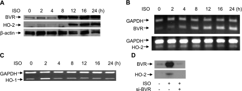
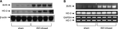
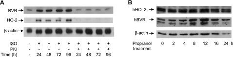
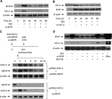
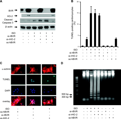
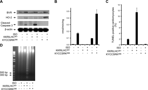
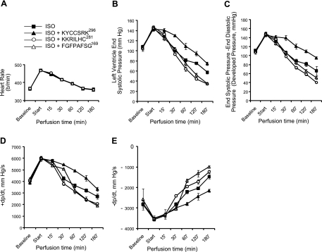
HO-2 oxidizes heme to CO and biliverdin; the latter is reduced to bilirubin by biliverdin reductase (BVR). In addition, HO-2 is a redox-sensitive K/Ca(2)-associated protein, and BVR is an S/T/Y kinase. The two enzymes are components of cellular defense mechanisms. This is the first reporting of regulation of HO-2 by BVR and that their coordinated increase in isolated myocytes and intact heart protects against cardiotoxicity of β-adrenergic receptor activation by isoproterenol (ISO). The induction of BVR mRNA, protein, and activity and HO-2 protein was maintained for ≥ 96 h; increase in HO-1 was modest and transient. In isolated cardiomyocytes, experiments with cycloheximide, proteasome inhibitor MG-132, and siBVR suggested BVR-mediated stabilization of HO-2. In both models, activation of BVR offered protection against the ligand's stimulation of apoptosis. Two human BVR-based peptides known to inhibit and activate the reductase, KKRILHC(281) and KYCCSRK(296), respectively, were tested in the intact heart. Perfusion of the heart with the inhibitory peptide blocked ISO-mediated BVR activation and augmented apoptosis; conversely, perfusion with the activating peptide inhibited apoptosis. At the functional level, peptide-mediated inhibition of BVR was accompanied by dysfunction of the left ventricle and decrease in HO-2 protein levels. Perfusion of the organ with the activating peptide preserved the left ventricular contractile function and was accompanied by increased levels of HO-2 protein. Finding that BVR and HO-2 levels, myocyte apoptosis, and contractile function of the heart can be modulated by small human BVR-based peptides offers a promising therapeutic approach for treatment of cardiac dysfunctions.


Figures

Figure 1.
Figure 1.
ISO increases expression of BVR and HO-2 in rat neonatal cardiomyocytes. A) Effects of ISO on HO-2 and BVR protein levels. Cells were treated with 10 μM ISO (ISO) for the times indicated and lysed; proteins were resolved by SDS-PAGE. Western blot was probed sequentially with antibodies to rat BVR, rat HO-2, and β-actin, as the control. B) HO-2 and BVR mRNA expression (RT-PCR) in ISO-stimulated rat neonatal cardiomyocytes. RNA was isolated at the indicated times and reverse transcribed. The rat BVR and HO-2 sequences were amplified by PCR, and products were resolved by agarose gel electrophoresis. GAPDH was included as a control. C) ISO causes a transient induction of HO-1. Cardiomyocytes were treated with 10 μM ISO for the times indicated; RNA was isolated, and the levels of HO-1 and GAPDH mRNAs determined as in B. D) Induction of HO-2 synthesis by ISO requires induction of BVR. Cardiomyocytes were treated with siBVR retrovirus, and 12 h later with 10 μM ISO for 24 h; siBVR was replenished every 12 h; proteins were resolved by gel electrophoresis and analyzed by Western blot.
Figure 2.
Figure 2.
Increased expression of BVR and HO-2 after continuous infusion of ISO in vivo. A) Levels of HO-2 and BVR protein in hearts from 4 individual rats after 4 d of continuous infusion with 400 μg ISO/kg/h, or from 4 sham-operated rats were determined by Western blot. β-Actin is included as control. B) HO-2 and BVR mRNA expression in ISO-infused rat heart in vivo, determined by RT-PCR, as in Fig. 1. Animals were infused with ISO, as in A. GAPDH is included as a control.
Figure 3.
Figure 3.
Increase in BVR and HO-2 protein levels is mediated via PKA. A) Cardiomyocytes were infected with an adenovirus expressing PKA-I or a control gene (lacZ), then treated with 10 μM ISO for the indicated times. BVR and HO-2 were detected in cell lysates by a Western blot, using β-actin as a control. B) HEK293A cells were treated with 10 μM propranolol for the times indicated and lysed; cell lysates were analyzed by gel electrophoresis and Western blotting; blot was sequentially probed with antibodies to HO-2, BVR, and the control β-actin.
Figure 4.
Figure 4.
Ablation of BVR protein prevents ISO-induced HO-2 protein expression. A) Cardiomyocytes were infected with retrovirus expressing siBVR, 6 h later, cells were treated with 10 μM ISO. Samples were collected at 24, 48, 72, and 96 h posttreatment. Cell lysates were analyzed by gel electrophoresis and Western blot analysis. Blots were probed with antibodies against rat BVR and HO-2. β-Actin served as a control. B) Cardiomyocytes were infected with a retrovirus expressing siHO-2. Expression of BVR and HO-2 was measured as in A. C) BVR expression regulates the stability of the HO-2 protein. In the first experiment, HEK293A cells were cotransfected with pcDNA-hBVR and pcDNA-hHO-2, then stimulated with ISO for 24 h, followed by treatment with the protein synthesis inhibitor, cycloheximide (10 μg/ml), for the indicated intervals. Expression of hHO-2 and hBVR in HEK293A cells was measured by Western blot. In the second series, the cells were transfected with pcDNA-hHO-2 and treated with retrovirus expressing sihBVR. These data were replicated in ≥2 independent experiments. D) Cardiomyocytes were treated with 10 μM ISO, infected with siBVR virus, or treated with the proteasome inhibitor MG132 (50 μg/ml). HO-2 and BVR protein expression were detected by Western blot. β-Actin served as a control.
Figure 5.
Figure 5.
Inhibition of BVR and HO-2 protein expression during ISO stimulation increases cardiomyocyte apoptosis. A) Cardiomyocytes were treated with siRNA against rat BVR, HO-2, or both, after which ISO was added at a final concentration of 10 μM, and the cells were incubated for 24 h. A randomized form of sihBVR (scBVR) was added to some cells as a control. Expression of BVR, HO-2, and cleaved caspase-3 was determined by Western blot. β-Actin served as a loading control. B) Quantitative analysis of the number of apoptotic cells detected, in cultures treated with siBVR, siHO-2, or both; scBVR served as control. Number of TUNEL-positive cells that also stained with EA-53 anti-α-actinin antibody was measured as a fraction of all cardiomyocytes. C) Detection of apoptosis in cardiomyocytes by microscopy. Cardiomyocytes were characterized by their staining with antibody against α-actinin. Apoptosis was detected by TUNEL staining; nuclei were visualized with DAPI. D) Total genomic DNA was isolated from cardiomyocytes that had been treated with ISO and siBVR, siHO-2, or both. Fragmentation of nuclear DNA is detectable in ISO-treated cardiomyocytes that were also treated with siBVR, siHO-2, or both. The 1-kb-plus ladder was used as DNA marker.
Figure 6.
Figure 6.
Peptide-mediated inhibition of BVR activation in perfused heart increases cardiomyocyte apoptosis, whereas BVR activation is protective. A) Three groups of 4 rats were injected subcutaneously with ISO (0.01 mg/kg), twice, with a 6-h interval between injections prior to perfusion of the isolated hearts. As indicated, isolated hearts were perfused with the peptides (25 μM) KKRILHC281 or KYCCSRK296 in the presence or absence of 0.1 μM ISO, for 3 h. Levels of BVR, HO-2, and cleaved caspase-3 were determined by Western blot, using β-actin as a control for equal loading. B) BVR activity was measured at pH 6.7, using NADH as cofactor. Rate of conversion of biliverdin to bilirubin was determined from the increase in absorbance at 450 nm at 25°C. Specific activity is expressed as nanomoles of bilirubin per minute per milligram of protein. C) Detection of apoptosis in perfused heart cardiomyocytes. Tissue from the perfused hearts was analyzed using the TUNEL assay, as described in Fig. 5B. D) Genomic DNA was isolated from perfused heart, and resolved by agarose gel electrophoresis; fragmentation is detectable only in hearts perfused with ISO and the inhibitory peptide, KKRILHC281. The 1-kb-plus ladder DNA was used as a marker.
Figure 7.
Figure 7.
Inhibition of BVR activation exacerbates ISO-mediated decline in the contractile function of the heart; BVR activation protects against loss of heart function. A) Heart rate was measured during perfusion with ISO and peptides (25 μM) KKRILHC281 (n=4), KYCCSRK296 (n=4) or FGFPAFSG169 (n=3), or with ISO alone (n=7). B) Left ventricular systolic pressure was measured simultaneously with heart rate shown in A. C) Developed pressure, calculated as the difference between the end systolic pressure and end diastolic pressure for the left ventricle, for the same animals as in A. D, E) First derivatives of left ventricular systolic pressure during compression, dp/dt (D), and relaxation −dp/dt (E), for the data shown in A–C.

Similar articles

Cited by

References

    1. Olivetti G., Abbi R., Quaini F., Kajstura J., Cheng W., Nitahara J. A., Quaini E., Di Loreto C., Beltrami C. A., Krajewski S., Reed J. C., Anversa P. (1997) Apoptosis in the failing human heart. N. Engl. J. Med. 336, 1131–1141 - PubMed
    1. Tomita H., Nazmy M., Kajimoto K., Yehia G., Molina C. A., Sadoshima J. (2003) Inducible cAMP early repressor (ICER) is a negative-feedback regulator of cardiac hypertrophy and an important mediator of cardiac myocyte apoptosis in response to beta-adrenergic receptor stimulation. Circ. Res. 93, 12–22 - PubMed
    1. Zaugg M., Xu W., Lucchinetti E., Shafiq S. A., Jamali N. Z., Siddiqui M. A. (2000) Beta-adrenergic receptor subtypes differentially affect apoptosis in adult rat ventricular myocytes. Circulation 102, 344–350 - PubMed
    1. Bristow M. R., Ginsburg R., Umans V., Fowler M., Minobe W., Rasmussen R., Zera P., Menlove R., Shah P., Jamieson S., Stinson E. B. (1986) Beta 1- and beta 2-adrenergic-receptor subpopulations in nonfailing and failing human ventricular myocardium: coupling of both receptor subtypes to muscle contraction and selective beta 1-receptor down-regulation in heart failure. Circ. Res. 59, 297–309 - PubMed
    1. Del Monte F., Kaumann A. J., Poole-Wilson P. A., Wynne D. G., Pepper J., Harding S. E. (1993) Coexistence of functioning beta 1- and beta 2-adrenoceptors in single myocytes from human ventricle. Circulation 88, 854–863 - PubMed

Publication types

MeSH terms

LinkOut - more resources