. 2011 Jan;25(1):348-57.
doi: 10.1096/fj.10-169789. Epub 2010 Sep 27.

PECAM-targeted delivery of SOD inhibits endothelial inflammatory response

Affiliations

PECAM-targeted delivery of SOD inhibits endothelial inflammatory response

Vladimir V Shuvaev et al. FASEB J. 2011 Jan.

Abstract

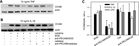
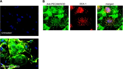
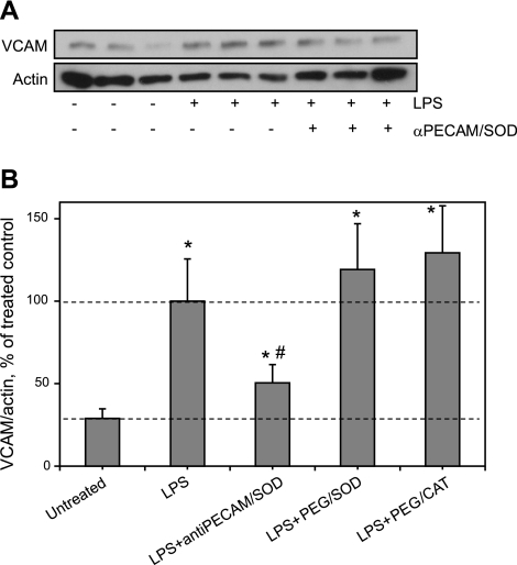
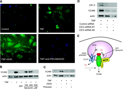
Elevated generation of reactive oxygen species (ROS) by endothelial enzymes, including NADPH-oxidase, is implicated in vascular oxidative stress and endothelial proinflammatory activation involving exposure of vascular cell adhesion molecule-1 (VCAM-1). Catalase and superoxide dismutase (SOD) conjugated with antibodies to platelet/endothelial cell adhesion molecule 1 (PECAM-1) bind specifically to endothelium and inhibit effects of corresponding ROS, H(2)O(2), and superoxide anion. In this study, anti-PECAM/SOD, but not anti-PECAM/catalase or nontargeted enzymes, including polyethylene glycol (PEG)-SOD, inhibited 2- to 3-fold VCAM expression caused by tumor necrosis factor (TNF), interleukin-1β, and lipopolysaccharide. Anti- PECAM/SOD, but not nontargeted counterparts, accumulated in vascular endothelium after intravenous injection, localized in endothelial endosomes, and inhibited by 70% lipopolysaccharide-caused VCAM-1 expression in mice. Anti-PECAM/SOD colocalized with EEA-1-positive endothelial vesicles and quenched ROS produced in response to TNF. Inhibitors of NADPH oxidase and anion channel ClC3 blocked TNF-induced VCAM expression, affirming that superoxide produced and transported by these proteins, respectively, mediates inflammatory signaling. Anti-PECAM/SOD abolished VCAM expression caused by poly(I:C)-induced activation of toll-like receptor 3 localized in intracellular vesicles. These results directly implicate endosomal influx of superoxide in endothelial inflammatory response and suggest that site-specific interception of this signal attained by targeted delivery of anti-PECAM/SOD into endothelial endosomes may have anti-inflammatory effects.


Figures

Figure 1.
Figure 1.
Kinetics of endothelial cell activation by proinflammatory agents. A–C) HUVECs were treated with TNF (10 ng/ml; A), IL-1β (10 ng/ml; B), or LPS (0.5 μg/ml; C) for indicated times, and samples were subjected to Western blot analysis of VCAM. D) Quantification of VCAM expression level normalized by actin and to TNF effect according to relative extent of VCAM expression (inset).
Figure 2.
Figure 2.
Anti-PECAM/SOD inhibits VCAM expression in endothelial cells activated by cytokines and LPS. A, B) Confluent human endothelial cells were treated with native SOD or catalase vs. anti-PECAM/SOD or anti-PECAM/catalase for 1 h before the addition of TNF (A), IL-1β (B), or LPS. Four hours later, VCAM expression level was assessed by Western blot analysis, with normalization per actin level in the samples. Representative images are shown. C) Summarized results of ≥3 independent experiments. Data are shown as percentage of maximal activation by cytokine or LPS alone in control cells (n≥3). *P < 0.05.
Figure 3.
Figure 3.
Endothelial endocytosis and endosomal delivery of anti-PECAM/SOD. A) Binding and uptake of anti-PECAM/SOD. Cells were treated with anti-PECAM/SOD for 1 h, washed, fixed, stained with Alexa Fluor 594-labeled antibody to rat IgG (common component of all conjugates used in the study), permeabilized, and stained with Alexa Fluor 488-labeled antibody to rat IgG. In such a dual-labeling assay, green and yellow colors depict internalized and surface-bound conjugates, respectively. Nuclei are stained with DAPI (blue). B) Endosomal localization of anti-PECAM/SOD. Cells were pretreated with the conjugate for 15 min, fixed, permeabilized, and stained for anti-PECAM/SOD (green), endosomal marker EEA-1 (red), and nuclei (DAPI, blue).
Figure 4.
Figure 4.
Pulmonary localization of anti-PECAM/SOD in vivo. Anesthetized mice were injected intravenously with 150 μg of anti-PECAM/SOD or control IgG/SOD. At 1 h postinjection, lungs were harvested and fixed, and tissue cryosections were immunostained as described in Materials and Methods. Colocalization of anti-PECAM/SOD (A) and IgG/SOD (B) conjugates with endosomal marker EEA-1. Scale bars = 30 μm. Arrows on phase-contrast images indicate capillaries.
Figure 5.
Figure 5.
Anti-PECAM/SOD inhibits elevation of pulmonary VCAM in LPS-challenged animals. Anti-PECAM/SOD or PEG-modified enzymes were injected in tail vein followed by LPS challenge (50 μg/kg). After 5 h, lungs were harvested for VCAM level analysis by Western blot (A; representative image is shown) and normalized per actin level for quantitative analysis, presented as a percentage of maximal LPS-induced VCAM level (B; analysis of ≥3 independent experiments); n ≥ 3. *P < 0.05 vs. untreated group; #P < 0.05 vs. LPS group.
Figure 6.
Figure 6.
Mechanism of anti-inflammatory effects of anti-PECAM/SOD. A) Anti-PECAM/SOD, but not naked SOD, quenches endosomal superoxide. Endothelial cells were pretreated with membrane nonpermeable dye OxyBURST Green and SOD (bottom left panel) or anti-PECAM/SOD (bottom right panel) and activated with TNF (except top left panel). Cells were fixed, and endosomal ROS generation was visualized by fluorescence microscopy. B–D) Nox2 is the source and ClC3 is the transmitter for endosomal superoxide in TNF-induced endothelial expression of VCAM (analysis by Western blot analysis as in Fig. 1). B) Inhibition of NADPH oxidases by DPI (20 μM) and apocynin (100–500 μM). C) Inhibition of anion channels by DIDS (25 μM) and phloretin (30 μM). D) Inhibition by ClC3 silencing by two types of ClC3 siRNA vs. control siRNA. E) Schematic representation of endosomal superoxide signaling in endothelial cells and its antagonists.
Figure 7.
Figure 7.
Anti-PECAM/SOD blocks TLR3-mediated endothelial cell activation by poly(I:C). A) Effect of antioxidant enzymes on poly(I:C) activation of VCAM protein level determined by Western blotting. B) Quantitative analysis of 3 independent experiments.

References

    1. Rhee S. G. (2006) Cell signaling. H2O2, a necessary evil for cell signaling. Science 312, 1882–1883 - PubMed
    1. Janssen-Heininger Y. M., Mossman B. T., Heintz N. H., Forman H. J., Kalyanaraman B., Finkel T., Stamler J. S., Rhee S. G., van der Vliet A. (2008) Redox-based regulation of signal transduction: principles, pitfalls, and promises. Free Radic. Biol. Med. 45, 1–17 - PMC - PubMed
    1. Yun J., Rocic P., Pung Y. F., Belmadani S., Carrao A. C., Ohanyan V., Chilian W. M. (2009) Redox-dependent mechanisms in coronary collateral growth: the “redox window” hypothesis. Antioxid. Redox Signal. 11, 1961–1974 - PMC - PubMed
    1. Rudolph T. K., Freeman B. A. (2009) Transduction of redox signaling by electrophile-protein reactions. Sci. Signal. 2, re7. - PMC - PubMed
    1. Gow A. J., Farkouh C. R., Munson D. A., Posencheg M. A., Ischiropoulos H. (2004) Biological significance of nitric oxide-mediated protein modifications. Am. J. Physiol. Lung Cell. Mol. Physiol. 287, L262–L268 - PubMed

Publication types

MeSH terms

LinkOut - more resources