. 2010 Oct 4;191(1):141-54.
doi: 10.1083/jcb.201007026. Epub 2010 Sep 27.

Coa3 and Cox14 are essential for negative feedback regulation of COX1 translation in mitochondria

Affiliations

Coa3 and Cox14 are essential for negative feedback regulation of COX1 translation in mitochondria

David U Mick et al. J Cell Biol. .

Abstract

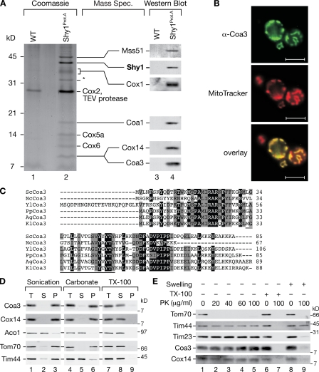
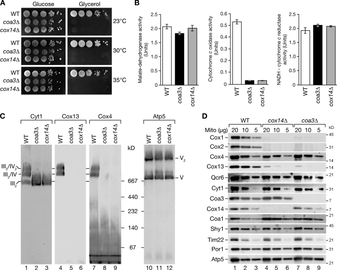
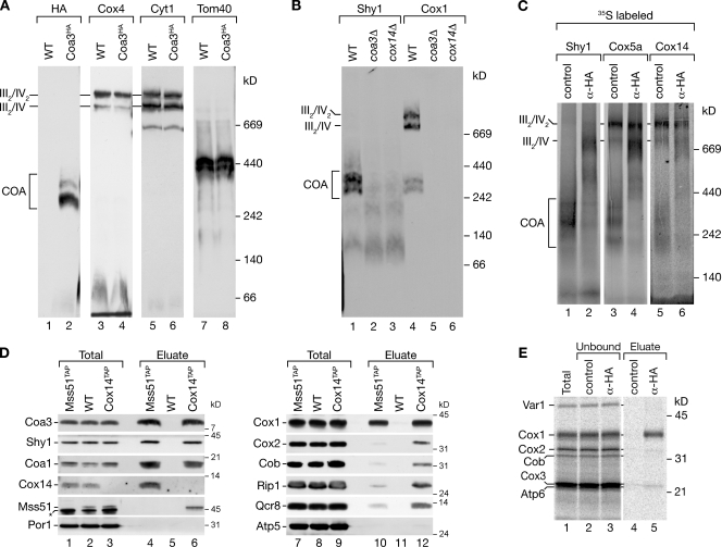
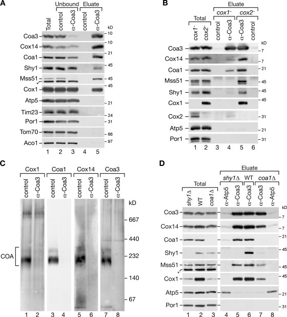
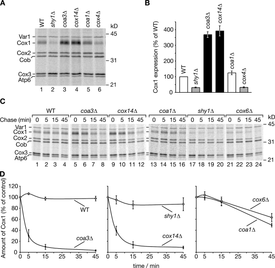
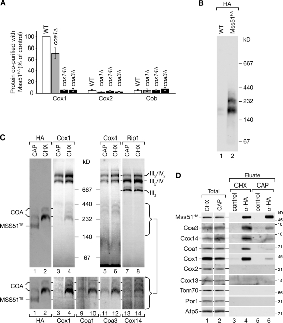
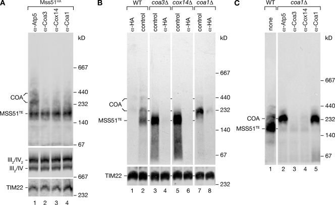
Regulation of eukaryotic cytochrome oxidase assembly occurs at the level of Cox1 translation, its central mitochondria-encoded subunit. Translation of COX1 messenger RNA is coupled to complex assembly in a negative feedback loop: the translational activator Mss51 is thought to be sequestered to assembly intermediates, rendering it incompetent to promote translation. In this study, we identify Coa3 (cytochrome oxidase assembly factor 3; Yjl062w-A), a novel regulator of mitochondrial COX1 translation and cytochrome oxidase assembly. We show that Coa3 and Cox14 form assembly intermediates with newly synthesized Cox1 and are required for Mss51 association with these complexes. Mss51 exists in equilibrium between a latent, translational resting, and a committed, translation-effective, state that are represented as distinct complexes. Coa3 and Cox14 promote formation of the latent state and thus down-regulate COX1 expression. Consequently, lack of Coa3 or Cox14 function traps Mss51 in the committed state and promotes Cox1 synthesis. Our data indicate that Coa1 binding to sequestered Mss51 in complex with Cox14, Coa3, and Cox1 is essential for full inactivation.


Figures

Figure 1.
Figure 1.
Coa3 is a mitochondrial inner membrane protein facing the IMS. (A) Isolated wild-type (WT) and Shy1ProtA mitochondria were solubilized and subjected to IgG chromatography. After tobacco etch virus (TEV) protease cleavage, eluates were analyzed by SDS-PAGE, Western blotting, or peptide LC/tandem MS. The asterisk marks ADP/ATP carrier, an impurity of purification. Cytochrome bc1 complex components identified are not depicted (Mick et al., 2007). (B) Immunofluorescence analysis using MitoTracker red and Coa3-specific antibodies. Bars, 5 µm. (C) Alignment of Coa3 homologues (ClustalW 2.0.11). Black boxes indicate identical residues in at least five species; gray boxes indicate similar amino acids. Black underlining indicates the predicted transmembrane segment. Sc, S. cerevisiae; Nc, Neurospora crassa; Yl, Yarrowia lipolytica; Pp, Pichia pastoris; Ag, Ashbya gossypii; and Kl, Kluyveromyces lactis. (D and E) Membrane association and submitochondrial localization of Coa3 as described in Materials and methods. T indicates the total; S and P indicate the supernatant and pellet, respectively, after ultracentrifugation. TX-100, Triton X-100; PK, proteinase K.
Figure 2.
Figure 2.
coa3Δ yeast cells are respiratory deficient and lack cytochrome oxidase. (A) Wild-type (WT), coa3Δ, and cox14Δ yeast cells were spotted in serial 10-fold dilutions on fermentable (glucose) and nonfermentable (glycerol) media. (B) Enzyme assays of isolated mitochondria. Means of four independent experiments (SEM; n = 4). (C) Solubilized mitochondria were separated by BN-PAGE and analyzed by Western blotting. (D) Mitochondria (Mito) were separated by SDS-PAGE and analyzed by Western blotting.
Figure 3.
Figure 3.
Coa3 associates with newly synthesized Cox1. (A) Wild-type (WT) and Coa3HA mitochondria were solubilized and analyzed by BN-PAGE and Western blotting. (B) Wild-type and mutant mitochondria were analyzed as in A. (C) Radiolabeled Shy1, Cox5a, and Cox14 were imported into isolated mitochondria containing Coa3HA. Mitochondria were solubilized and incubated with Flag (control) or HA antibodies before separation by BN-PAGE and digital autoradiography. (D) IgG chromatography of isolated mitochondria from wild type, Mss51TAP, or Cox14TAP. Samples were analyzed by SDS-PAGE and Western blotting. The asterisk indicates a cross-reactive signal detected by Mss51 antiserum. (E) In organello translation of isolated Coa3HA mitochondria was performed, and the sample was split and subjected to coimmunoprecipitations with anti-Flag (control) or anti-HA antibodies. Samples were analyzed by SDS-PAGE and digital autoradiography. (D and E) The amounts of protein loaded in the total samples correspond to 2% of the eluate.
Figure 4.
Figure 4.
Formation of COA complexes strictly depends on Cox1; sequestration of Mss51 is independent of Cox2, Shy1, and Coa1. (A) Coimmunoprecipitations of Coa3 from digitonin-solubilized mitochondria. After solubilization, samples were incubated with Coa3-specific or preimmune (control) antisera. Bound material was eluted and analyzed by SDS-PAGE and Western blotting. The amounts of protein loaded in the total and unbound samples correspond to 6% of the eluate. (B) Coimmunoprecipitation experiments as in A were performed in cox1 and cox2 mitochondria. (C) Isolated cox2 mitochondria were solubilized in digitonin and subjected to immunoprecipitation with Coa3 or preimmune (control) antisera. Lysates were depleted from antibody-bound complexes with protein A–Sepharose, and unbound material was separated by BN-PAGE, followed by Western blot analysis. (D) Coimmunoprecipitation experiments from wild-type (WT), shy1Δ, and coa1Δ mitochondria were performed as in A except that anti-Atp5 antiserum was used as a control. (A and D) Asterisks indicate a cross-reactive signal detected by Mss51 antiserum.
Figure 5.
Figure 5.
coa3Δ and cox14Δ yeast cells display increased COX1 expression and reduced stability of Cox1. (A) In vivo labeling of mitochondrial translation products was performed according to Materials and methods. After 5-min pulse, cells were lysed and subjected to TCA precipitation, and labeled proteins were analyzed by SDS-PAGE and digital autoradiography. (B) Three independent experiments as in A were quantified using ImageQuant TL software (GE Healthcare). Values represent the mean ratios of Cox1/Var1, relative to wild type (WT; 100%). Error bars indicate SEM (n = 3). (C) In vivo labeling of mitochondrial translation products was performed as in A. After 5-min pulse, translations were stopped by the addition of excess unlabeled methionine and chloramphenicol. Samples were taken after 5-, 15-, and 45-min chase and analyzed as in A. (D) Four independent experiments as in C were quantified using ImageQuant TL software, and Cox1 signals were plotted against chase times. Values represent mean ratios of Cox1/Cob, relative to 0-min chase (100%). Error bars indicate SEM (n = 4).
Figure 6.
Figure 6.
Mss51 complexes are dynamic in nature. (A) In organello labeling of mitochondrial translation products was performed in the indicated strains, expressing Mss51HA. Coimmunoprecipitation experiments were analyzed as in Fig. 3 E. Autoradiograph signals were quantified using ImageQuant TL software. The ratios of signals for Cox1, Cox2, and Cob between eluate and total were calculated and normalized to the Cox1 eluate/total ratio in wild type (WT; 100%). Means of three independent experiments (SEM; n = 3) are shown. (B) Mss51HA and wild-type mitochondria were solubilized and analyzed by BN-PAGE and Western blotting using anti-HA antibodies. (C) Mitochondria isolated from Mss51HA yeast cells treated with chloramphenicol (CAP) or cycloheximide (CHX) were analyzed as in B. Brackets indicate the molecular mass range of the Mss51 complexes. Lanes 1–4 in the bottom panels are cropped from the top panels and displayed with increased contrast for Cox1. (D) Solubilized mitochondria from pretreated cells were subjected to coimmunoprecipitation with anti-HA and control antibodies and analyzed by SDS-PAGE and Western blotting.
Figure 7.
Figure 7.
Lack of Coa1 stalls Mss51 in a 250-kD complex. (A) Mss51HA mitochondria were solubilized, and samples were split and incubated with indicated antibodies. Antibodies with bound material were precipitated with protein A–Sepharose, and unbound material was analyzed by BN-PAGE and Western blotting using anti-HA antibodies. Respiratory chain supercomplexes and carrier translocase (TIM22) were detected with anti-Cox1 or anti-Tim54 antibodies, respectively. (B) Mitochondria from the indicated mutants expressing Mss51HA were analyzed as in A. (C) Antibody depletion experiments were performed in coa1Δ mitochondria as in A. For comparison, untreated wild-type (WT) mitochondria were analyzed in lane 1.

Similar articles

Cited by

References

    1. Arlt H., Tauer R., Feldmann H., Neupert W., Langer T. 1996. The YTA10-12 complex, an AAA protease with chaperone-like activity in the inner membrane of mitochondria. Cell. 85:875–885 10.1016/S0092-8674(00)81271-4 - DOI - PubMed
    1. Barrientos A., Korr D., Tzagoloff A. 2002. Shy1p is necessary for full expression of mitochondrial COX1 in the yeast model of Leigh’s syndrome. EMBO J. 21:43–52 10.1093/emboj/21.1.43 - DOI - PMC - PubMed
    1. Barrientos A., Zambrano A., Tzagoloff A. 2004. Mss51p and Cox14p jointly regulate mitochondrial Cox1p expression in Saccharomyces cerevisiae. EMBO J. 23:3472–3482 10.1038/sj.emboj.7600358 - DOI - PMC - PubMed
    1. Barrientos A., Gouget K., Horn D., Soto I.C., Fontanesi F. 2009. Suppression mechanisms of COX assembly defects in yeast and human: insights into the COX assembly process. Biochim. Biophys. Acta. 1793:97–107 10.1016/j.bbamcr.2008.05.003 - DOI - PMC - PubMed
    1. Bonnefoy N., Fiumera H.L., Dujardin G., Fox T.D. 2009. Roles of Oxa1-related inner-membrane translocases in assembly of respiratory chain complexes. Biochim. Biophys. Acta. 1793:60–70 10.1016/j.bbamcr.2008.05.004 - DOI - PMC - PubMed

Publication types

MeSH terms