. 2010 Oct 4;191(1):31-43.
doi: 10.1083/jcb.201001160. Epub 2010 Sep 27.

The p400 ATPase regulates nucleosome stability and chromatin ubiquitination during DNA repair

Affiliations

The p400 ATPase regulates nucleosome stability and chromatin ubiquitination during DNA repair

Ye Xu et al. J Cell Biol. .

Abstract

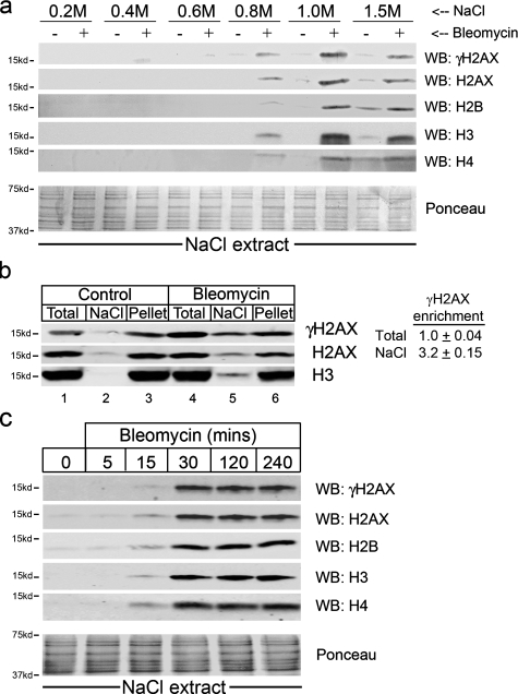
The complexity of chromatin architecture presents a significant barrier to the ability of the DNA repair machinery to access and repair DNA double-strand breaks (DSBs). Consequently, remodeling of the chromatin landscape adjacent to DSBs is vital for efficient DNA repair. Here, we demonstrate that DNA damage destabilizes nucleosomes within chromatin regions that correspond to the γ-H2AX domains surrounding DSBs. This nucleosome destabilization is an active process requiring the ATPase activity of the p400 SWI/SNF ATPase and histone acetylation by the Tip60 acetyltransferase. p400 is recruited to DSBs by a mechanism that is independent of ATM but requires mdc1. Further, the destabilization of nucleosomes by p400 is required for the RNF8-dependent ubiquitination of chromatin, and for the subsequent recruitment of brca1 and 53BP1 to DSBs. These results identify p400 as a novel DNA damage response protein and demonstrate that p400-mediated alterations in nucleosome and chromatin structure promote both chromatin ubiquitination and the accumulation of brca1 and 53BP1 at sites of DNA damage.


Figures

Figure 1.
Figure 1.
DNA damage reduces the interaction between DNA and histones. (a) 293T cells were exposed to 5 µM bleomycin for 30 min. Isolated nuclei were extracted in buffer containing the indicated concentration of NaCl. γ-H2AX, H2AX, H2B, H3, and H4 detected by Western blot. Membranes were stained with ponceau S to ensure equal loading. (b) 293T cells were untreated (control) or exposed to 5 µM bleomycin for 30 min. Total chromatin-associated histones were obtained by acid extraction. In parallel, cells were first extracted with 1.0 M NaCl and the nuclei collected by centrifugation. The histones remaining in the nuclear pellet were then purified by acid extraction. γ-H2AX, H2AX, and H3 were detected by Western blot. Table: Relative enrichment of γ-H2AX in the Total and NaCl extracts was calculated by normalizing γ-H2AX scan intensities to H2AX signal in each extract, expressed as [γ-H2AXTotal]/[H2AXTotal] and [γ-H2AXNaCl]/[H2AXNaCl]. Results from three independent replicates, ± SD. (c) 293T cells were treated with 5 µM bleomycin and extracted in 1.0 M NaCl. γ-H2AX, H2AX, H2B, H3, and H4 were detected by Western blot. Equal loading confirmed by ponceau S staining.
Figure 2.
Figure 2.
ATM is not required for altered nucleosome stability after DNA damage. (a) 293T cells were irradiated (10Gy), fractionated in 1.0 M NaCl, and released histones detected by Western blot. (b) 293T cells were irradiated (10Gy) and whole-cell extracts examined by Western blot for total γ-H2AX, H2AX, ATM, and pATM. (c) GM5849 A-T cells complemented with either vector (A-T) or full-length ATM (A-TATM) were exposed to 5 µM bleomycin, fractionated in 1.0 M NaCl, and released histones detected by Western blot. (d) 293T cells were incubated with 10 µM of the ATM kinase inhibitor Ku55933 for 60 min, followed by 5 µM bleomycin. Cells were fractionated in 1.0 M NaCl, and released histones detected by Western blot.
Figure 3.
Figure 3.
Tip60, p400, and Trrap are required for altered nucleosome stability. (a) 293T cells expressing a nonspecific shRNA (vector) or shRNA targeting p400 were exposed to 5 µM bleomycin. Cells were fractionated in 1.0 M NaCl, and released histones detected by Western blot. (b) HeLa cells expressing vector or shRNA targeting Trrap were exposed to bleomycin. Cells were fractionated in 1.0 M NaCl, and released histones detected by Western blot. (c) 293T cells expressing either HA-Tip60 or catalytically inactive HA-Tip60HD were incubated with 5 µM bleomycin. Cells were fractionated in 1.0 M NaCl, and released histones detected by Western blot. Where indicated, cells were pretreated with 300 nM TSA for 2 h. (d) 293T cells expressing either HA-p400 or HA-p400ATPase were incubated with 5 µM bleomycin. Cells were fractionated in 1.0 M NaCl, and released histones detected by Western blot. Where indicated, cells were pretreated with 300 nM TSA for 2 h.
Figure 4.
Figure 4.
Loss of p400’s ATPase activity increases radiosensitivity and chromosome aberrations. (a) HeLaS3 cells expressing either p400 (○) or p400ATPase (●) were irradiated, and the number of surviving colonies measured 12 d later. Each data point represents the average of three independent assays, results ± SD. (b) p400 or p400ATPase cells were untreated (0) or irradiated (2Gy) and allowed to recover for 14 h. Metaphase spreads were subsequently scored visually for chromosome aberrations. Results expressed as aberrations per cell (n = 50 cells), and P values determined using t test.
Figure 5.
Figure 5.
p400 is recruited to DSBs generated by the p84-ZFN. (a) 293T cells were transiently transfected with p84-ZFN, DNA extracted and examined by qPCR to monitor the fraction of cells with DSBs. (b) 293T cells were transiently transfected with vector (−) or p84-ZFN (+). 18 h later, cells were processed for ChIP analysis using anti–γ-H2AX antibody and primer pairs located left and right of the DSB. ChIP data points were calculated as IP DNA/input DNA (see Materials and methods). Fold enrichment is the relative fold increase in signal from the ZFN samples compared with the vector samples, which were normalized to give a value of 1. Results ± SE (n = 3). (c) 293T cells were transiently transfected with vector (−) or p84-ZFN (+) as described in b. Cells were processed for ChIP analysis using anti-p400 antibody. Results ± SE (n = 3). (d) 293T cells expressing p400 or p400ATPase were transiently transfected with vector (−) or p84-ZFN (+). 18 h later, cells were preextracted in 1.0 M NaCl to release histones, and the nuclear pellet collected and processed for ChIP analysis as described in Materials and methods. Samples were immunoprecipitated with anti-H3 antibody. qPCR was performed using primers located at +1.5 kb relative to the ZFN site on chromosome 19 (Chr19), or at nucleotide positions 122,788,208 and 122,788,440 on chromosome 6 (Chr 6). Results ± SE (n = 3).
Figure 6.
Figure 6.
Acetylation of histone H4 by the p400–Tip60 complex. (a) 293T cells expressing wild-type Tip60 (Tip60wt), catalytically inactive Tip60 (Tip60HD), wild-type p400 (p400), or p400 lacking ATPase activity (p400ATPase) were transiently transfected with vector (−) or p84-ZFN (+), and ChIP assays using anti-p400 antibody performed using primers located at +1.5 kb relative to the DSB. Results ± SE (n = 3). (b) 293T cells expressing Tip60wt, Tip60HD, p400, or p400ATPase were transiently transfected with vector (−) or p84-ZFN (+). 18 h later, ChIP assays were performed using anti-acetylH4 antibody (AcH4) and primers located +1.5 kb relative to the DSB. Results ± SE (n = 3). (c) p400 was immunopurified from 293T cells expressing either HA-p400, HA-p400ATPase, or cells in which p400 was silenced with shRNA and the associated Tip60 acetyltransferase activity measured. Cells were exposed to bleomycin (+: 5 µM for 40 min) where indicated. Inset: Immunoprecipitates were separated by SDS-PAGE, and p400 and coprecipitating Tip60 detected by Western blot.
Figure 7.
Figure 7.
Recruitment of p400 to DSBs requires mdc1. (a) H2AX−/− MEFs or H2AX−/− MEFs complemented with H2AX (H2AXwt) were exposed to 5 µM bleomycin. 20 µM Wortmannin was added 60 min before bleomycin. Cells were fractionated in 1.0 M NaCl, and released histones detected by Western blot. (b) 293T cells expressing vector or mdc1 shRNA (shmdc1) were exposed to 5 µM bleomycin. Cells were fractionated in 1.0 M NaCl, and released histones detected by Western blot. (c) 293T cells expressing GFP or mdc1 shRNA were transiently transfected with vector or p84-ZFN. 18 h later, ChIP assays using p400 antibody and primer pairs located at +1.5 kb were performed. Results ± SE (n = 3). (d) 293T cells stably expressing FLAG-RNF8 or RNF8 lacking the catalytic domain (FLAG-RNF8δring), which functions as a dominant-negative inhibitor of endogenous RNF8 function (Huen et al., 2007), were exposed to bleomycin. Cells were fractionated in 1.0 M NaCl, and released histones detected by Western blot.
Figure 8.
Figure 8.
p400 is required to recruit 53BP1 and brca1 to DSBs. 293T cells expressing p400 or p400ATPase were irradiated (2Gy). At the indicated times, cells were fixed and processed by immunofluorescent staining to detect either brca1 (a) or 53BP1 (b) foci. Cells with >5 foci were counted, with an average of 100 cells per slide. Experiments represent at least three independent replicates. Results ± SE (n = 100). (c) 293T cells expressing p400 or p400ATPase were irradiated (2Gy). At the indicated times, cells were fixed and processed by immunofluorescent staining to detect brca1, 53BP1 foci, and γ-H2AX. Bar, 5 µm.
Figure 9.
Figure 9.
p400 is required for RNF8-dependent ubiquitination of the chromatin. (a) 293T cells expressing p400 or p400ATPase were irradiated as indicated. Cells were fixed and processed by immunofluorescent staining using FK2 antibody. Bar, 5 µm. (b) 293T cells expressing p400 or p400ATPase were irradiated (2Gy). Immunofluorescent staining using the FK2 antibody was used to detect proteins ubiquitinated by RNF8. Cells with >5 foci per cell were counted, with an average of 100 cells per slide. Results ± SE (n = 100). (c) 293T cells expressing p400 or p400ATPase were transiently transfected with vector or p84-ZFN. 18 h later, ChIP assays using the FK2 antibody and primer pairs located at −3.5 kb, −1.5 kb, −0.5 kb, 0.5 kb, 1.5 kb, and 3.5 kb were performed. Results ± SE (n = 3). (d) 293T cells expressing p400 or p400ATPase were irradiated (2Gy), fixed, and immunofluorescent staining to detect RNF8 and FK2 performed. Bar, 5 µm. (e) Quantitation of FK2 and RNF8 foci in p400 and p400ATPase cells from d. Cells with >5 foci were counted, with an average of 100 cells per slide counted. Experiments represent at least three independent replicates. Results ± SE (n = 100).

Similar articles

Cited by

References

    1. Bird A.W., Yu D.Y., Pray-Grant M.G., Qiu Q., Harmon K.E., Megee P.C., Grant P.A., Smith M.M., Christman M.F.. 2002. Acetylation of histone H4 by Esa1 is required for DNA double-strand break repair. Nature. 419:411–415. 10.1038/nature01035 - DOI - PubMed
    1. Bonner W.M., Redon C.E., Dickey J.S., Nakamura A.J., Sedelnikova O.A., Solier S., Pommier Y.. 2008. GammaH2AX and cancer. Nat. Rev. Cancer. 8:957–967. 10.1038/nrc2523 - DOI - PMC - PubMed
    1. Botuyan M.V., Lee J., Ward I.M., Kim J.E., Thompson J.R., Chen J., Mer G.. 2006. Structural basis for the methylation state-specific recognition of histone H4-K20 by 53BP1 and Crb2 in DNA repair. Cell. 127:1361–1373. 10.1016/j.cell.2006.10.043 - DOI - PMC - PubMed
    1. Brower-Toland B., Wacker D.A., Fulbright R.M., Lis J.T., Kraus W.L., Wang M.D.. 2005. Specific contributions of histone tails and their acetylation to the mechanical stability of nucleosomes. J. Mol. Biol. 346:135–146. 10.1016/j.jmb.2004.11.056 - DOI - PubMed
    1. Brunet E., Simsek D., Tomishima M., DeKelver R., Choi V.M., Gregory P., Urnov F., Weinstock D.M., Jasin M.. 2009. Chromosomal translocations induced at specified loci in human stem cells. Proc. Natl. Acad. Sci. USA. 106:10620–10625. 10.1073/pnas.0902076106 - DOI - PMC - PubMed

Publication types