. 2010 Dec;31(12):2049-57.
doi: 10.1093/carcin/bgq192. Epub 2010 Sep 27.

Oncogenic HER2{Delta}16 suppresses miR-15a/16 and deregulates BCL-2 to promote endocrine resistance of breast tumors

Affiliations

Oncogenic HER2{Delta}16 suppresses miR-15a/16 and deregulates BCL-2 to promote endocrine resistance of breast tumors

Diana M Cittelly et al. Carcinogenesis. 2010 Dec.

Abstract

Tamoxifen is the most commonly prescribed therapy for patients with estrogen receptor (ER)α-positive breast tumors. Tumor resistance to tamoxifen remains a serious clinical problem especially in patients with tumors that also overexpress human epidermal growth factor receptor 2 (HER2). Current preclinical models of HER2 overexpression fail to recapitulate the clinical spectrum of endocrine resistance associated with HER2/ER-positive tumors. Here, we show that ectopic expression of a clinically important oncogenic isoform of HER2, HER2Δ16, which is expressed in >30% of ER-positive breast tumors, promotes tamoxifen resistance and estrogen independence of MCF-7 xenografts. MCF-7/HER2Δ16 cells evade tamoxifen through upregulation of BCL-2, whereas mediated suppression of BCL-2 expression or treatment of MCF-7/HER2Δ16 cells with the BCL-2 family pharmacological inhibitor ABT-737 restores tamoxifen sensitivity. Tamoxifen-resistant MCF-7/HER2Δ16 cells upregulate BCL-2 protein levels in response to suppressed ERα signaling mediated by estrogen withdrawal, tamoxifen treatment or fulvestrant treatment. In addition, HER2Δ16 expression results in suppression of BCL-2-targeting microRNAs miR-15a and miR-16. Reintroduction of miR-15a/16 reduced tamoxifen-induced BCL-2 expression and sensitized MCF-7/HER2Δ16 to tamoxifen. Conversely, inhibition of miR-15a/16 in tamoxifen-sensitive cells activated BCL-2 expression and promoted tamoxifen resistance. Our results suggest that HER2Δ16 expression promotes endocrine-resistant HER2/ERα-positive breast tumors and in contrast to wild-type HER2, preclinical models of HER2Δ16 overexpression recapitulate multiple phenotypes of endocrine-resistant human breast tumors. The mechanism of HER2Δ16 therapeutic evasion, involving tamoxifen-induced upregulation of BCL-2 and suppression of miR-15a/16, provides a template for unique therapeutic interventions combining tamoxifen with modulation of microRNAs and/or ABT-737-mediated BCL-2 inhibition and apoptosis.


Figures

Fig. 1.
Fig. 1.
HER2Δ16 expression promotes estrogen independent and tamoxifen-resistant growth. (ac) Graphs representing xenograft tumor kinetics of at least five nude mice per group injected with (a) MCF-7/Vector, (b) MCF-7/HER2 or (c) MCF-7/HER2Δ16 cells. With the exception of the E2 treatments, all mice were primed with E2 pellets and after 21 days, mice with established tumors were left untreated or implanted with TAM pellets. (d and e) MCF-7/HER2Δ16 cells are E2 independent and TAM resistant in vitro. (d and e) MCF-7/HER2Δ16 cells are estrogen independent and tamoxifen resistant in vitro. (d) Each cell line was untreated or treated for 5 days with 100 pM E2 alone or in combination with 1.0 μM TAM. 3-(4,5-dimethylthiazole-2-yl)-2,5-diphenyl tetrazolium bromide assay was used to quantitate cell growth. Results represent mean ± SE percent growth inhibition relative to 100 pM E2 alone. (e) Each cell line was cultured for 24 h in CS-MEM and treated for 72 h with 100 pM E2 alone or in combination with 1.0 μM TAM. Apoptosis was quantitated using a Cell Death Detection ELISA. Results represent apoptosis relative to cells treated with 100 pM E2 alone.
Fig. 2.
Fig. 2.
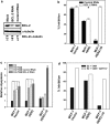
BCL-2 upregulation mediates tamoxifen resistance of MCF-7/HER2Δ16 cells. (a) Quantitation of estrogen-induced BCL-2 mRNA expression. Each cell line was treated the indicated time with 100 pM E2 alone or in combination with 1.0 μM TAM. Each RNA sample was analyzed in triplicate, normalized to a β-actin internal control and BCL-2 mRNA expression is represented relative to the untreated MCF-7/Vector. (b) BCL-2 protein is upregulated in tamoxifen-treated MCF-7/HER2Δ16 cells. Each cell line was cultured for 48 h in phenol red-free modified Eagle's medium containing 5% charcoal-stripped fetal bovine serum and then treated as above and cell lysates were analyzed by western blot for BCL-2 expression 24, 48 and 72 h after treatment. (c) Inactivation of ERα signaling induces BCL-2 protein upregulation in MCF-7/HER2Δ16 cells. The MCF-7/HER2Δ16 cell line was cultured for 48 h in and left untreated (-E2) or treated with 1.0 μM TAM or 100 nM ICI 18278 (ICI) for the indicated time. Cell lysates were prepared and analyzed by western blot for BCL-2 expression. In all cases, western blot analysis of α-tubulin was included as a loading control and images were quantitated using the Odyssey Infrared Imaging System software.
Fig. 3.
Fig. 3.
Suppression of BCL-2 restores tamoxifen sensitivity. (a) RNAi-mediated suppression of BCL-2 expression. The MCF-7/HER2Δ16 cell line was cultured for 24 h in phenol red-free modified Eagle's medium containing 5% charcoal-stripped fetal bovine serum and treated with non-specific control or BCL-2-specific RNAi. Immediately after transfection, each sample was treated with 1.0 μM 4-hydroxytamoxifen for an additional 48 h. Cell lysates were analyzed for BCL-2 expression by western blot. Western blot analysis of α-tubulin was included as a loading control and images were quantitated using the Odyssey Infrared Imaging System software. (b) Suppression of BCL-2 restores tamoxifen sensitivity to MCF-7/HER2Δ16 cells. Each cell line was treated with non-specific control or BCL-2 RNAi. Each sample was treated with 100 pM E2 alone or in combination with 1.0 μM TAM for 72 h. 3-(4,5-dimethylthiazole-2-yl)-2,5-diphenyl tetrazolium bromide assay was used to quantitate cell growth. Results represent mean ± SE percent growth inhibition relative to 100 pM E2 alone. Asterisks indicate significant difference by paired Student’s t-test (P = 0.007). (c) Suppression of BCL-2 sensitizes MCF-7/HER2Δ16 cells to tamoxifen-induced apoptosis. Each cell line was treated as above and apoptosis was quantitated using a Cell Death Detection ELISA. Results represent mean ± SE apoptosis relative to MCF-7/Vector treated with 100 pM E2 alone of three independent experiments of samples prepared in triplicate. Asterisks indicate samples with significant differences by paired Student’s t-test (P < 0.001). (d) The BCL-2 inhibitor ABT-737 sensitizes MCF-7/HER2Δ16 cells to tamoxifen. Each cell line was cultured for 48 h in CS-MEM and then treated with 100 pM E2, 100 pM E2 and 1.0 μM TAM alone or in combination with 5 μM of the BCL-2 family inhibitor ABT-737 for 5 days. Results represent mean percent growth inhibition of triplicate samples relative to cells treated with 100 pM E2 alone.
Fig. 4.
Fig. 4.
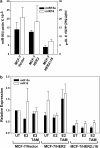
HER2Δ16 expression suppresses miR-15a and miR-16. (a) Expression of miR-15a and miR-16 is suppressed in MCF-7/HER2Δ16 cells. Total RNA was extracted and analyzed for miR-15a or miR-16 expression by qRT–PCR. Results from three independent RNA extractions are represented as mean ± SE expression relative to β-actin. The lower levels of miR-15a and miR-16 expression in the MCF-7/HER2Δ16 cells failed to reach significance (paired Student’s t-test; P = 0.07 and P = 0.08, respectively). (b) Expression of miR-15a and miR-16 is not altered by estrogen or tamoxifen. Each cell line was cultured for 48 h in phenol red-free modified Eagle;s medium containing 5% charcoal-stripped fetal bovine serum and then left untreated or treated for 16 h with 100 pM E2 alone or in combination with 1.0 μM TAM. Three independent total RNA extractions from each cell line were analyzed in triplicate for miR-15a and miR-16 expression by qRT–PCR. Results were normalized to β-actin and represented as mean ± SE expression relative to untreated MCF-7/Vector cells. Differences failed to obtain significance as determined by paired Student’s t-test.
Fig. 5.
Fig. 5.
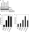
Expression of pre-miR-15a/16 sensitizes MCF-7/HER2Δ16 cells to tamoxifen. (a) Pre-miR-15a and/or pre-miR-16 suppresses BCL-2 expression. The MCF-7/HER2Δ16 cell line was untreated or treated with 30 nM of the indicated pre-miR and treated with 100 pM E2 and 1.0 μM TAM for 48 h. Cell lysates were analyzed by western blot and BCL-2 expression relative to the untreated control was quantitated by densitometry. (b and c) Pre-miR-15a and/or pre-miR-16 sensitizes MCF-7/HER2Δ16 cells to tamoxifen. The MCF-7/HER2Δ16 cell line was transfected with pre-miRs as above, treated with 100 pM E2 alone or in combination with 1.0 μM TAM for 72 h. (b) 3-(4,5-dimethylthiazole-2-yl)-2,5-diphenyl tetrazolium bromide assay was used to quantitate cell growth or (c) apoptosis was quantitated using a Cell Death Detection ELISA. Data represents mean ± SE of three independent experiments performed with triplicate samples relative to mock and E2/TAM-treated cells. Asterisks indicate samples with significant differences as determined by paired Student’s t-test (b, P < 0.001; c, pre-miR-15a, P = 0.029; pre-miR16, P = 0.004; pre-miR15a/16, P = 0.017).
Fig. 6.
Fig. 6.
Suppressed miR-15a/16 expression promotes tamoxifen resistance. (a) Inhibition of miR-15a/16 results in enhanced BCL-2 expression. Each cell line was treated with 50 nM of the indicated miR inhibitor for 48 h and BCL-2 expression was analyzed by western blot. Analysis of α-tubulin was included as a loading control and images were quantitated using the Odyssey Infrared Imaging System software. (b and c) Suppression of miR-15a/16 promotes tamoxifen resistance. Each cell line was untreated or treated with the indicated anti-miR and treated with 100 pM E2 alone or in combination with 1.0 μM TAM for five days. (b) 3-(4,5-dimethylthiazole-2-yl)-2,5-diphenyl tetrazolium bromide assay was used to quantitate cell growth and (c) apoptosis was quantitated using a Cell Death Detection ELISA. Data are represented as mean ± SE of three independent experiments performed with triplicate samples relative to mock anti-miR and E2/TAM-treated MCF-7/Vector cells. Asterisks indicate samples with significant differences as determined by paired Student’s t-test (P < 0.008).

Similar articles

Cited by

References

    1. Fabbri M, et al. MicroRNAs. Cancer J. 2008;14:1–6. - PubMed
    1. Esquela-Kerscher A, et al. Oncomirs—microRNAs with a role in cancer. Nat. Rev. Cancer. 2006;6:259–269. - PubMed
    1. Verghese ET, et al. Small is beautiful: microRNAs and breast cancer-where are we now? J. Pathol. 2008;215:214–221. - PubMed
    1. Tavazoie SF, et al. Endogenous human microRNAs that suppress breast cancer metastasis. Nature. 2008;451:147–152. - PMC - PubMed
    1. Miller TE, et al. MicroRNA-221/222 confers tamoxifen resistance in breast cancer by targeting p27(Kip1) J. Biol. Chem. 2008;283:29897–29903. - PMC - PubMed

Publication types