. 2010 Dec;78(12):5062-73.
doi: 10.1128/IAI.00516-10. Epub 2010 Sep 27.

Listeria monocytogenes uses Listeria adhesion protein (LAP) to promote bacterial transepithelial translocation and induces expression of LAP receptor Hsp60

Affiliations

Listeria monocytogenes uses Listeria adhesion protein (LAP) to promote bacterial transepithelial translocation and induces expression of LAP receptor Hsp60

Kristin M Burkholder et al. Infect Immun. 2010 Dec.

Abstract

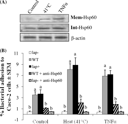
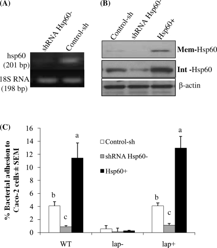
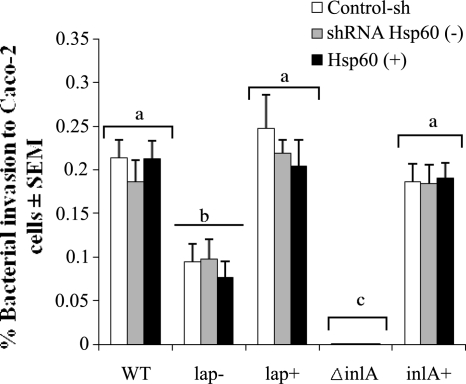
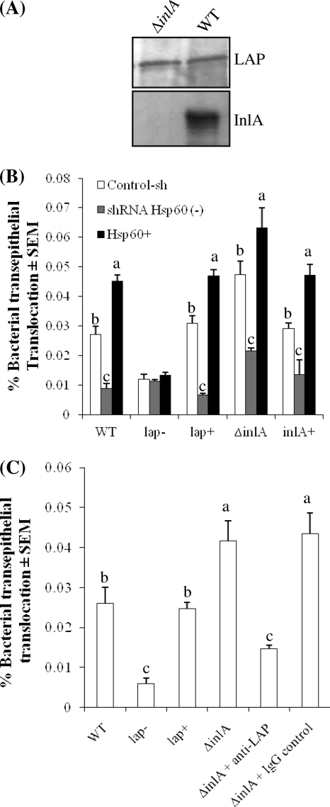
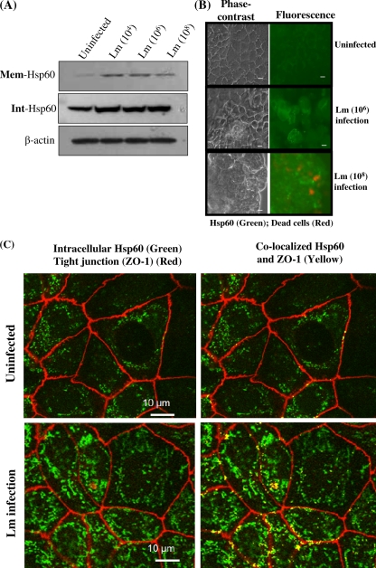
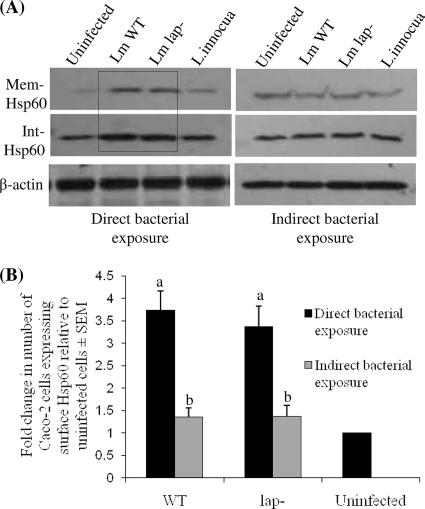
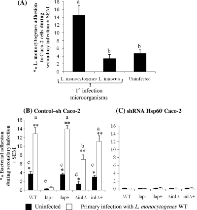
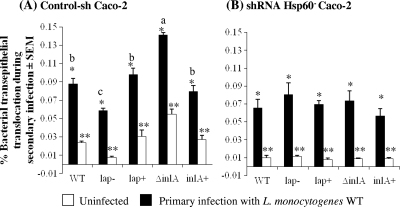
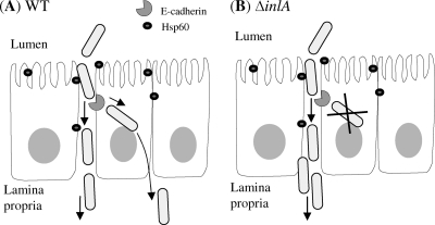
Listeria monocytogenes interaction with the intestinal epithelium is a key step in the infection process. We demonstrated that Listeria adhesion protein (LAP) promotes adhesion to intestinal epithelial cells and facilitates extraintestinal dissemination in vivo. The LAP receptor is a stress response protein, Hsp60, but the precise role for the LAP-Hsp60 interaction during Listeria infection is unknown. Here we investigated the influence of physiological stressors and Listeria infection on host Hsp60 expression and LAP-mediated bacterial adhesion, invasion, and transepithelial translocation in an enterocyte-like Caco-2 cell model. Stressors such as heat (41°C), tumor necrosis factor alpha (TNF-α) (100 U), and L. monocytogenes infection (10(4) to 10(6) CFU/ml) significantly (P < 0.05) increased plasma membrane and intracellular Hsp60 levels in Caco-2 cells and consequently enhanced LAP-mediated L. monocytogenes adhesion but not invasion of Caco-2 cells. In transepithelial translocation experiments, the wild type (WT) exhibited 2.7-fold more translocation through Caco-2 monolayers than a lap mutant, suggesting that LAP is involved in transepithelial translocation, potentially via a paracellular route. Short hairpin RNA (shRNA) suppression of Hsp60 in Caco-2 cells reduced WT adhesion and translocation 4.5- and 3-fold, respectively, while adhesion remained unchanged for the lap mutant. Conversely, overexpression of Hsp60 in Caco-2 cells enhanced WT adhesion and transepithelial translocation, but not those of the lap mutant. Furthermore, initial infection with a low dosage (10(6) CFU/ml) of L. monocytogenes increased plasma membrane and intracellular expression of Hsp60 significantly, which rendered Caco-2 cells more susceptible to subsequent LAP-mediated adhesion and translocation. These data provide insight into the role of LAP as a virulence factor during intestinal epithelial infection and pose new questions regarding the dynamics between the host stress response and pathogen infection.


Figures

FIG. 1.
FIG. 1.
Influence of Caco-2 Hsp60 expression on LAP-mediated adhesion of L. monocytogenes. (A) Immunoblot showing the level of Hsp60 expression in Caco-2 plasma membrane (Mem-Hsp60) and intracellular (Int-Hsp60) fractions after heat stress (41°C, 1 h) and TNF-α exposure (100 U/ml, 1 h). β-Actin (43 kDa) was used as an internal control. Proteins were loaded at 15 μg/well. (B) Adhesion of WT, lap (mutant), and lap+ (lap-complemented) L. monocytogenes strains to Caco-2 monolayers following Caco-2 heat stress (41°C) and TNF-α (100 U/ml) exposure and after treatment with anti-Hsp60 PAb (1 μg/ml). Adhesion data are averages for at least three independent experiments performed in quadruplicate (n ≥ 12). Error bars represent standard errors of the means (SEM). Lowercase letters (a and b) represent significant differences (P < 0.05) in adhesion of bacterial strains.
FIG. 2.
FIG. 2.
Analysis of L. monocytogenes adhesion to Caco-2 cells following Hsp60 knockdown by shRNA (shRNA Hsp60−) or Hsp60 overexpression (Hsp60+) or adhesion to control-sh-transfected cells (Caco-2 cells transfected with noncoding shRNA). (A) RT-PCR analysis of hsp60 expression (23 cycles) in Caco-2 cells following shRNA-Hsp60 knockdown. 18S RNA was used as an internal control. (B) Immunoblot showing Hsp60 suppression (shRNA Hsp60−) and overexpression (Hsp60+) in Caco-2 plasma membrane (Mem-Hsp60) and intracellular (Int-Hsp60) fractions. β-Actin (43 kDa) was used as an internal control. Proteins were loaded at 15 μg/well. (C) Adhesion of L. monocytogenes WT, lap (mutant), and lap+ (lap-complemented) strains to control-sh, shRNA-Hsp60, and Hsp60+ Caco-2 monolayers. Adhesion data are averages for at least three independent experiments performed in quadruplicate (n ≥ 12) and are presented with SEM. Lowercase letters (a, b, and c) indicate significant differences (P < 0.05) in adhesion of individual bacterial strains to Caco-2 monolayers expressing varied levels of Hsp60.
FIG. 3.
FIG. 3.
Analysis of L. monocytogenes WT, lap, lap+, ΔinlA, and inlA+ strain invasion of Caco-2 cells following Hsp60 knockdown by shRNA (shRNA Hsp60−) or Hsp60 overexpression (Hsp60+) or of control-sh-transfected cells (Caco-2 cells with scrambled shRNA). Invasion results are averages for at least three independent experiments performed in quadruplicate (n ≥ 12) and are presented with SEM. Lowercase letters (a to c) indicate significant differences (P < 0.05) in invasion among bacterial strains.
FIG. 4.
FIG. 4.
Confocal microscopic analysis of tight junction integrity in uninfected (A) and L. monocytogenes-infected (2 h) (B) Caco-2 monolayers grown on Transwell filters. Monolayers were labeled with antibodies specific for the tight junction proteins ZO-1, occludin, and claudin (red) and for Hsp60 (green) and with a nuclear stain (blue). The images reveal visible tight junction borders in both uninfected and infected Caco-2 monolayers, with no alteration in cellular distribution of tight junction proteins or in cellular damage.
FIG. 5.
FIG. 5.
Analysis of transepithelial translocation of L. monocytogenes through polarized Caco-2 cell monolayers grown on a Transwell filter insert (see the text for further details). (A) Immunoblot showing LAP and InlA expression in L. monocytogenes WT and ΔinlA strains. Each lane was loaded with 15 μg of total cell protein. (B) Transepithelial translocation of L. monocytogenes WT, lap, lap+, ΔinlA, and inlA+ cells through Caco-2 monolayers following Hsp60 knockdown by shRNA [shRNA Hsp60(−)] or Hsp60 overexpression (Hsp60+) or through control-sh-transfected cells (Caco-2 cells transfected with noncoding shRNA). (C) Translocation of WT, lap, lap+, and ΔinlA cells and of ΔinlA cells pretreated with anti-LAP MAb (1 μg/ml) or an IgG control (MAb C11E9) (1 μg/ml) through control-sh-transfected Caco-2 monolayers. Data are averages for at least three independent translocation experiments performed in quadruplicate (n ≥ 12) and are presented with SEM. Lowercase letters (a to c) indicate significant differences (P < 0.05) in translocation of individual bacterial strains through Caco-2 monolayers expressing various levels of Hsp60.
FIG. 6.
FIG. 6.
Influence of L. monocytogenes infection on Hsp60 expression in Caco-2 cells. (A) Immunoblot analysis of the level of Hsp60 expression in Caco-2 plasma membrane (Mem-Hsp60) and intracellular (Int-Hsp60) fractions after exposure to L. monocytogenes (Lm) at 1 × 10⁴, 1 × 10⁶, or 1 × 10⁸ CFU/ml. β-Actin was used as an internal control. Proteins were loaded at 15 μg/well. Blots are representative of at least three individual experiments. (B) Microscopic analysis of Hsp60 (green) on the surfaces of uninfected or L. monocytogenes-infected (106 CFU/ml [MOI of 10:1] and 108 CFU/ml [MOI of 1,000:1]) Caco-2 cells. Caco-2 cells were labeled with anti-Hsp60 antibody and an FITC-conjugated secondary antibody. Propidium iodide was used to stain dead cells (red). Samples were viewed on a Leica fluorescence microscope with a 40× objective. Bars, 10 μm. (C) Confocal microscopic analysis of intracellular Hsp60 (FITC; green) expression in Caco-2 cells left uninfected (top panels) or infected with 106 CFU/ml L. monocytogenes (bottom panels). The tight junction protein ZO1 was labeled (Cy5; red) for the purpose of visualizing cell borders. Yellow highlighting indicates areas where Hsp60 and ZO-1 are colocalized (right panels). Hsp60 expression was more abundant in L. monocytogenes-infected cells than in uninfected cells.
FIG. 7.
FIG. 7.
Hsp60 expression in Caco-2 cells following direct versus indirect exposure to Listeria. (A) Immunoblot analysis of Hsp60 levels in Caco-2 plasma membrane (Mem-Hsp60) and intracellular (Int-Hsp60) fractions following direct or indirect (via separation by a 0.4-μm-pore-size filter) exposure to the L. monocytogenes (Lm) WT, the L. monocytogenes lap strain, or L. innocua at 106 CFU/ml (MOI, 10:1). β-Actin was used as an internal control. Blots are representative of three individual experiments. Proteins were loaded at 15 μg/well. Lanes marked with an open box indicate increased Hsp60 expression compared to that of controls. (B) Flow cytometric analysis of Hsp60 expression on the surfaces of Caco-2 cells following direct or indirect exposure to L. monocytogenes WT or lap strain. Data are averages for 3 experiments, with treatments run in quadruplicate (n = 12), and are presented as fold changes in the number of Caco-2 cells expressing surface Hsp60 compared to uninfected cells. Data are presented with SEM. Lowercase letters (a and b) represent significant differences (P < 0.05) in surface Hsp60 expression in Caco-2 cells due to direct versus indirect bacterial exposure.
FIG. 8.
FIG. 8.
Influence of primary Listeria infection on Caco-2 cell susceptibility to subsequent (secondary) L. monocytogenes adhesion. (A) Secondary adhesion of L. monocytogenes WT to normal Caco-2 cells after primary infection with L. monocytogenes WT or L. innocua compared to that with previously uninfected cells. Data are averages for at least three independent experiments performed in quadruplicate (n ≥ 12) and are presented with SEM. Lowercase letters (a and b) represent significant differences (P < 0.05) in adhesion of bacterial strains. (B and C) Adhesion of L. monocytogenes WT, lap, lap+, ΔinlA, and inlA+ strains to control-sh-transfected Caco-2 cells (Caco-2 cells transfected with noncoding shRNA) (B) and to Hsp60 knockdown Caco-2 cells (shRNA Hsp60) (C) following primary infection with L. monocytogenes WT for 1 h. After primary infection, cells were incubated for 3 h in gentamicin (50 μg/ml)-containing cell culture medium (recovery period) prior to secondary infection for 1 h with L. monocytogenes. Adhesion data are averages for at least three independent experiments performed in quadruplicate (n ≥ 12) and are presented with SEM. Bars marked with asterisks indicate significant differences in secondary bacterial adhesion between previously uninfected and infected Caco-2 monolayers (P < 0.05). Bars marked with lowercase letters (a to e) represent significant (P < 0.05) differences in LAP-mediated adhesion between bacterial strains.
FIG. 9.
FIG. 9.
Influence of Listeria-induced Hsp60 expression on subsequent LAP-mediated translocation through Caco-2 monolayers. Translocation of L. monocytogenes WT, lap, lap+, ΔinlA, and inlA+ strains is shown for control-sh-transfected cells (Caco-2 cells transfected with noncoding shRNA) (A) and shRNA-Hsp60-transfected Caco-2 cells (B) following primary infection with L. monocytogenes WT. Caco-2 cell preparation and treatment were the same as those described in the legend to Fig. 7, except that the translocation assay during secondary infection was performed for 2 h. Translocation assays were repeated at least three times in quadruplicate wells (n ≥ 12), and data are presented with SEM. Bars marked with asterisks indicate significant differences in secondary bacterial adhesion between previously uninfected and infected Caco-2 monolayers (P < 0.05), while bars marked with lowercase letters (a to c) represent significant (P < 0.05) differences in LAP-mediated adhesion between bacterial strains.
FIG. 10.
FIG. 10.
Proposed model of LAP-mediated paracellular translocation in Caco-2 intestinal epithelial monolayers, which occurs independently of InlA-mediated invasion of L. monocytogenes. (A) In WT L. monocytogenes, the interaction of InlA with the epithelial receptor E-cadherin promotes invasion of Caco-2 cells, while interaction of LAP with the epithelial receptor Hsp60 mediates paracellular transepithelial translocation. (B) In an L. monocytogenes ΔinlA strain, the absence of InlA-specific E-cadherin interaction facilitates greater interaction of LAP-Hsp60 and promotes increased paracellular bacterial translocation through Caco-2 monolayers.

Similar articles

Cited by

References

    1. Alvarez-Dominguez, C., J. A. Vazquez-Boland, E. Carrasco-Marin, P. Lopez-Mato, and F. Leyva-Cobian. 1997. Host cell heparan sulfate proteoglycans mediate attachment and entry of Listeria monocytogenes, and the listerial surface protein ActA is involved in heparan sulfate receptor recognition. Infect. Immun. 65:78-88. - PMC - PubMed
    1. Bansal, T., R. C. Alaniz, T. K. Wood, and A. Jayaraman. 2010. The bacterial signal indole increases epithelial-cell tight-junction resistance and attenuates indicators of inflammation. Proc. Natl. Acad. Sci. U. S. A. 107:228-233. - PMC - PubMed
    1. Barbour, A. H., A. Rampling, and C. E. Hormaeche. 2001. Variation in the infectivity of Listeria monocytogenes isolates following intragastric inoculation of mice. Infect. Immun. 69:4657-4660. - PMC - PubMed
    1. Belles, C., A. Kuhl, R. Nosheny, and S. R. Carding. 1999. Plasma membrane expression of heat shock protein 60 in vivo in response to infection. Infect. Immun. 67:4191-4200. - PMC - PubMed
    1. Bocharov, A. V., T. G. Vishnyakova, I. N. Baranova, A. T. Remaley, A. P. Patterson, and T. L. Eggerman. 2000. Heat shock protein 60 is a high-affinity high-density lipoprotein binding protein. Biochem. Biophys. Res. Commun. 277:228-235. - PubMed

Publication types

MeSH terms