. 2010 Dec;78(12):5054-61.
doi: 10.1128/IAI.00299-10. Epub 2010 Sep 27.

Uptake of Helicobacter pylori outer membrane vesicles by gastric epithelial cells

Affiliations

Uptake of Helicobacter pylori outer membrane vesicles by gastric epithelial cells

Heather Parker et al. Infect Immun. 2010 Dec.

Abstract

Helicobacter pylori bacteria colonize the human stomach where they stimulate a persistent inflammatory response. H. pylori is considered noninvasive; however, lipopolysaccharide (LPS)-enriched outer membrane vesicles (OMV), continuously shed from the surface of this bacterium, are observed within gastric epithelial cells. The mechanism of vesicle uptake is poorly understood, and this study was undertaken to examine the roles of bacterial VacA cytotoxin and LPS in OMV binding and cholesterol and clathrin-mediated endocytosis in vesicle uptake by gastric epithelial cells. OMV association was examined using a fluorescent membrane dye to label OMV, and a comparison was made between the associations of vesicles from a VacA(+) strain and OMV from a VacA(-) isogenic mutant strain. Within 20 min, essentially all associated OMV were intracellular, and vesicle binding appeared to be facilitated by the presence of VacA cytotoxin. Uptake of vesicles from the VacA(+) strain was inhibited by H. pylori LPS (58% inhibition with 50 μg/ml LPS), while uptake of OMV from the VacA(-) mutant strain was less affected (25% inhibition with 50 μg/ml LPS). Vesicle uptake did not require cholesterol. However, uptake of OMV from the VacA(-) mutant strain was inhibited by a reduction in clathrin-mediated endocytosis (42% with 15 μg/ml chlorpromazine), while uptake of OMV from the VacA(+) strain was less affected (25% inhibition with 15 μg/ml chlorpromazine). We conclude that VacA toxin enhances the association of H. pylori OMV with cells and that the presence of the toxin may allow vesicles to exploit more than one pathway of internalization.


Figures

FIG. 1.
FIG. 1.
Flow cytometry assessment of the association of DiO-labeled OMV (strain 60190) with AGS cells. To confirm that trypan blue quenches DiO-OMV fluorescence, cells were incubated with labeled OMV for 4 h then fixed and permeabilized (A) or not permeabilized (B). Fluorescence was measured before and after the addition of trypan blue. (C) The proportion of intracellular OMV was monitored over time. Cells were incubated with DiO-labeled 60190 OMV for 1 to 4 h, and then fluorescence was measured before (total associated) and after (intracellular) the addition of trypan blue. (D) The proportion of intracellular OMV during the first hour incubation. (E) Inhibition of de novo protein synthesis does not inhibit the association of OMV with AGS cells. Cells were pretreated with cycloheximide for 15 min prior to incubation with labeled 60190 OMV for 2 h, and then fluorescence was measured by flow cytometry. Controls were incubated with OMV in the absence of cycloheximide. Results shown in panels C to E are means ± SE for three independent experiments.
FIG. 2.
FIG. 2.
OMV internalization occurs in the presence and absence of VacA. OMV from strain 60190 (A) and its VacA isogenic mutant 60190:v1 (B) within AGS cells. Green, OMV; red, actin cytoskeleton. Images are a side view of composite Z-stack series. (C) To confirm that DiO fluorescence was OMV associated, AGS cells were incubated with DiO-labeled OMV for 5 h, fixed, and immunolabeled with an antibody to H. pylori OMV detected with a phycoerythrin (PE)-conjugated secondary antibody. No fluorescence was observed when cells were incubated without OMV (Control). Image shows two adjacent cells whose cytoplasm contains labeled OMV (n, nucleus; c, cytoplasm). Each image is a representative of three independent experiments.
FIG. 3.
FIG. 3.
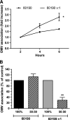
The presence of VacA increases vesicle association with AGS cells. (A) Cells were incubated for up to 6 h with DiO-labeled OMV from strain 60190 or 60190:v1, and the increase in cell-associated vesicle fluorescence was measured by flow cytometry. Data are presented as the fold increase of the fluorescence measured at 2 h for each experiment. *, results are statistically different at the 6-h time point (P < 0.01). (B) AGS cells incubated for 4 h with DiO-labeled wild-type OMV (black bar), DiD-labeled VacA mutant OMV (gray bar), or a 50:50 mix of labeled OMV (hatched bars) from both strains. Measurement of DiO fluorescence is shown over the bar designated 60190, while measurement of DiD fluorescence is shown over the bar designated 60190:v1. Results are means ± SE for three (A) or four (B) independent experiments. **, significantly fewer VacA OMV are internalized when mixed 50:50 with wild-type OMV (P = 0.002).
FIG. 4.
FIG. 4.
H. pylori LPS competitively inhibits OMV uptake by AGS cells. LPS (3.125 to 50 μg/ml) was added to cells at the same time as labeled OMV from either wild-type or VacA mutant strains. Cells were then incubated for 4 h, and fluorescence was measured after the addition of trypan blue. Results are means ± SE for three independent experiments performed in triplicate. Overall, purified LPS had a significant effect on wild-type and VacA mutant OMV uptake (P < 0.0001 and P = 0.0002, respectively). *, results are statistically significant compared to controls not treated with purified LPS (P < 0.05).
FIG. 5.
FIG. 5.
Effect of MβCD and nystatin on vacuole formation and vesicle uptake in AGS cells. (A) Cells were pretreated with MβCD (1 to 5 mg/ml) for 1 h and then incubated with 60190 OMV for 6 h. Vacuolation was measured by neutral red assay. (B) Treatment with MβCD increased the uptake of 60190 OMV with AGS cells. Cells were pretreated with MβCD for 1 h and then incubated with DiO-labeled OMV for 4 h. Fluorescence was measured after the addition of trypan blue. (C) Cells were pretreated with nystatin (5 to 50 μg/ml) for 1 h and then incubated with OMV for 6 h. Vacuolation was measured by neutral red assay. (D) Cells were pretreated with nystatin for 1 h and then incubated with DiO-labeled 60190 OMV or labeled 60190:v1 OMV for 4 h. Fluorescence was measured after the addition of trypan blue. Results for panels A and C are means ± SE for three separate experiments performed in triplicate and, for panels B and D, are means ± SE for three separate experiments. Controls were incubated with OMV in the absence of MβCD or nystatin. Overall, MβCD and nystatin had a significant effect on vacuole formation (P < 0.0001 and P = 0.027, respectively). *, **, and ***, results are statistically significant from controls not treated with MβCD or nystatin (P < 0.05, P < 0.01, and P < 0.001, respectively).
FIG. 6.
FIG. 6.
Effect of chlorpromazine on vacuole formation and vesicle uptake in AGS cells. (A) Cells were pretreated with chlorpromazine (5 to 15 μg/ml) for 30 min and then incubated with 60190 OMV for 6 h. Vacuolation was measured by neutral red assay. (B and C) Chlorpromazine reduces OMV uptake. Cells were pretreated with chlorpromazine for 30 min prior to 4 h of incubation with labeled OMV from either strain 60190 (B) or 60190:v1 (C) or with fluorescently labeled LDL (D). Fluorescence was measured after the addition of trypan blue. Results shown in panel A are means ± SE for three independent experiments performed in triplicate. Overall, vacuolation decreased in 60190 OMV-treated cells in response to a dose-dependent increase in chlorpromazine (P < 0.0001). Results are means ± SE for six independent experiments for OMV for panels B and C and for three separate experiments for LDL for panel D. Controls were incubated with OMV in the absence of chlorpromazine. Overall, there was a significant decrease in 60190 (P = 0.011), 60190:v1 (P = 0.003), and LDL (P = 0.002) uptake in response to increasing concentrations of chlorpromazine. *, and ***, results are statistically significant from controls not treated with chlorpromazine (P < 0.05 and P < 0.001, respectively).

Similar articles

Cited by

References

    1. Abrami, L., S. Liu, P. Cosson, S. H. Leppla, and F. G. van der Goot. 2003. Anthrax toxin triggers endocytosis of its receptor via a lipid raft-mediated clathrin-dependent process. J. Cell Biol. 160:321-328. - PMC - PubMed
    1. Atherton, J. C., P. Cao, R. M. Peek, Jr., M. K. Tummuru, M. J. Blaser, and T. L. Cover. 1995. Mosaicism in vacuolating cytotoxin alleles of Helicobacter pylori. Association of specific vacA types with cytotoxin production and peptic ulceration. J. Biol. Chem. 270:17771-17777. - PubMed
    1. Ayala, G., L. Torres, M. Espinosa, G. Fierros-Zarate, V. Maldonado, and J. Melendez-Zajgla. 2006. External membrane vesicles from Helicobacter pylori induce apoptosis in gastric epithelial cells. FEMS Microbiol. Lett. 260:178-185. - PubMed
    1. Bauman, S. J., and M. J. Kuehn. 2009. Pseudomonas aeruginosa vesicles associate with and are internalized by human lung epithelial cells. BMC Microbiol. 9:26. - PMC - PubMed
    1. Bayerdorffer, E., A. Neubauer, B. Rudolph, C. Thiede, N. Lehn, S. Eidt, and M. Stolte. 1995. Regression of primary gastric lymphoma of mucosa-associated lymphoid tissue type after cure of Helicobacter pylori infection. MALT Lymphoma Study Group. Lancet 345:1591-1594. - PubMed

Publication types

MeSH terms

Substances