. 2010 Dec;30(23):5562-71.
doi: 10.1128/MCB.00618-10. Epub 2010 Sep 27.

Why Dom34 stimulates growth of cells with defects of 40S ribosomal subunit biosynthesis

Affiliations

Why Dom34 stimulates growth of cells with defects of 40S ribosomal subunit biosynthesis

Arpita Bhattacharya et al. Mol Cell Biol. 2010 Dec.

Abstract

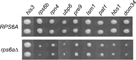
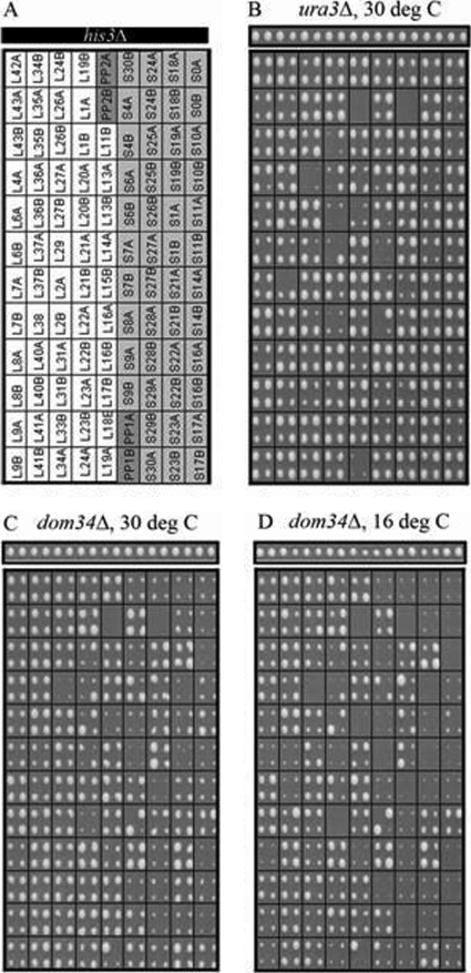
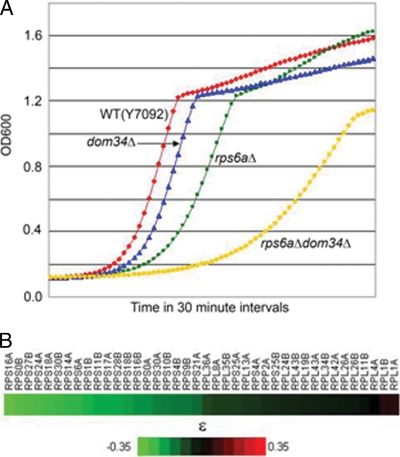
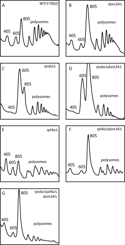
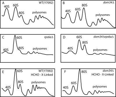
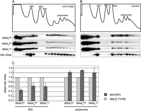
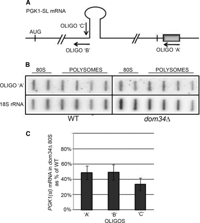
A set of genome-wide screens for proteins whose absence exacerbates growth defects due to pseudo-haploinsufficiency of ribosomal proteins in Saccharomyces cerevisiae identified Dom34 as being particularly important for cell growth when there is a deficit of 40S ribosomal subunits. In contrast, strains with a deficit of 60S ribosomal proteins were largely insensitive to the loss of Dom34. The slow growth of cells lacking Dom34 and haploinsufficient for a protein of the 40S subunit is caused by a severe shortage of 40S subunits available for translation initiation due to a combination of three effects: (i) the natural deficiency of 40S subunits due to defective synthesis, (ii) the sequestration of 40S subunits due to the large accumulation of free 60S subunits, and (iii) the accumulation of ribosomes "stuck" in a distinct 80S form, insensitive to the Mg(2+) concentration, and at least temporarily unavailable for further translation. Our data suggest that these stuck ribosomes have neither mRNA nor tRNA. We postulate, based on our results and on previously published work, that the stuck ribosomes arise because of the lack of Dom34, which normally resolves a ribosome stalled due to insufficient tRNAs, to structural problems with its mRNA, or to a defect in the ribosome itself.


Figures

FIG. 1.
FIG. 1.
Genetic interactions between rps6aΔ and different nonessential gene deletions. Shown is a composite image taken from the final plates of the SGA screen, comparing nine deletion strains (in duplicate). (Top) The query strain is the ura3Δ strain. (Bottom) The query strain is the rps6aΔ strain. The deletion strains are listed at the top, with the His3Δ strain as a control.
FIG. 2.
FIG. 2.
SGA screens using a miniarray of RP gene deletion strains. A miniarray in quadruplicate of strains carrying G418-resistant deletions of individual RP genes (A) was crossed to clon-NAT-resistant ura3Δ (B) and dom34Δ (C and D) query strains. Genetic interactions of haploid double-drug-resistant strains were scored according to standard SGA protocols (34). The figures show the final selection plates incubated at either 30°C or 16°C. The narrow panels above show the his3Δ controls on the same plates. The ura3Δ screen served as a control. Strains lacking one of the two copies of an RP gene have a relatively high probability of pseudoreversion through the duplication of the remaining gene. This is evident for the rps6aΔ strain (B to D) as well as from the occasional substantial variations in colony size among the quadruplicates.
FIG. 3.
FIG. 3.
(A) Growth curves from the Bioscreen C analysis. The growths of the wild-type (Y7092), dom34Δ, rps6aΔ, and dom34Δ rps6aΔ strains were determined from light-scattering measurements at 30-min intervals (see Materials and Methods). (B) Quantitation of genetic interactions between the DOM34 and RP genes. Doubling times of dom34Δ, rpxΔ, and dom34Δ rpxΔ mutants measured by Bioscreen C analysis at 30°C were used to calculate strain fitness. The fitness (W) of a strain deleted for a given gene, “x,” is defined as the ratio of the doubling time (D) of the wild-type strain to the deletion strain (Wx = Dwt/Dx). The value of ɛ is the deviation in the fitness phenotype of the double mutant (Wxy) from Wx × Wy, as predicted for noninteracting gene pairs by the multiplicative model (20). The values of ɛ for strains representing double mutants of dom34Δ and 40 individual RP deletion mutants are presented as a “heat map.” ɛ values of <0 indicate synthetic sick genetic interactions.
FIG. 4.
FIG. 4.
Polysome profiles of extracts of various strains. (A) Y7092; (B) dom34Δ; (C) rps6aΔ; (D) rps6aΔ dom34Δ; (E) rpl4aΔ; (F) rpl4aΔ dom34Δ; (G) rps6aΔ rpl4aΔ dom34Δ (in the presence of 12 mM MgCl2). Cultures of the six strains were grown at 30°C to mid-log phase. Cell extracts were prepared and polysome profiles were analyzed as described in Materials and Methods. Sedimentation is from left to right.
FIG. 5.
FIG. 5.
Polysome profiles of extracts of various strains. (A and E) Y7092; (B and F) dom34Δ; (C) rps6aΔ; (D) rps6aΔ dom34Δ (in the presence of 1.5 mM MgCl2). A to D are as described in the legend of Fig. 4 except that the extraction and the sucrose gradient buffers contained 1.5 mM MgCl2. E and F were prepared using HCHO but without cycloheximide (see Materials and Methods).
FIG. 6.
FIG. 6.
Distribution of several tRNAs in 80S and polysomes. Extracts of Y7092 (A) and dom34Δ (B) were analyzed on sucrose gradients in the presence of 1.5 mM MgCl2. RNA was prepared from individual fractions and separated on a 1.5% agarose gel. Northern analysis was done with 32P-end-labeled oligonucleotides complementary to sequences specific for formula image, formula image, and tRNAGln. (C) The ratio of tRNA/18S rRNA present in a particular fraction was calculated as a percentage of the total in the entire gradient. The values shown represent the ratio of tRNA/18SrRNA in 80S and polysome fractions of the dom34Δ strain normalized to that in the wild-type strain.
FIG. 7.
FIG. 7.
Distribution of “no-go” mRNA in 80S and polysomes. (A) The wild-type or dom34Δ strain was transformed with plasmids containing the PGK1(sl) gene under the control of the GAL1 promoter and tagged at the 3′ end with a string of G residues (gray box) and containing a strong stem-loop within the ORF (5). The sites of oligonucleotides A, B, and C are indicated. (B) RNA was prepared from fractions of 1.5 mM Mg2+ sucrose gradients of each strain and was applied onto slot blots, which were probed with oligonucleotide A, complementary to the tagged PGK1(sl) transcripts, and subsequently with an oligonucleotide complementary to 18S rRNA as a measure of ribosome content. The intensity of the individual bands was quantitated with a phosphorimager, and the values for the 80S and polysomal regions were individually totaled. (C) The proportion of PGK1(sl) mRNA in 80S monosomes, normalized to 18S rRNA, is shown compared to that present in the combined 80S and polysome fractions, with values for the wild-type strain set to 1.0. The slot blot was also probed with oligonucleotides complementary to the sequences within (oligonucleotide C) and just upstream of (oligonucleotide B) the stem-loop. Note that the latter will identify not only the no-go transcripts but also those from the endogenous PGK1.

Similar articles

Cited by

References

    1. Carr-Schmid, A., C. Pfund, E. A. Craig, and T. G. Kinzy. 2002. Novel G-protein complex whose requirement is linked to the translational status of the cell. Mol. Cell. Biol. 22:2564-2574. - PMC - PubMed
    1. Cole, S. E., F. J. LaRiviere, C. N. Merrikh, and M. J. Moore. 2009. A convergence of rRNA and mRNA quality control pathways revealed by mechanistic analysis of nonfunctional rRNA decay. Mol. Cell 34:440-450. - PMC - PubMed
    1. Costanzo, M., A. Baryshnikova, J. Bellay, Y. Kim, E. D. Spear, C. S. Sevier, H. Ding, J. L. Koh, K. Toufighi, S. Mostafavi, J. Prinz, R. P. St. Onge, B. VanderSluis, T. Makhnevych, F. J. Vizeacoumar, S. Alizadeh, S. Bahr, R. L. Brost, Y. Chen, M. Cokol, R. Deshpande, Z. Li, Z. Y. Lin, W. Liang, M. Marback, J. Paw, B. J. San Luis, E. Shuteriqi, A. H. Tong, N. van Dyke, I. M. Wallace, J. A. Whitney, M. T. Weirauch, G. Zhong, H. Zhu, W. A. Houry, M. Brudno, S. Ragibizadeh, B. Papp, C. Pal, F. P. Roth, G. Giaever, C. Nislow, O. G. Troyanskaya, H. Bussey, G. D. Bader, A. C. Gingras, Q. D. Morris, P. M. Kim, C. A. Kaiser, C. L. Myers, B. J. Andrews, and C. Boone. 2010. The genetic landscape of a cell. Science 327:425-431. - PMC - PubMed
    1. Davis, L., and J. Engebrecht. 1998. Yeast dom34 mutants are defective in multiple developmental pathways and exhibit decreased levels of polyribosomes. Genetics 149:45-56. - PMC - PubMed
    1. Doma, M. K., and R. Parker. 2006. Endonucleolytic cleavage of eukaryotic mRNAs with stalls in translation elongation. Nature 440:561-564. - PMC - PubMed

Publication types

MeSH terms

LinkOut - more resources