. 2010 Nov 1;185(9):5486-94.
doi: 10.4049/jimmunol.0903596. Epub 2010 Sep 27.

Heparan sulfate, including that in Bruch's membrane, inhibits the complement alternative pathway: implications for age-related macular degeneration

Affiliations

Heparan sulfate, including that in Bruch's membrane, inhibits the complement alternative pathway: implications for age-related macular degeneration

Una Kelly et al. J Immunol. .

Abstract

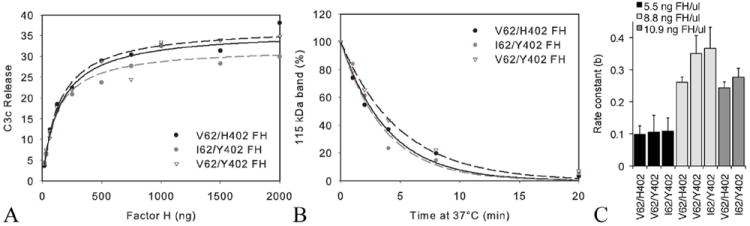
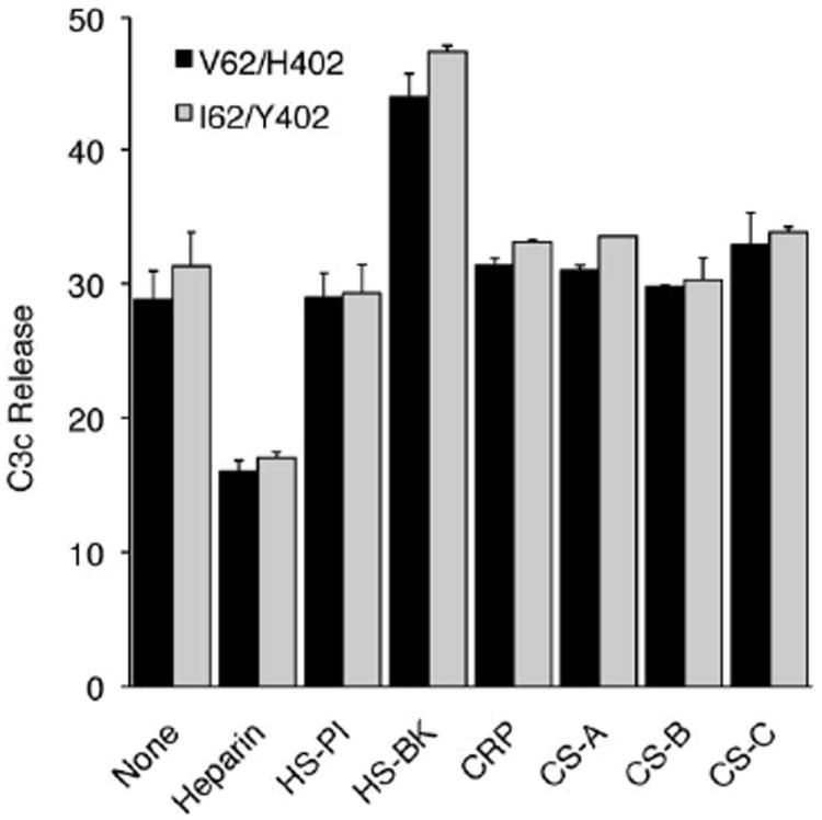
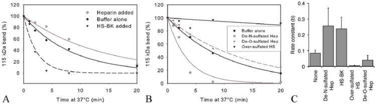
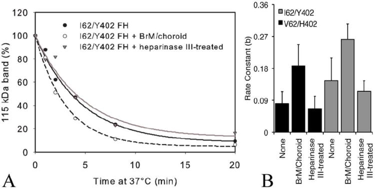
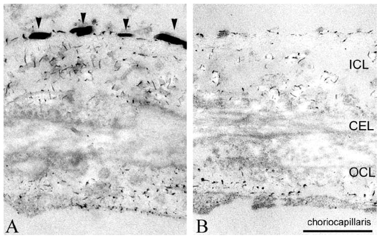
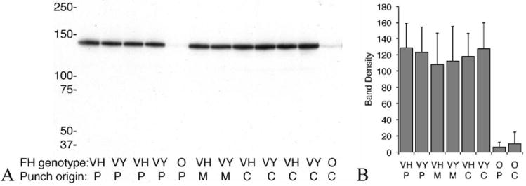
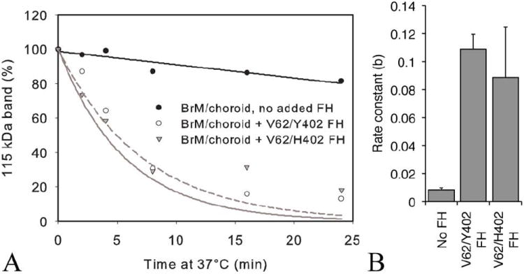
An imbalance between activation and inhibition of the complement system has been implicated in the etiologies of numerous common diseases. Allotypic variants of a key complement fluid-phase regulatory protein, complement factor H (CFH), are strongly associated with age-related macular degeneration (AMD), a leading cause of worldwide visual dysfunction, although its specific role in AMD pathogenesis is still not clear. CFH was isolated from individuals carrying combinations of two of the nonsynonymous coding variants most strongly associated with AMD risk, V62/H402 (risk haplotype variants), I62/Y402 (nonrisk haplotype variants), and V62/Y402. These proteins were used in two functional assays (cell surface- and fluid-phase-based) measuring cofactor activity of CFH in the factor I-mediated cleavage of C3b. Although no variant-specific differences in the cofactor activity were detected, when heparan sulfate (HS) was added to these assays, it accelerated the rate of C3b cleavage, and this effect could be modulated by degree of HS sulfation. Bruch's membrane/choroid, a site of tissue damage in AMD, contains high concentrations of glycosaminoglycans, including HS. Addition of human Bruch's membrane/choroid to the fluid-phase assay accelerated the C3b cleavage, and this effect was lost posttreatment of the tissue with heparinase III. Binding of CFH variants to Bruch's membrane/choroid isolated from elderly, non-AMD donor eyes, was similar, as was the functional activity of bound CFH. These findings refine our understanding of interactions of HS and complement and support the hypothesis that these interactions play a role in the transition between normal aging and AMD in Bruch's membrane/choroid.


Figures

FIGURE 1
FIGURE 1
(A) Cofactor activities of allotypic variants of purified CFH (FH). In this graph of a representative assay, C3c release was measured (% cpm in supernatant) after 4 min at 37°C in cell-based assays of CFH cofactor activity with increasing concentrations of CFH (I62/Y402, V62/Y402 or V62/H402), factor I and 125I-EC3b cells. (B) Cleavage of C3b was measured in the fluid-phase assay using the I62/Y402, V62/Y402 or V62/H402 variants of purified CFH. Decay of the 115 kDa band (C3bα′) was determined by densitometry using ImageJ, expressed as a % of the 115 kDa band at time zero and plotted using SigmaPlot. (C) The rate constant for the exponential decay was measured using the equation y=a*exp(-b*x). There was no statistically significant difference in the rate constants between variants of CFH used. This held true over a range of CFH concentrations tested (5.5, 8.8 and 10.9 ng FH/μl) (p>0.07 for all comparisons of the different variants). [5.5 ng FH/μl], n=7; [8.8 ng FH/μl], n=3; and [10.9 ng FH/μl], n=5.
FIGURE 2
FIGURE 2
The effect of various molecules on cofactor activity of allotypic variants of purified CFH in the cell surface-based assay. CFH, factor I and heparin, heparan sulfate (HS) isolated from porcine intestinal mucosa (HS-PI), HS isolated from bovine kidney (HS-BK), chondroitin sulfate (CS)-A, CS-B, CS-C or CRP were preincubated at room temperature before 125I- EC3b cells were added and incubated for at 37°C (mean ± SD). Differences in between the allotypic variants are not statistically significant, but differences between the effect of heparin and HS-BK on the modulation of the co-factor activity are. Heparin decreased C3c release (***, p< 0.001) and HS-BK increased C3c release (p< 0.001). Data depicted are representative of two independent experiments, each done in triplicate.
FIGURE 3
FIGURE 3
The rate of C3b cleavage is reduced by highly sulfated GAGs (heparin and over-sulfated HS) and accelerated by less N-sulfated GAGs (HS-BK and de-N-sulfated heparin). (A) Cleavage of C3b was measured in the fluid-phase assay using purified CFH with heparin (Hep) or HS-BK added. CFH (390 ng) and Factor I (200 ng) with or without 146 μg of a specific GAG were mixed and added to C3b (3.4 μg) in a total volume of 70 μl EDTA-VBS. At various time points, 10μl aliquots were removed and proteins separated on a gel as previously described. Decay of the 115 kDa band (C3bα′) was determined by densitometry using ImageJ, expressed as a % of the 115 kDa band at time zero and plotted using SigmaPlot. (B) Cleavage of C3b measured in the fluid-phase assay using purified CFH with sulfate-modified heparin (de-N- or de-O-sulfated) or over-sulfated HS added. (C) The rate of the fluid phase reaction is greatly reduced by over-sulfated HS (***, p< 0.001) and to a lesser extent heparin and de-O-sulfated heparin (**, p< 0.01) whereas HS-BK and de-N-sulfated heparin significantly increase the rate of this reaction (p< 0.001). Similar results were obtained using V62/Y402 or V62/H402 (data not shown). None (no added GAG), n=18; de-N-sulfated heparin, n=18; HS-BK, n=6; over-sulfated HS, n=9; de-O-sulfated heparin, n=6
FIGURE 4
FIGURE 4
Addition of human Bruch’s membrane/choroid punches increases the rate of cleavage of C3b by factor I and variants of CFH. (A) Cleavage of C3b was measured in the presence of four 6 mm diameter punches of Bruch’s membrane/choroid from the peripheral (two) and central (two) regions of posterior eyecups from a pair of human eyes (68-year-old donor) per reaction. Punches (four treated or four untreated with heparinase III) were added to CFH and factor I in ice cold EDTA-VBS. C3b was added, and the tubes placed at 37°C. The control tube had no punches added. At various times 18 μl was removed and run on a gel as previously described. Representative results from one isoform, I62/Y402, are depicted. (B) I62/Y402 and V62/H402 CFH both showed statistically significant increases in the rate constant in the presence of tissue (p< 0.01; n=5 each). There were no statistically significant differences between the variants of CFH alone compared to reactions with tissue pre-treated with heparanase III V62/H402). Data depicted are representative of 5 independent experiments from 5 pairs of donor eyes.
FIGURE 5
FIGURE 5
Electron micrograph of human Bruch’s membrane/choroid stained for proteoglycans using Cupromeronic Blue. (A) HS [arrowheads in inner collagenous layer (ICL)], chondroitin sulfate and dermatan sulfate are stained, and can be distinguished based on their length/diameter and locations (Call and Hollyfield, 1990). (B) The HS associated with the basal lamina of pigment epithelium and choriocapillaris has disappeared following heparinase III treatment. CEL, central elastin layer; OCL, outer collagen layer. Scale bar = 500 nm.
FIGURE 6
FIGURE 6
CFH binding to Bruch’s membrane/choroid. Immunoblot of CFH variant binding to a single 6 mm Bruch’s membrane/choroid punches isolated from different regions of the human eye. Peripheral (P), central (C) or macula (M) punches were incubated with buffer alone (O) or in buffer with V62/Y402 (VY) or V62/H402 (VH) variants of CFH. Following incubation the punches were washed thoroughly and added to 35 μl of XT sample buffer. The sample was vortexed vigorously for 30 seconds, left at room temperature for 10 min, vortexed again, spun down and supernatant was run on a gel as previously described. The gel was transferred to nitrocellulose and probed with mouse monoclonal anti-human CFH (Serotec, OX24). Immunoreactive proteins were visualized with ECL and quantified by densitometry, n=18 for both VH and VY on peripheral punches, n=13 for both on central punches and n=3 for macular punches. (B) No statistical difference in the amount of each added variant of CFH bound to the Bruch’s membrane/choroid tissue was detected (p> 0.07), no matter where the punches were taken from. Very little endogenous CFH is detected in the punches with no added CFH.
FIGURE 7
FIGURE 7
Functional cofactor activity of CFH variant prebound to a single 6 mm Bruch’s membrane/choroid punch. (A) Cleavage of C3b measured in the fluid-phase assay using one Bruch’s membrane/choroid 6 mm punch preincubated with or without CFH (V62/Y402 or V62/H402) and washed, then added to VBS/EDTA, factor I and C3b. Aliquots were removed at 0, 2, 4, 8, 16, and 24 min and these samples were boiled and run with reducing buffer on 10% Bis-tris Criterion XT gels with MOPS buffer. After silver staining, the gel was scanned and the density of the 115 kDa band measured with ImageJ software, and SigmaPlot 9 used to create the graphs. The example shown is representative of 6 separate experiments. (B) Rate constants showed no significant differences between CFH variants independent of region tissue punch was taken from (i.e. periphery (as shown) or central) (p> 0.2).

Similar articles

Cited by

References

    1. Rodriguez de Cordoba S, Esparza-Gordillo J, Goicoechea de Jorge E, Lopez-Trascasa M, Sanchez-Corral P. The human complement factor H: functional roles, genetic variations and disease associations. Molecular immunology. 2004;41:355–367. - PubMed
    1. Sharma AK, Pangburn MK. Identification of three physically and functionally distinct binding sites for C3b in human complement factor H by deletion mutagenesis. Proceedings of the National Academy of Sciences of the United States of America. 1996;93:10996–11001. - PMC - PubMed
    1. DiScipio RG. Ultrastructures and interactions of complement factors H and I. J Immunol. 1992;149:2592–2599. - PubMed
    1. Soames CJ, Sim RB. Interactions between human complement components factor H, factor I and C3b. The Biochemical journal. 1997;326(Pt 2):553–561. - PMC - PubMed
    1. Meri S, Pangburn MK. Discrimination between activators and nonactivators of the alternative pathway of complement: regulation via a sialic acid/polyanion binding site on factor H. Proceedings of the National Academy of Sciences of the United States of America. 1990;87:3982–3986. - PMC - PubMed

Publication types

MeSH terms