. 2010 Oct;136(4):389-405.
doi: 10.1085/jgp.201010475.

Activation of the K(ATP) channel by Mg-nucleotide interaction with SUR1

Affiliations

Activation of the K(ATP) channel by Mg-nucleotide interaction with SUR1

Peter Proks et al. J Gen Physiol. 2010 Oct.

Abstract

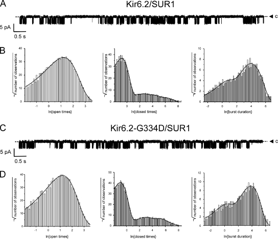
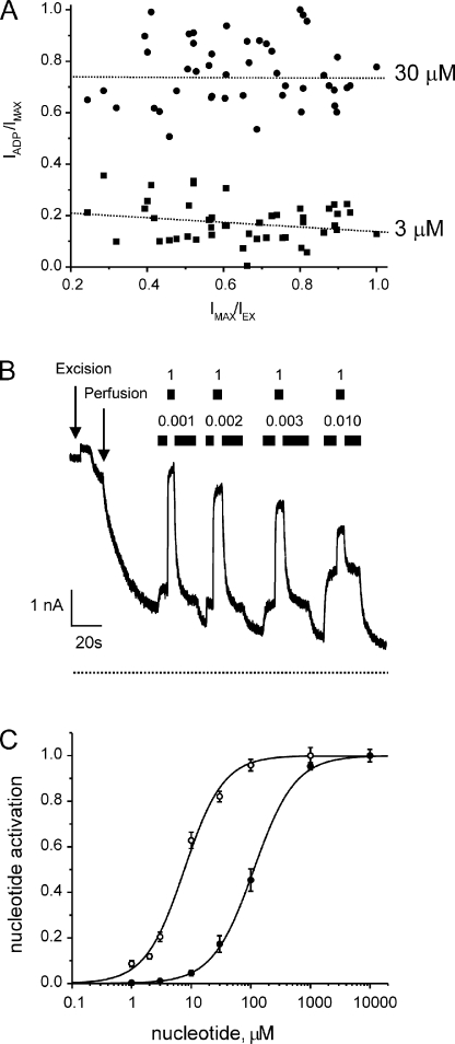
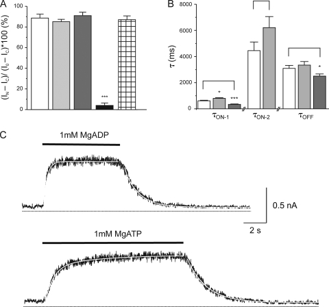
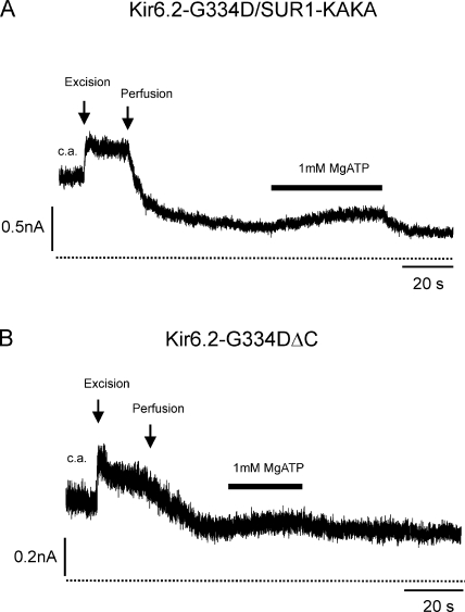
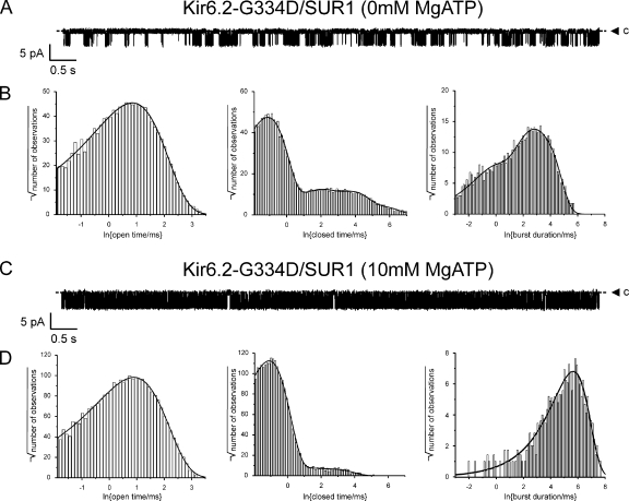
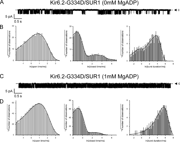
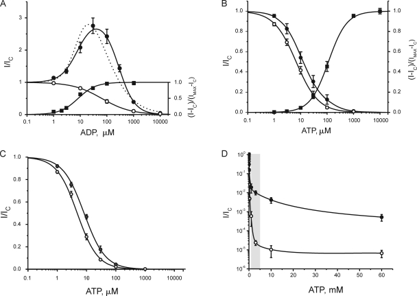
The mechanism of adenosine triphosphate (ATP)-sensitive potassium (K(ATP)) channel activation by Mg-nucleotides was studied using a mutation (G334D) in the Kir6.2 subunit of the channel that renders K(ATP) channels insensitive to nucleotide inhibition and has no apparent effect on their gating. K(ATP) channels carrying this mutation (Kir6.2-G334D/SUR1 channels) were activated by MgATP and MgADP with an EC(50) of 112 and 8 µM, respectively. This activation was largely suppressed by mutation of the Walker A lysines in the nucleotide-binding domains of SUR1: the remaining small (∼10%), slowly developing component of MgATP activation was fully inhibited by the lipid kinase inhibitor LY294002. The EC(50) for activation of Kir6.2-G334D/SUR1 currents by MgADP was lower than that for MgATP, and the time course of activation was faster. The poorly hydrolyzable analogue MgATPγS also activated Kir6.2-G334D/SUR1. AMPPCP both failed to activate Kir6.2-G334D/SUR1 and to prevent its activation by MgATP. Maximal stimulatory concentrations of MgATP (10 mM) and MgADP (1 mM) exerted identical effects on the single-channel kinetics: they dramatically elevated the open probability (P(O) > 0.8), increased the mean open time and the mean burst duration, reduced the frequency and number of interburst closed states, and eliminated the short burst states. By comparing our results with those obtained for wild-type K(ATP) channels, we conclude that the MgADP sensitivity of the wild-type K(ATP) channel can be described quantitatively by a combination of inhibition at Kir6.2 (measured for wild-type channels in the absence of Mg(2+)) and activation via SUR1 (determined for Kir6.2-G334D/SUR1 channels). However, this is not the case for the effects of MgATP.


Figures

Figure 1.
Figure 1.
Data segments used for noise analysis of macroscopic Kir6.2-G334D/SUR1 currents. Representative example of activation of Kir6.2-G334D/SUR1 channels by 10 mM MgATP. Nucleotide application is indicated by the bar. The parallel vertical lines denote 1-s data segments during which the current reaches a quasi–steady-state level. The dotted horizontal line indicates the zero current level. PO and N in the absence of nucleotides (open triangles in Fig. 4, B–D) were calculated from segment 1, and in the presence of 10 mM MgATP (open circles in Fig. 4, B–D) from segment 2.
Figure 2.
Figure 2.
Rundown of Kir6.2/SUR1 and Kir6.2-G334D/SUR1 channels in macropatches. (A) Macroscopic Kir6.2/SUR1 currents at −60 mV recorded from a giant patch. Patch excision and the start of the perfusion with nucleotide-free intracellular solution are indicated by the arrows. The dotted line represents the zero current level. (B) Macroscopic Kir6.2-G334D/SUR1 currents recorded at −60 mV. Patch excision and the start of the perfusion with nucleotide-free solution are indicated by the arrows. The dotted line represents the zero current level. (C) Mean macroscopic KATP current at −60 mV during the first 60 s of rundown, expressed as a fraction of the maximum value reached after excision, is plotted as a function of the time after patch excision for Kir6.2/SUR1 (open circles; n = 10) and Kir6.2-G334D/SUR1 channels (filled circles; n = 10). The lines are the best fit of a double-exponential function to the mean data (solid line, Kir6.2/SUR1; dotted line, Kir6.2-G334D/SUR1).
Figure 3.
Figure 3.
Kinetics of Kir6.2/SUR1 and Kir6.2-G334D/SUR1 channels. (A) Single Kir6.2/SUR1 currents recorded at −60 mV in nucleotide-free solution. c, the closed current level. (B) Distributions of Kir6.2/SUR1 open times (left), closed times (middle), and burst times (right) in nucleotide-free solution. The PO(0) of the channel illustrated was 0.36. The distributions were fit with the following parameters. Open times: τO = 3.39 ms (τO,COR = 2.46 ms); closed times: τC1 = 0.44 ms, aC1 = 0.949; τC2 = 9.6 ms, aC2 = 0.022; τC3 = 53 ms, aC3 = 0.020; τC4 = 363 ms, aC4 = 0.009; burst times: τB1 = 93 ms, aB1 = 0.526, τB2 = 31 ms, aB2 = 0.328, τB3 = 1.7 ms, aB3 = 0.145. The mean burst duration (τB) was 59.3 ms. (C) Single Kir6.2-G334D/SUR1 currents recorded at −60 mV in nucleotide-free solution. c, the closed current level. (D) Distributions of Kir6.2-G334D/SUR1 open times (left), closed times (middle), and burst times (right) in nucleotide-free solution. The PO(0) of the channel illustrated was 0.27. The distributions were fit with the following parameters. Open times: τO = 3.32 ms (τO,COR = 2.32 ms); closed times: τC1 = 0.39 ms, aC1 = 0.940; τC2 = 16.9 ms, aC2 = 0.030; τC3 = 139 ms, aC3 = 0.023; τC4 = 415 ms, aC4 = 0.007; burst times: τB1 = 61 ms, a1 = 0.651; τB2 = 20 ms, a2 = 0.187; τB3 = 1.0 ms, a3 = 0.162. The mean burst duration (τB) was 43.6 ms.
Figure 4.
Figure 4.
Rundown and reactivation of Kir6.2-G334D/SUR1 channels by MgATP and MgADP. (A) Representative record showing repetitive activation of Kir6.2-G334D/SUR1 current by 10 mM MgATP. Nucleotide application is indicated by the bars. Patch excision and the start of the perfusion with nucleotide-free solution are indicated by the arrows. The dotted line indicates the zero current level. (B–D) Estimates of NrPO (B), PO (C), and the fraction of functional channels (N) (D) calculated from noise analysis of current records obtained during repetitive application of 10 mM MgATP (open circles) or 1 mM MgADP (open squares), or in control solution before the application of nucleotide (open triangles). Data are mean ± SEM from 10 patches. The lines are drawn by eye.
Figure 5.
Figure 5.
Nucleotide activation of Kir6.2-G334D/SUR1 channels. (A) Kir6.2-G334D/SUR1 current magnitude (IADP) in the presence of 3 µM MgADP (filled squares) or 30 µM MgADP (filled circles), expressed as a fraction of that in the presence of a maximal stimulatory ADP concentration (1 mM; IMAX). This is plotted against the current magnitude in the presence of 1 mM MgADP (IMAX), expressed as a fraction of the current immediately after excision (IEX). The dotted lines represent the best linear fit to the data at 3 µM (lower) or 30 µM (upper) MgADP. (B) Repetitive activation of Kir6.2-G334D/SUR1 channels by different concentrations of MgADP. Nucleotide application is indicated by the bars, and the numbers above the bars give the nucleotide concentration in millimolars. The dotted line indicates the zero current level. (C) Concentration–response relationships for activation of Kir6.2-G334D/SUR1 channels by MgATP (filled circles; n = 8) or MgADP (open circles; n = 8). The lines are the best fit of Eq. 1 to the mean data with EC50 = 7.7 µM and h = 1.30 (MgADP) and EC50 = 112 µM and h = 1.25 (MgATP).
Figure 6.
Figure 6.
Effect of other Mg-nucleotides. (A) Mean increase in Kir6.2-G334D/SUR1 current in the presence of different nucleotides (refer to Materials and methods). White bar, 1 mM MgATP (n = 13); pale gray bar, 1 mM MgATPγS (n = 6); dark gray bar, 1 mM MgADP (n = 7); black bar, 1 mM MgAMP-PNP (n = 6); crosshatched bar, 1 mM MgAMP-PNP plus 1 mM MgATP (n = 6). ***, P < 0.001 (against MgATP). (B) Mean time constants (τ) of current activation (τON-1 and τON-2) and deactivation (τOFF) by MgATP (white bars, n = 13), MgATPγS (pale gray bars, n = 6), and MgADP (dark gray bars, n = 7). *, P < 0.5; ***, P < 0.001 (against MgATP). (C) Time course of current activation and deactivation by 1 mM MgADP (top) and 1 mM MgATP (bottom). Nucleotide application is indicated by the bar, and the dotted line represents the zero current level. The white lines are the best fit of one (MgADP) or two (MgATP) exponential components for activation (A1*et/τ1+A2*et/τ2) and one for deactivation (A*e−t/τ). For MgADP: AON-1 = 680 pA, τON-1 = 330 ms; AOFF = 680 pA, τOFF = 1,320 ms. For MgATP: AON-1 = 490 pA, τON-1 = 730 ms; AON-2 = 180 pA, τON-2 = 4,060 ms; AOFF = 670 pA; τOFF = 1,835 ms.
Figure 7.
Figure 7.
NBD-independent nucleotide activation. Representative examples of the effect of 1 mM MgATP on Kir6.2-G334D/SUR-KAKA channels (A) or Kir6.2-G334DΔC channels (B). Patch excision and the start of the perfusion with nucleotide-free solution are indicated by the arrows and MgATP application by the bars. The dotted line indicates the zero current level.
Figure 8.
Figure 8.
Effect of 10 mM MgATP on Kir6.2-G334D/SUR1 kinetics. (A and C) Single Kir6.2-G334D/SUR1 currents recorded at −60 mV from the same patch first in nucleotide-free solution (A), and then in the presence of 10 mM MgATP (C). c, the closed current level. (B and D) Distributions of open times (left), closed times (middle), and burst durations (right) in 0 mM MgATP (B; PO = 0.26) and in 10 mM MgATP (D; PO = 0.82). The distributions were fit with the following parameters. For 0 mM MgATP (B): open times: τO = 2.34 ms (τO,COR = 1.57 ms); closed times: τC1 = 0.33 ms, aC1 = 0.901; τC2 = 5.5 ms, aC2 = 0.043; τC3 = 39 ms, aC3 = 0.049; τC4 = 192 ms, aC4 = 0.007; burst times: τB1 = 33 ms, aB1 = 0.53; τB2 = 9.3 ms, aB2 = 0.34; τB3 = 0.63 ms, aB3 = 0.13. The mean burst duration (τB) was 20.8 ms. For 10 mM MgATP (D): open times: τO = 2.73 ms (τO,COR = 1.75 ms); closed times: τC1 = 0.33 ms, aC1 = 0.994; τC2 = 2.5 ms, aC2 = 0.004; τC3 = 14 ms, aC3 = 0.003; burst times: τB ≡ τB1 = 282 ms.
Figure 9.
Figure 9.
Effect of 1 mM MgADP on Kir6.2-G334D/SUR1 kinetics. (A and C) Single Kir6.2-G334D/SUR1 currents recorded at −60 mV from the same patch first in nucleotide-free solution (A), and then in 1 mM MgADP (C). (B and D) Distributions of open times (left), closed times (middle), and burst durations (right) in 0 mM MgADP (B; PO = 0.39) and 1 mM MgADP (D; PO = 0.82). The distributions were fit with the following parameters. For 0 mM MgADP (B): open times: τO = 2.31 ms (τO,COR = 1.51 ms); closed times: τC1 = 0.33 ms, aC1 = 0.942; τC2 = 7.3 ms, aC2 = 0.021; τC3 = 36 ms, aC3 = 0.034; τC4 = 230 ms, aC4 = 0.003; burst times: τB1 = 41 ms, aB1 = 0.770; τB2 = 5.6 ms, aB2 = 0.115; τB3 = 0.46 ms, aB3 = 0.115. The mean burst duration (τ) was 32.3 ms. For 1 mM MgADP (D): open times: τO = 2.61 ms (τO,COR = 1.77 ms); closed times: τC1 = 0.38 ms, aC1 = 0.990; τC2 = 3.8 ms, aC2 = 0.001; τC3 = 14 ms, aC3 = 0.009; burst times: τB ≡ τB1 = 263 ms.
Figure 10.
Figure 10.
Comparison of the activatory and inhibitory effects of Mg-nucleotides. (A) Concentration–response relationships for activation and inhibition of macroscopic KATP currents by ADP. Inhibition was measured for Kir6.2/SUR1 currents in the absence of Mg2+ (open circles; n = 6). Activation was measured for Kir6.2-G334D/SUR1 currents in the presence of Mg2+ (filled squares; n = 8; data from Fig. 5 C). Filled circles (n = 15) indicate data obtained for Kir6.2/SUR1 currents in the presence of Mg2+, where activation and inhibition are simultaneously present. The solid lines are the best fit of Eqs. 1–3 to the mean data. For Kir6.2-G334D/SUR1 channels, data were fit with Eq. 1, EC50 = 7.7 µM and h = 1.30. For Kir6.2/SUR1 channels, data in Mg-free solution were fit with Eq. 2, IC50 = 62 µM and h = 0.81, and data in Mg-containing solution were fit with Eq. 3 using fixed activation parameters (EC50 = 8 µM and h2 = 1.3) and a = 2; IC50 = 280 µM and h1 = 1.4 for inhibition. The dotted line is best fit of the data for Kir6.2/SUR1 in Mg-containing solution assuming no interaction between the activatory and inhibitory sites (IC50 was fixed at 62 µM and EC50 at 8 µM) and a = 2.5, h1 = 1.3, and h2 = 1.3. (B) Concentration–response relationships for activation and inhibition of macroscopic KATP currents by ATP. Inhibition was measured for Kir6.2/SUR1 currents in the absence of Mg2+ (open circles; n = 6). Activation was measured for Kir6.2-G334D/SUR1 currents in the presence of Mg2+ (filled squares; n = 8; data from Fig. 5 C). Filled circles (n = 8) indicate data obtained for Kir6.2/SUR1 currents in the presence of Mg2+, where activation and inhibition are simultaneously present. For Kir6.2-G334D/SUR1 channels, the line through the mean data were fit with Eq. 1, EC50 = 112 µM and h = 1.25. For Kir6.2/SUR1 channels in the absence of Mg2+, the line through the mean data are the best fit of Eq. 2, with IC50 = 7.1 µM and h = 1.1. For Kir6.2/SUR1 channels, data in Mg-containing solution were fit with Eq. 2, with IC50 = 14 µM and h = 1.0. (C) Concentration–inhibition relationships for Kir6.2/SUR1-KAKA currents in the absence (open circles; n = 8) and presence of Mg2+ (gray circles; n = 8). The lines through the mean data are the best fit of Eq. 2, with IC50 = 4.8 µM and h = 1.2 for Mg2+-free solution, and IC50 = 8.3 µM and h = 1.2 for Mg2+ solution. (D) Concentration–response inhibition relationships for Kir6.2/SUR1 currents in the absence (open circles; n = 8) or presence of Mg2+ (filled circles; n = 8). The physiological range of [ATP]i is shown by the bar. The lines are drawn by eye.

Similar articles

Cited by

References

    1. Abraham M.R., Selivanov V.A., Hodgson D.M., Pucar D., Zingman L.V., Wieringa B., Dzeja P.P., Alekseev A.E., Terzic A. 2002. Coupling of cell energetics with membrane metabolic sensing. Integrative signaling through creatine kinase phosphotransfer disrupted by M-CK gene knock-out. J. Biol. Chem. 277:24427–24434 10.1074/jbc.M201777200 - DOI - PubMed
    1. Aittoniemi J., Fotinou C., Craig T.J., de Wet H., Proks P., Ashcroft F.M. 2009. Review. SUR1: a unique ATP-binding cassette protein that functions as an ion channel regulator. Philos. Trans. R. Soc. Lond. B Biol. Sci. 364:257–267 10.1098/rstb.2008.0142 - DOI - PMC - PubMed
    1. Alekseev A.E., Brady P.A., Terzic A. 1998. Ligand-insensitive state of cardiac ATP-sensitive K+ channels. Basis for channel opening. J. Gen. Physiol. 111:381–394 10.1085/jgp.111.2.381 - DOI - PMC - PubMed
    1. Antcliff J.F., Haider S., Proks P., Sansom M.S.P., Ashcroft F.M. 2005. Functional analysis of a structural model of the ATP-binding site of the KATP channel Kir6.2 subunit. EMBO J. 24:229–239 10.1038/sj.emboj.7600487 - DOI - PMC - PubMed
    1. Ashcroft F.M. 2007. The Walter B. Cannon Physiology in Perspective Lecture, 2007. ATP-sensitive K+ channels and disease: from molecule to malady. Am. J. Physiol. Endocrinol. Metab. 293:E880–E889 10.1152/ajpendo.00348.2007 - DOI - PubMed

Publication types

MeSH terms