Incidental findings in imaging research: evaluating incidence, benefit, and burden
- PMID: 20876402
- PMCID: PMC3721142
- DOI: 10.1001/archinternmed.2010.317
Incidental findings in imaging research: evaluating incidence, benefit, and burden
Abstract
Background: Little information exists concerning the frequency and medical significance of incidental findings (IFs) in imaging research.
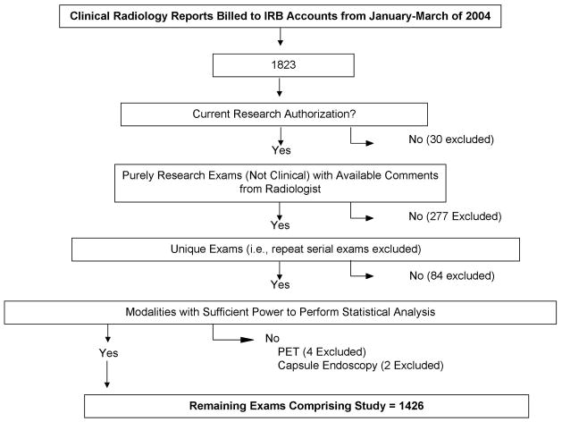
Methods: Medical records of research participants undergoing a research imaging examination interpreted by a radiologist during January through March 2004 were reviewed, with 3-year clinical follow-up. An expert panel reviewed all IFs generating clinical action to determine medical benefit/burden on the basis of predefined criteria. The frequency of IFs that generated further clinical action was estimated by modality, body part, age, and sex, along with net medical benefit or burden.
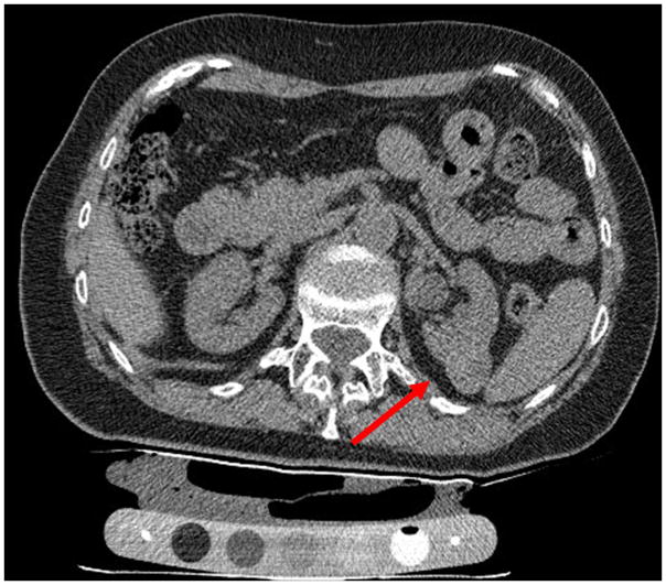
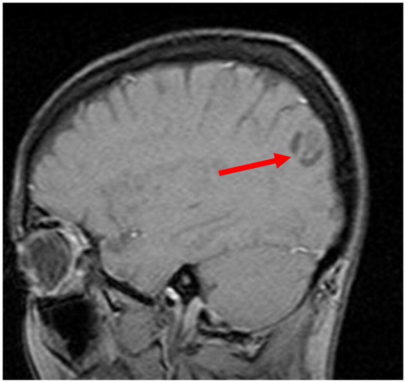
Results: Of 1426 research imaging examinations, 567 (39.8%) had at least 1 IF (1055 total). Risk of an IF increased significantly by age (odds ratio [OR], 1.5; 95% confidence interval, 1.4-1.7 per decade increase). Abdominopelvic computed tomography generated more IFs than other examinations (OR, 18.9 vs ultrasonography; 9.2% with subsequent clinical action), with computed tomography of the thorax and magnetic resonance imaging of the head next (OR, 11.9 and 5.9; 2.8% and 2.2% with action, respectively). Of the 567 examinations with an IF, 35 (6.2%) generated clinical action, resulting in clear medical benefit in 1.1% (6 of 567) and clear medical burden in 0.5% (3 of 567). Medical benefit/burden was usually unclear (26 of 567 [4.6%]).
Conclusions: Frequency of IFs in imaging research examinations varies significantly by imaging modality, body region, and age. Research imaging studies at high risk for generating IFs can be identified. Routine evaluation of research images by radiologists may result in identification of IFs in a high number of cases and subsequent clinical action to address them in a small but significant minority. Such clinical action can result in medical benefit to a small number of patients.
Figures

Comment in
-
Responding to incidental findings on research imaging studies: now what?Arch Intern Med. 2010 Sep 27;170(17):1522-4. doi: 10.1001/archinternmed.2010.306. Arch Intern Med. 2010. PMID: 20876401 No abstract available.
-
Another point of view.Arch Intern Med. 2011 Apr 11;171(7):707; author reply 707-8. doi: 10.1001/archinternmed.2011.119. Arch Intern Med. 2011. PMID: 21482853 No abstract available.
References
-
- Gluecker TM, Johnson CD, Wilson LA, et al. Extracolonic findings at CT colonography: evaluation of prevalence and cost in a screening population. Gastroenterology. 2003 Apr;124(4):911–916. - PubMed
-
- Hara AK. Extracolonic findings at CT colonography. Semin Ultrasound CT MR. 2005 Feb;26(1):24–27. - PubMed
-
- Hara AK, Johnson CD, MacCarty RL, Welch TJ. Incidental extracolonic findings at CT colonography. Radiology. 2000 May;215(2):353–357. - PubMed
Publication types
MeSH terms
Grants and funding
LinkOut - more resources
Full Text Sources
Medical
Miscellaneous