. 2010 Dec 9;116(24):5191-9.
doi: 10.1182/blood-2010-03-275529. Epub 2010 Sep 27.

Aluminum adjuvants elicit fibrin-dependent extracellular traps in vivo

Affiliations

Aluminum adjuvants elicit fibrin-dependent extracellular traps in vivo

Michael W Munks et al. Blood. .

Abstract

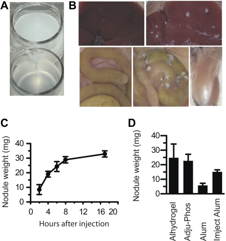
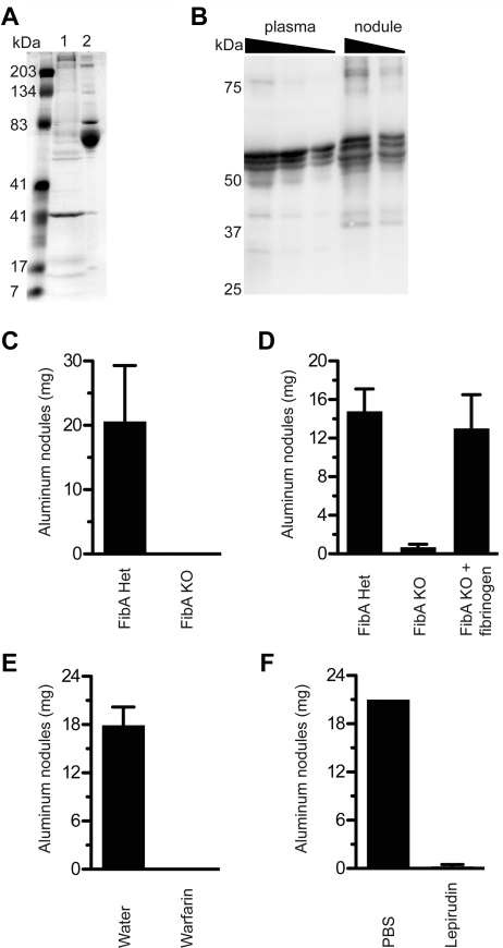
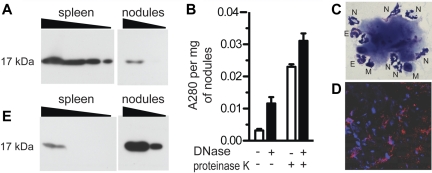
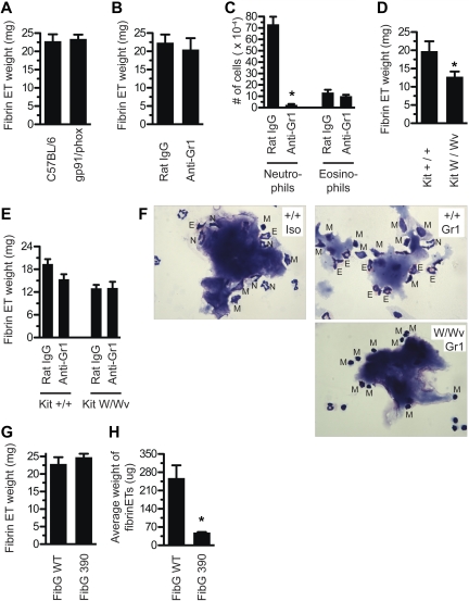
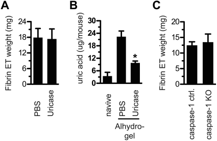
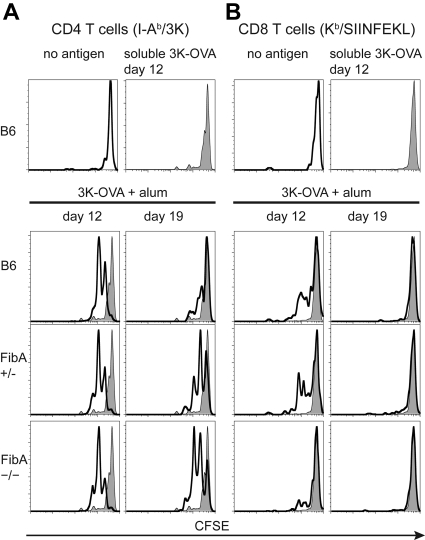
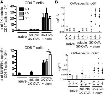
It has been recognized for nearly 80 years that insoluble aluminum salts are good immunologic adjuvants and that they form long-lived nodules in vivo. Nodule formation has long been presumed to be central for adjuvant activity by providing an antigen depot, but the composition and function of these nodules is poorly understood. We show here that aluminum salt nodules formed within hours of injection and contained the clotting protein fibrinogen. Fibrinogen was critical for nodule formation and required processing to insoluble fibrin by thrombin. DNase treatment partially disrupted the nodules, and the nodules contained histone H3 and citrullinated H3, features consistent with extracellular traps. Although neutrophils were not essential for nodule formation, CD11b(+) cells were implicated. Vaccination of fibrinogen-deficient mice resulted in normal CD4 T-cell and antibody responses and enhanced CD8 T-cell responses, indicating that nodules are not required for aluminum's adjuvant effect. Moreover, the ability of aluminum salts to retain antigen in the body, the well-known depot effect, was unaffected by the absence of nodules. We conclude that aluminum adjuvants form fibrin-dependent nodules in vivo, that these nodules have properties of extracellular traps, and the nodules are not required for aluminum salts to act as adjuvants.


Figures

Figure 1
Figure 1
Aluminum adjuvants rapidly form into nodules in vivo. (A) A suspension of Alhydrogel in PBS. (B) Top left, a normal mouse liver; top right, liver of a mouse injected with Alhydrogel; bottom left, intestines of an uninjected mouse; bottom middle, intestines of a mouse injected with Alhydrogel; bottom right, leg muscle of a mouse injected with Alhydrogel. Photos taken 4 hours after injection. (C) At various times after intraperitoneal injection of 4 mg of Alhydrogel, nodules were collected and weighed. (D) At 4 hours after injection of 4 mg of Alhydrogel, Adju-Phos, aluminum potassium sulfate (Alum), or Imject Alum, nodules all form nodules within 4 hours in vivo. All experiments included at least 3 mice per group. Panel B was repeated more than 5 times. Panels C and D were repeated twice each.
Figure 2
Figure 2
Fibrinogen and thrombin are required for aluminum nodule formation. (A) At 4 hours after Alhydrogel injection, nodules were collected and protein was eluted by boiling in SDS, separation by SDS-PAGE, and analysis with Coomassie staining. Lane 1, protein eluted from aluminum nodules; lane 2, normal mouse serum. (B) Plasma (0.1, 0.05, 0.025 μL), and proteins eluted from 4-hour Alhydrogel nodules (4 μg, 2 μg), were analyzed by the use of Western blot for fibrinogen under reducing conditions. Note that the fibrinogen bands in the plasma samples are probably compressed by albumin, which is very abundant but not visualized. (C) FibA KO mice were tested for the ability to form aluminum nodules in vivo compared with littermate controls. (D) FibA KO mice were injected with 1 mg of human fibrinogen 10 minutes before an injection of Alhydrogel. At 5 hours later, mice were evaluated for nodule formation. (E) Mice given 2.5 μg/mL warfarin in their drinking water for 7 days were injected with Alhydrogel and evaluated 5 hours later for the presence of aluminum nodules. (F) Mice were injected intraperitoneally with lepirudin 10 minutes before injection with Alhydrogel, then evaluated 5 hours later for the presence of aluminum nodules. All experiments included at least 3 mice per group and were performed at least 3 times, except panel D, which was performed twice.
Figure 3
Figure 3
Aluminum nodules have features consistent with ETs. (A) Alhydrogel nodules (10 μg, 3 μg) were analyzed by Western blot for the presence of histone H3, compared with spleen cells (1.0, 0.3, 0.1, 0.03 × 10⁶ cell equivalents). (B) Alhydrogel nodules were digested in the presence of DNase alone, proteinase K alone, or both. Absorbance of released material was measured at 280 nm. (C) Aluminum nodules were analyzed by Wright-Giemsa staining at ×1000 magnification. N indicates neutrophil; E, eosinophil; M, mononuclear cell. (D) Aluminum nodules were analyzed by immunofluorescence staining for DNA with DAPI (blue) and myeloperoxidase (red). (E) A gel identical to that shown in panel A (and run in parallel) was analyzed by Western blot for the presence of citH3. All experiments used Alhydrogel nodules collected 4-5 hours after injection and were performed at least 3 times.
Figure 4
Figure 4
Fibrinogen interacts with CD11b+ cells but there are redundant ET-releasing cell types. (A) Mice deficient for gp91/phox were injected with Alhydrogel and evaluated for fibrin ET formation 5 hours later. (B) Mice were depleted of neutrophils with 200 μg of anti–Gr-1 intravenously at day −1, or injected with isotype control Ab, then tested for fibrin ET formation 5 hours after Alhydrogel injection. (C) Peritoneal lavage was collected from mice in Figure 4B. Cells were stained by Wright-Giemsa and neutrophils and eosinophils were counted. *P < .001 compared with Rat IgG. (D) KitW/Wv mice, or littermate controls, were injected with Alhydrogel and evaluated for fibrin ET formation 5 hours later. Note that fibrin ET formation was decreased by 35% (P = .03). (E) KitW/Wv mice, or littermate controls, were depleted of neutrophils with 200 μg of anti–Gr-1 intravenously at day −1 or given isotype control Ab. On day 0, mice were injected with Alhydrogel and evaluated for fibrin ET formation 5 hours later. (F) Nodules from the peritoneal lavage of mice in part E were stained by Wright-Giemsa. Top left, Kit+/+ mice receiving Rat IgG. Top right, Kit+/+ mice receiving anti–Gr-1. Bottom right, KitW/Wv mice receiving anti–Gr-1. N indicates neutrophil; E, eosinophil; M, mononuclear cell. (G) FibG 390-396A mice were injected with Alhydrogel and evaluated for nodule formation 5 hours later. (H) Nodules from FibG 390-396A mice or controls were counted and the average weight determined (*P = .02). All experiments included at least 3 mice per group and were performed at least 3 times, except panel E, which was performed twice with 4 mice per group and shows the average from all 8 mice.
Figure 5
Figure 5
Uric acid and caspase-1 are dispensable for fibrin ET formation. (A) Mice were treated with uricase or PBS on day −1 and also 10 minutes before Alhydrogel injection. After 5 hours, fibrin ETs were weighed. (B) Peritoneal lavage fluid from each mouse in panel A was centrifuged to remove cells, and then the supernatant was tested for uric acid. Naive indicates mice that were never injected with PBS, uricase, or Alhydrogel. *P < .05 compared with PBS alone. (C) At 5 hours after Alhydrogel injection, caspase-1 KO mice and littermate controls were tested for fibrin ET formation. All experiments included at least 3 mice per group and were performed at least 3 times.
Figure 6
Figure 6
Aluminum adjuvant fibrin ETs are not required to maintain an antigen depot. C57BL/6 mice, and FibA Het (+/−), or FibA KO (−/−) recipient mice were vaccinated with 3K-OVA plus alum. Spleen and lymph node cells from 508 mice and OT-I mice, containing TCR Tg T cells specific for I-Ab-3K, and Kb-SIINFEKL, respectively, were isolated from TCR Tg mice, pooled, labeled with CFSE, and adoptively transferred into recipients. Two days later, mice were killed and splenic TCR Tg T cells were analyzed by fluorescence-activated cell sorting for proliferation of Tg CD4 T cells (A) and Tg CD8 T cells (B). The genotype of the recipient mouse is indicated in each histogram. The gray histogram indicates staining of T cells transferred to control mice that were vaccinated 12 days previously with soluble 3K-OVA. Similar results were obtained from the inguinal lymph node (not shown). There were 3 mice per group and the mouse with median CFSE dilution is shown from each group. Transfers were done on 3 days, 2 of which are shown (day 12 and day 19).
Figure 7
Figure 7
Aluminum adjuvant fibrin ETs are not required for adaptive immunity. C57BL/6, FibA Het, or FibA KO mice were vaccinated with 3K-OVA plus aluminum adjuvant. (A) Nine days later, the number of 3K-specific CD4 T cells and SIINFEKL-specific CD8 T cells per spleen were determined with IAb-3K and Kb-SIINFEKL tetramers, respectively (*P = .03). (B) Also at day 9, serum OVA-specific IgG1 and IgG2c antibodies were measured by ELISA. Small diamonds indicate individual mice and large circles indicate the geometric mean. Data are pooled from 3 experiments, n = 6-10 mice per nonadjuvant group and 8-13 mice per adjuvant group, n.d. indicates not determined.

Similar articles

Cited by

References

    1. Glenny AT, Pope CG, Waddington H, Wallace U. Immunological notes: XVII-XXIV. J Pathol Bacteriol. 1926;29:31–40.
    1. Glenny AT, Buttle GAH, Stevens MF. Rate of disappearance of diphtheria toxoid injected into rabbits and guinea-pigs: toxoid precipitated with alum. J Pathol Bacteriol. 1931;34:267–287.
    1. Harrison WT. Some observations on the use of alum precipitated diphtheria toxoid. Am J Public Health Nations Health. 1935;25(3):298–300. - PMC - PubMed
    1. White RG, Coons AH, Connolly JM. Studies on antibody production. III. The alum granuloma. J Exp Med. 1955;102(1):73–82. - PMC - PubMed
    1. Suh TT, Holmback K, Jensen NJ, et al. Resolution of spontaneous bleeding events but failure of pregnancy in fibrinogen-deficient mice. Genes Dev. 1995;9(16):2020–2033. - PubMed

Publication types