Review
. 2010 Oct;41(10 Suppl):S139-43.
doi: 10.1161/STROKEAHA.110.596056.

Leukoaraiosis and stroke

Affiliations
Review

Leukoaraiosis and stroke

Eric E Smith. Stroke. 2010 Oct.

Abstract

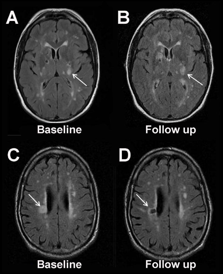
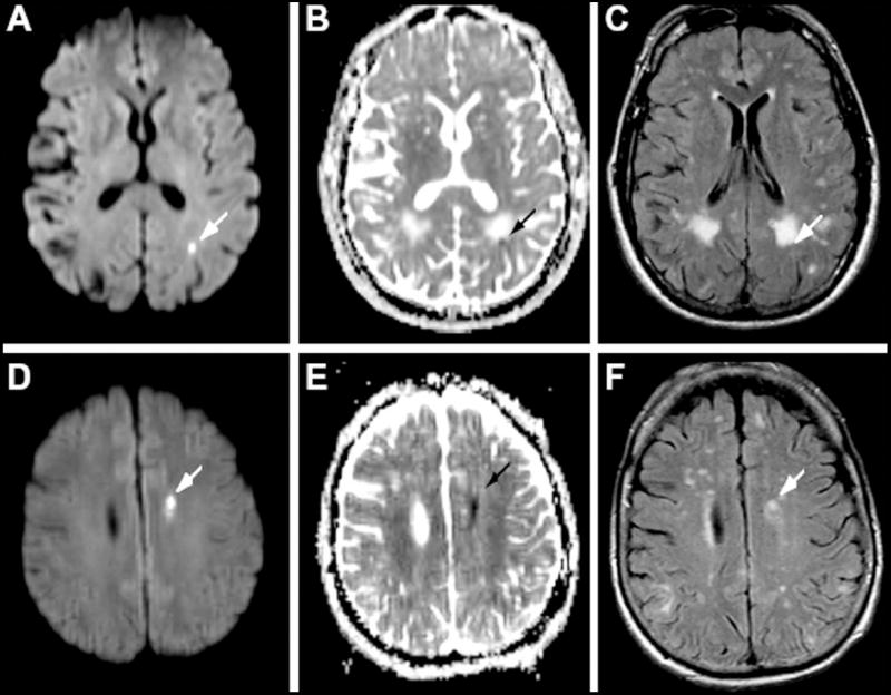
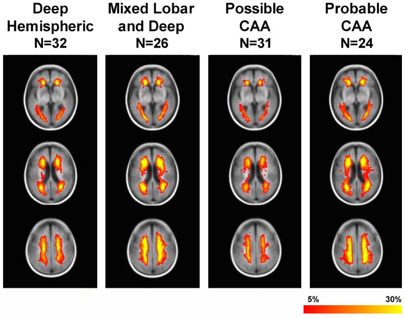
Leukoaraiosis is a common finding in stroke patients and has been strongly associated with risk of incident stroke and dementia. Leukoaraiosis may also be an independent predictor of stroke outcomes. There is increasing evidence from neuroimaging to support the concept that some leukoaraiosis is caused by white matter infarcts, which may be particularly frequent in patients with aggressive small vessel diseases such as cerebral amyloid angiopathy. The relatively similar distribution of leukoaraiosis regardless of the distribution of vascular pathology suggests a conserved vulnerability to white matter injury across various vascular diseases, possibly related to resting patterns of blood flow. More insights into the pathophysiology of leukoaraiosis are sorely needed to reduce the burden of disability associated with this common condition.


Conflict of interest statement

There are no commercial conflicts of interest.

Figures

Figure 1
Figure 1. Acute Small Vessel Infarction May Evolve Into White Matter Lesions
Recent data suggest that the majority of acute small artery infarctions on MRI evolve into T2-hyperintense white matter lesions (Panels A and B, fluid attenuated inversion recovery (FLAIR) MRI at 4 days and 65 days after acute infarction in the left internal capsule) rather than chronic cavitated lesions (Panels C and D, FLAIR MRI at 17 days and 7 months after acute infarction in the right corona radiata). This figure was graciously provided by Dr. Gillian M. Potter and Dr. Joanna M. Wardlaw.
Figure 2
Figure 2. Incidental Small Infarcts in Cerebral Amyloid Angiopathy
Incidental small acute cerebral infarcts can be detected with surprisingly high frequency in patients with cerebral amyloid angiopathy. Here, small lesions are seen in the periventricular white matter in 2 patients (panels A-C and D-F) with hyperintensity on diffusion-weighted sequence (A and D), hypointensity on apparent diffusion coefficient sequence (B and E) and hyperintensity on fluid attenuated inversion recovery (FLAIR, C and F). Reproduced with permission from Neurology.
Figure 3
Figure 3. Distribution of Leukoaraiosis in Patients with Hypertensive Deep Hemispheric Intracerebral Hemorrhage Compared to Cerebral Amyloid Angiopathy-Associated Hemorrhage
The distribution of leukoaraiosis is substantially similar despite the near inverse distribution of vascular pathology in hypertensive arteriopathy (maximally affecting the penetrating arteries to the deep hemispheric brain structures) compared to cerebral amyloid angiopathy (affecting the cortex and meninges). The color code (red to yellow) is proportional to the frequency of leukoaraiosis at that voxel.

References

    1. Debette S, Beiser A, DeCarli C, Au R, Himali JJ, Kelly-Hayes M, Romero JR, Kase CS, Wolf PA, Seshadri S. Association of MRI markers of vascular brain injury with incident stroke, mild cognitive impairment, dementia, and mortality: The Framingham Offspring Study. Stroke. 2010;41:600–606. - PMC - PubMed
    1. Vermeer SE, Prins ND, den Heijer T, Hofman A, Koudstaal PJ, Breteler MM. Silent brain infarcts and the risk of dementia and cognitive decline. N Engl J Med. 2003;348:1215–1222. - PubMed
    1. Longstreth WT, Jr, Arnold AM, Beauchamp NJ, Jr, Manolio TA, Lefkowitz D, Jungreis C, Hirsch CH, O’Leary DH, Furberg CD. Incidence, manifestations, and predictors of worsening white matter on serial cranial magnetic resonance imaging in the elderly: the Cardiovascular Health Study. Stroke. 2005;36:56–61. - PubMed
    1. Kissela B, Lindsell CJ, Kleindorfer D, Alwell K, Moomaw CJ, Woo D, Flaherty ML, Air E, Broderick J, Tsevat J. Clinical prediction of functional outcome after ischemic stroke: the surprising importance of periventricular white matter disease and race. Stroke. 2009;40:530–536. - PMC - PubMed
    1. Arsava EM, Rahman R, Rosand J, Lu J, Smith EE, Rost NS, Singhal AB, Lev MH, Furie KL, Koroshetz WJ, Sorensen AG, Ay H. Severity of leukoaraiosis correlates with clinical outcome after ischemic stroke. Neurology. 2009;72:1403–1410. - PMC - PubMed

Publication types