

Antagonistic roles of PP2A-Pab1 and Etd1 in the control of cytokinesis in fission yeast
- PMID: 20876564
- PMCID: PMC2998309
- DOI: 10.1534/genetics.110.121368
Antagonistic roles of PP2A-Pab1 and Etd1 in the control of cytokinesis in fission yeast
Abstract
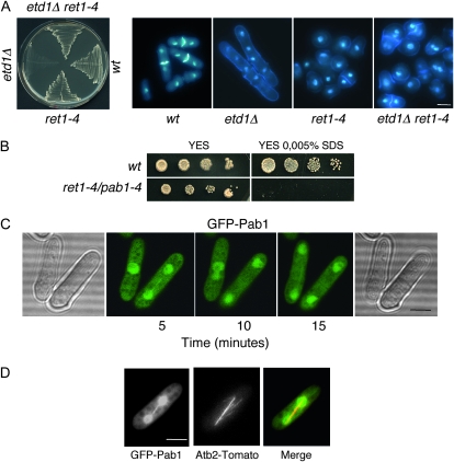
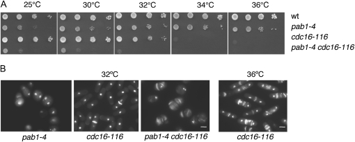
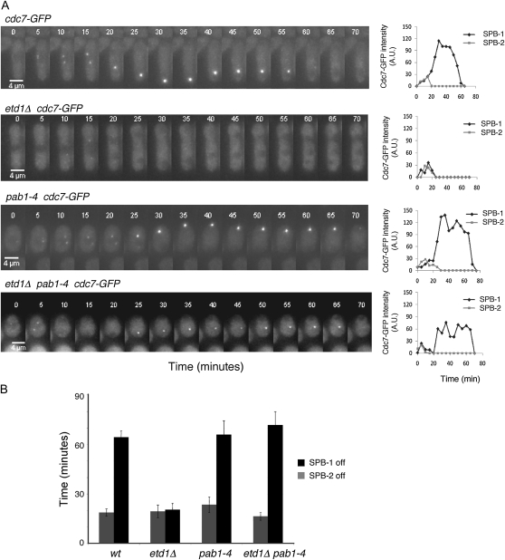
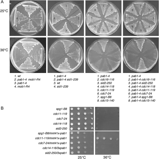
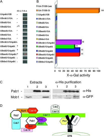
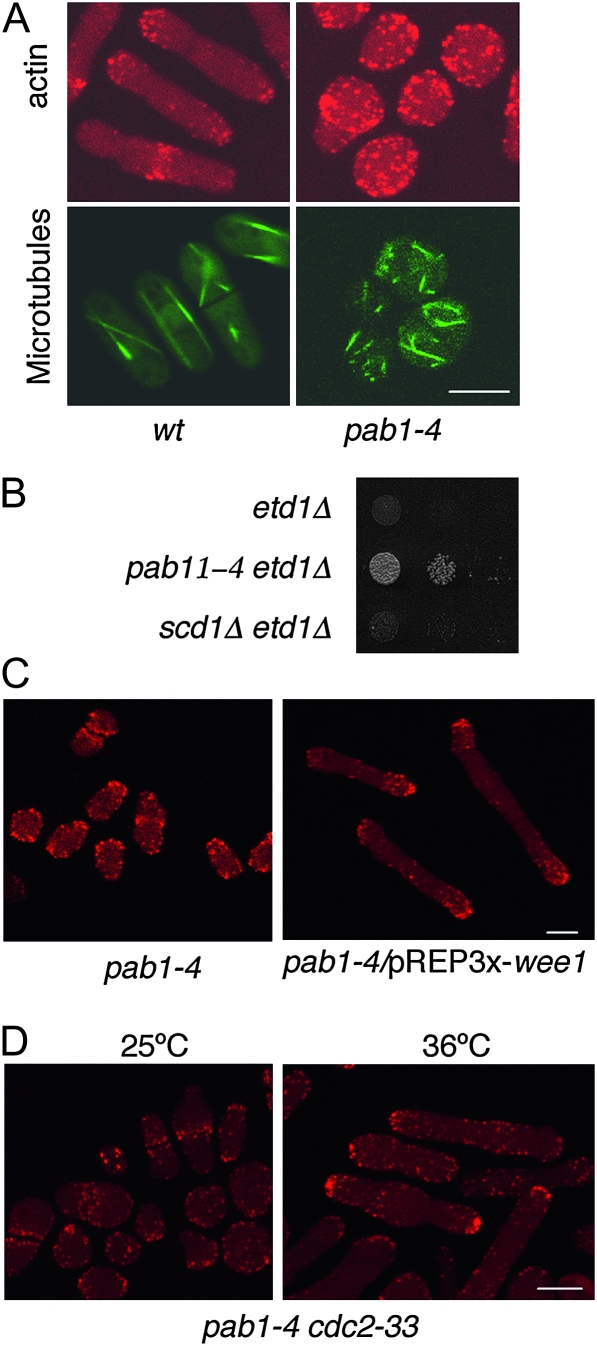
In Schizosaccharomyces pombe, Etd1 is a positive regulator of the septation initiation network (SIN), a conserved GTPase-regulated kinase cascade that triggers cytokinesis. Here we show that a mutation in the pab1 gene, which encodes the B-regulatory subunit of the protein phosphatase 2A (PP2A), suppresses mutations in the etd1 gene. Etd1 is required for the function of the GTPase Spg1, a key regulator of SIN signaling. Interestingly, the loss of Pab1 function restored the activity of Spg1 in Etd1-deficient cells. This result suggests that PP2A-Pab1-mediated dephosphorylation inhibits Spg1, thus antagonizing Etd1 function. The loss of pab1 function also rescues the lethality of mutants of other genes in the SIN cascade such as mob1, sid1, and cdc11. Two-hybrid assays indicate that Pab1 physically interacts with Mob1, Sid1, Sid2, and Cdc11, suggesting that the phosphatase 2A B-subunit is a component of the SIN complex. Together, our results indicate that PP2A-Pab1 plays a novel role in cytokinesis, regulating SIN activity at different levels. Pab1 is also required to activate polarized cell growth. Thus, PP2A-Pab1 may be involved in coordinating polar growth and cytokinesis.
Figures
Similar articles
-
Feedback regulation of SIN by Etd1 and Rho1 in fission yeast.Genetics. 2014 Feb;196(2):455-70. doi: 10.1534/genetics.113.155218. Epub 2013 Dec 13. Genetics. 2014. PMID: 24336750 Free PMC article.
-
Proper timing of cytokinesis is regulated by Schizosaccharomyces pombe Etd1.J Cell Biol. 2009 Sep 7;186(5):739-53. doi: 10.1083/jcb.200902116. J Cell Biol. 2009. PMID: 19736319 Free PMC article.
-
Specific detection of fission yeast primary septum reveals septum and cleavage furrow ingression during early anaphase independent of mitosis completion.PLoS Genet. 2018 May 29;14(5):e1007388. doi: 10.1371/journal.pgen.1007388. eCollection 2018 May. PLoS Genet. 2018. PMID: 29813053 Free PMC article.
-
Polar opposites: Fine-tuning cytokinesis through SIN asymmetry.Cytoskeleton (Hoboken). 2012 Oct;69(10):686-99. doi: 10.1002/cm.21044. Epub 2012 Jul 11. Cytoskeleton (Hoboken). 2012. PMID: 22786806 Free PMC article. Review.
-
An overview of the fission yeast septation initiation network (SIN).Biochem Soc Trans. 2008 Jun;36(Pt 3):411-5. doi: 10.1042/BST0360411. Biochem Soc Trans. 2008. PMID: 18481970 Review.
Cited by
-
An Extended, Boolean Model of the Septation Initiation Network in S.Pombe Provides Insights into Its Regulation.PLoS One. 2015 Aug 5;10(8):e0134214. doi: 10.1371/journal.pone.0134214. eCollection 2015. PLoS One. 2015. PMID: 26244885 Free PMC article.
-
The fission yeast septation initiation network (SIN) kinase, Sid2, is required for SIN asymmetry and regulates the SIN scaffold, Cdc11.Mol Biol Cell. 2012 May;23(9):1636-45. doi: 10.1091/mbc.E11-09-0792. Epub 2012 Mar 14. Mol Biol Cell. 2012. PMID: 22419817 Free PMC article.
-
Dual Regulation of the mitotic exit network (MEN) by PP2A-Cdc55 phosphatase.PLoS Genet. 2013;9(12):e1003966. doi: 10.1371/journal.pgen.1003966. Epub 2013 Dec 5. PLoS Genet. 2013. PMID: 24339788 Free PMC article.
-
The phosphatase inhibitor Sds23 promotes symmetric spindle positioning in fission yeast.Cytoskeleton (Hoboken). 2020 Dec;77(12):544-557. doi: 10.1002/cm.21648. Epub 2020 Dec 14. Cytoskeleton (Hoboken). 2020. PMID: 33280247 Free PMC article.
-
Identification of novel genes involved in DNA damage response by screening a genome-wide Schizosaccharomyces pombe deletion library.BMC Genomics. 2012 Nov 23;13:662. doi: 10.1186/1471-2164-13-662. BMC Genomics. 2012. PMID: 23173672 Free PMC article.
References
-
- Bahler, J., J. Q. Wu, M. S. Longtine, N. G. Shah, A. McKenzie, III et al., 1998. Heterologous modules for efficient and versatile PCR-based gene targeting in Schizosaccharomyces pombe. Yeast 14 943–951. - PubMed
-
- Bardin, A. J., R. Visintin and A. Amon, 2000. A mechanism for coupling exit from mitosis to partitioning of the nucleus. Cell 102 21–31. - PubMed
-
- Cerutti, L., and V. Simanis, 1999. Asymmetry of the spindle pole bodies and Spg1 GAP segregation during mitosis in fission yeast. J. Cell Sci. 112(Pt. 14): 2313–2321. - PubMed
Publication types
MeSH terms
Substances
LinkOut - more resources
Full Text Sources
Molecular Biology Databases
