. 2010 Dec;186(4):1337-44.
doi: 10.1534/genetics.110.120840. Epub 2010 Sep 27.

Tyrosinase is the modifier of retinoschisis in mice

Affiliations

Tyrosinase is the modifier of retinoschisis in mice

Britt A Johnson et al. Genetics. 2010 Dec.

Abstract

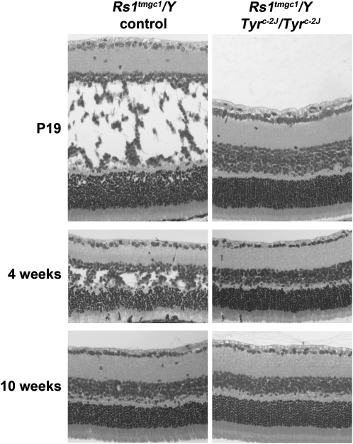
X-linked retinoschisis (XLRS) is a form of macular degeneration with a juvenile onset. This disease is caused by mutations in the retinoschisin (RS1) gene. The major clinical pathologies of this disease include splitting of the retina (schisis) and a loss in synaptic transmission. Human XLRS patients display a broad range in phenotypic severity, even among family members with the same mutation. This variation suggests the existence of genetic modifiers that may contribute to disease severity. Previously, we reported the identification of a modifier locus, named Mor1, which affects severity of schisis in a mouse model of XLRS (the Rs1tmgc1 mouse). Homozygosity for the protective AKR allele of Mor1 restores cell adhesion in Rs1tmgc1 mice. Here, we report our study to identify the Mor1 gene. Through collecting recombinant mice followed by progeny testing, we have localized Mor1 to a 4.4-Mb region on chromosome 7. In this genetic region, the AKR strain is known to carry a mutation in the tyrosinase (Tyr) gene. We observed that the schisis phenotype caused by the Rs1 mutation is rescued by a Tyr mutation in the C57BL/6J genetic background, strongly suggesting that Tyr is the Mor1 gene.


Figures

F<sc>igure</sc> 1.—
Figure 1.—
Genetic refinement of the Mor1 locus. (A) Critical recombinant chromosomes obtained in F2 (left) and F3 (right) mice used for progeny testing. Marker positions are indicated between F2 and F3 chromosomes. (B) Progeny testing for B9378 and B9379 that define the new Mor1 minimal genetic region. For both progeny tests, progeny that are homozygous for AKR across Chr 7 do not have schisis (rescued) and progeny that are heterozygous across Chr 7 have schisis (affected). Recombinant progeny from B9379 (left) do not have schisis, while recombinant progeny derived from B9378 (right) have schisis. Therefore Mor1 is located within the new minimal genetic region flanked by markers D7Mit62 and D7Mit123.
F<sc>igure</sc> 2.—
Figure 2.—
Histological examination of the retina of Rs1tmgc1 mice with homozygous Tyrc-2J mutation. Images are shown of H&E-stained retinal sections from Rs1tmgc1/Y control (left) and Rs1tmgc1/Y Tyrc-2J/Tyrc-2J (right) mice at P19 (top), 4 weeks (middle), and 16 weeks (bottom) of age. The schisis phenotype is not observed in the Rs1tmgc1/Y Tyrc-2J/Tyrc-2J retina throughout the ages examined.
F<sc>igure</sc> 3.—
Figure 3.—
Rescue of ectopic synaptic localization by the Tyrc-2J mutation. PKCα-labeled (green) bipolar cell dendrites and PSD95-labeled (red) photoreceptor synaptic terminals are localized in the outer plexiform layer (OPL) in B6 wild-type mice (A, left), while they are ectopically localized in the outer nuclear layer (ONL) in the Rs1tmgc1/Y control retina (A, center; asterisk). In the retina of Rs1tmgc1/Y Tyrc-2J/Tyrc-2J mice (A, right), the frequency of ectopically localized synapses is not statistically different from that in the B6 wild-type retina (B). Bar, 20 μm. Error bars represent SEM. Values with different superscripts differ with statistical significance (P < 0.05). INL, inner nuclear layer.
F<sc>igure</sc> 4.—
Figure 4.—
GFAP upregulation in the retina of Rs1tmgc1 mice and its rescue by the Tyrc-2J mutation. (A) Immunohistochemistry for GFAP. While GFAP expression is observed only in the ganglion cell and nerve fiber layers in 4-week-old B6 wild-type mice (A, left), it extends across the retinal layers and reaches the outer nuclear layer (ONL) in the Rs1tmgc1/Y control retina (A, center). In the retina of Rs1tmgc1/Y Tyrc-2J/ Tyrc-2J mice, upregulation and extension of GFAP signals are much reduced (A, right). (B) Quantification of GFAP immunostained area in the retina excluding the ganglion cell and nerve fiber layers. The level of GFAP signals in the Rs1tmgc1/Y Tyrc-2J/Tyrc-2J retina is not significantly different from that in the B6 control retina. Bar, 50 μm. GCL, ganglion cell layer; INL, inner nuclear layer; OPL, outer plexiform layer.
F<sc>igure</sc> 5.—
Figure 5.—
GFAP expression in the older Rs1tmgc1 mice. (A) Immunohistochemistry for GFAP in the retinas of 4-week-old (left, top) and 16-week-old (right, top) Rs1tmgc1 mice. While the schisis phenotype observed at 4 weeks of age (left, bottom) is rescued by 16 weeks of age (right, bottom), GFAP remains upregulated at 16 weeks of age. (B) The percentage of immunolabeled area in the green channel occurring in the retina excluding the ganglion cell and nerve fiber layers in 16-week-old Rs1tmgc1 retina is not significantly different from that in 4-week-old Rs1tmgc1 retina. Bar, 50 μm. GCL, ganglion cell layer; INL, inner nuclear layer; OPL, outer plexiform layer; ONL, outer nuclear layer.

References

    1. Baumgartner, S., K. Hofmann, R. Chiquet-Ehrismann and P. Bucher, 1998. The discoidin domain family revisited: new members from prokaryotes and a homology-based fold prediction. Protein Sci. 7 1626–1631. - PMC - PubMed
    1. Chang, B., J. R. Heckenlively, P. R. Bayley, N. C. Brecha, M. T. Davisson et al., 2006. The nob2 mouse, a null mutation in Cacna1f: anatomical and functional abnormalities in the outer retina and their consequences on ganglion cell visual responses. Vis. Neurosci. 23 11–24. - PMC - PubMed
    1. Dick, O., S. tom Dieck, W. D. Altrock, J. Ammermuller, R. Weiler et al., 2003. The presynaptic active zone protein bassoon is essential for photoreceptor ribbon synapse formation in the retina. Neuron 37 775–786. - PubMed
    1. Eksandh, L. C., V. Ponjavic, R. Ayyagari, E. L. Bingham, K. T. Hiriyanna et al., 2000. Phenotypic expression of juvenile X-linked retinoschisis in Swedish families with different mutations in the XLRS1 gene. Arch. Ophthalmol. 118 1098–1104. - PubMed
    1. George, N. D., J. R. Yates and A. T. Moore, 1995. X linked retinoschisis. Br. J. Ophthalmol. 79 697–702. - PMC - PubMed

Publication types