. 2010 Nov 26;285(48):37227-39.
doi: 10.1074/jbc.M110.132084. Epub 2010 Sep 28.

Suppressor of cytokine signaling 3 inhibits LPS-induced IL-6 expression in osteoblasts by suppressing CCAAT/enhancer-binding protein {beta} activity

Affiliations

Suppressor of cytokine signaling 3 inhibits LPS-induced IL-6 expression in osteoblasts by suppressing CCAAT/enhancer-binding protein {beta} activity

Chunguang Yan et al. J Biol Chem. .

Abstract

Suppressor of cytokine signaling 3 (SOCS3) is an important intracellular protein that inhibits cytokine signaling in numerous cell types and has been implicated in several inflammatory diseases. However, the expression and function of SOCS3 in osteoblasts are not known. In this study, we demonstrated that SOCS3 expression was transiently induced by LPS in osteoblasts, and apparently contributed to the inhibition of IL-6 induction by LPS treatment. We found that tyrosine 204 of the SOCS box, the SH2 domain, and the N-terminal kinase inhibitory region (KIR) of SOCS3 were all involved in its IL-6 inhibition. Furthermore, we demonstrated that CCAAT/enhancer-binding protein (C/EBP) β was activated by LPS (increased DNA binding activity), and played a key role in LPS-induced IL-6 expression in osteoblasts. We further provided the evidence that SOCS3 functioned as a negative regulator for LPS response in osteoblasts by suppressing C/EBPβ DNA binding activity. In addition, tyrosine 204 of the SOCS box, the SH2 domain, and the N-terminal kinase inhibitory region (KIR) of SOCS3 were all required for its C/EBPβ inhibition. These findings suggest that SOCS3 by interfering with C/EBPβ activation may have an important regulatory role during bone-associated inflammatory responses.


Figures

FIGURE 1.
FIGURE 1.
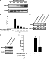
Silencing LPS-induced expression of SOCS3 enhances IL-6 secretion in MC3T3-E1 cells. MC3T3-E1 cells were incubated with 100 ng LPS/ml for indicated times. A, total cellular RNA was isolated for RT-PCR with primers for SOCS3 and GAPDH, respectively. The level of GAPDH was shown at the bottom as a loading control. B, total proteins were extracted to conduct Western blot using rabbit anti-SOCS3 antibody, and rabbit anti-GAPDH antibody, respectively. The level of GAPDH was shown at the bottom as a loading control. C, MC3T3-E1 cells were infected with Ad-Control-sh or Ad-SOCS3-sh at an MOI of 200. 48 h after infection, the cells were incubated with 100 ng LPS/ml for 1 h. RNAs were isolated, and RT-PCR was performed by using primers for SOCS3 and GAPDH, respectively. The level of GAPDH was shown at the bottom as a loading control. D, MC3T3-E1 cells were infected with Ad-Control-sh or Ad-SOCS3-sh at a MOI of 200. 48 h later, cells were stimulated by 100 ng LPS/ml for indicated times, and supernatants were harvested and subjected to ELISA. Data were means of six independent experiments ± S.E. **, p < 0.01; ***, p < 0.001 compared with Ad-Control-sh-infected group.
FIGURE 2.
FIGURE 2.
Overexpression of SOCS3 inhibits LPS-mediated IL-6 production in MC3T3-E1 cells. A, MC3T3-E1 cells were infected with pLP-Ad-SOCS3 at indicated MOI. 48 h after infection, the total protein extracts were subjected to Western blot using antibodies against SOCS3 and GAPDH, respectively. The level of GAPDH was shown at the bottom as a loading control. B, MC3T3-E1 cells were infected with Adeno-X-DsRed2 or pLP-Ad-SOCS3 at indicated MOI. 48 h later, the cells were stimulated either with or without 100 ng LPS/ml for 6 h. The supernatants were used to perform ELISA to determine IL-6 protein level. Data were expressed as means ± S.E., of six independent experiments. C, MC3T3-E1 cells were infected with Adeno-X-DsRed2 or pLP-Ad-SOCS3 at a MOI of 200. 48 h later, the cells were incubated with 100 ng/ml LPS for 4 h. Then proteins were harvested and subjected to Western blot using antibodies against SOCS3 and GAPDH, respectively. The level of GAPDH was shown at the bottom as a loading control. D, primary osteoblasts were infected with Adeno-X-DsRed2 and pLP-Ad-SOCS3 at 200 MOI, respectively. The total proteins were harvested 48 h after infection, and Western blot was conducted using antibodies against SOCS3 and GAPDH, respectively. The level of GAPDH was shown at the bottom as a loading control. E, primary osteoblasts were infected with Adeno-X-DsRed2 and pLP-Ad-SOCS3 at 200 MOI, respectively. 48 h later, the cells were stimulated either with or without 100 ng LPS/ml for 6 h. IL-6 secretion was measured by ELISA. Data were means of six independent experiments ± S.E. *** indicates a statistically significant difference (p < 0.001).
FIGURE 3.
FIGURE 3.
The KIR, SOCS box, and SH domain of SOCS3 are indispensable for its inhibitory role on IL-6 expression in MC3T3-E1 cells. A, MC3T3-E1 cells were transiently transfected with 0.5 μg of DNA comprised of IL-6 promoter-luciferase gene, thymidine kinase-luciferase gene and either SOCS3 expression plasmids or control vector. 24 h after transfection, the cells were treated or left untreated with 100 ng LPS/ml for 4 h. Cell lysates were used to perform luciferase activity measurement. B, MC3T3-E1 cells were transiently transfected with total of 0.5 μg plasmids as indicated. 24 h later, the cells were treated or left untreated with 100 ng LPS/ml. Cell lysates were harvested to conduct luciferase activity assay 4 h after LPS stimulation. SOCS3 Ctrl and mSOCS3 represent control vector and mouse SOCS3 expression plasmid, respectively. Y204F, Cis DC41, and Y211F are three of SOCS box mutants, in which Y204F and Y211F are mutants carrying a point mutation in each structure, Cis DC41 is a truncated mutant, 41 amino acids deletion from C terminus. L22D contains a point mutation in KIR. R71E is one of SH2 domain mutants. Luminometer values were normalized for expression from a co-transfected thymidine kinase reporter gene. The data were expressed as means of three experiments ± S.E. *** indicates a statistically significant difference (p < 0.001).
FIGURE 4.
FIGURE 4.
LPS induces C/EBPs and NF-κB binding to IL-6 promoter region. MC3T3-E1 cells were treated with 100 ng LPS/ml for the time as indicated. The nuclear proteins were extracted to perform EMSA to detect NF-κB activity (A) and C/EBP activation (B), respectively.
FIGURE 5.
FIGURE 5.
C/EBPβ/δ and NF-κB p65 synergistially stimulate IL-6 expression. A and B, MC3T3-E1 cells were transiently transfected with total of 0.5 μg indicated genes. 24 h later, luciferase activity was measured by using cell lysates. Luminometer values were normalized for expression from a co-transfected thymidine kinase-luciferase gene. C, MC3T3-E1 cells were transiently transfected with 0.5 μg of DNA consisting of thymidine kinase-luciferase gene, and either a wild type IL-6 promoter-luciferase construct or an IL-6 promoter-luciferase constructs harboring a mutation in either NF-κB binding site or C/EBP binding site. 24 h after transfection, the cells were incubated with 100 ng LPS/ml for 4 h. Cell lysates were used to perform luciferase activity assay. The data were expressed as means of three experiments ± S.E. *, **, and *** indicate a statistically significant difference, p < 0.05, p < 0.01, and p < 0.001, respectively.
FIGURE 6.
FIGURE 6.
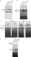
SOCS3 inhibits both LPS-induced and exogenously expressed-C/EBPs but not NF-κB binding to IL-6 promoter region in MC3T3-E1 cells. MC3T3-E1 cells were infected with Adeno-X-DsRed2 and pLP-Ad-SOCS3 at indicated MOI, respectively. 48 h later, the cells were treated or left untreated with 100 ng LPS/ml for 4 h. The nuclear proteins were harvested for EMSA to examine the influence of SOCS3 on NF-κB binding activity (A), and C/EBP binding activity (B), respectively. C, MC3T3-E1 cells were infected with Adeno-X-DsRed2 and pLP-Ad-SOCS3 at 200 MOI, respectively. 48 h later, the cells were treated or left untreated with 100 ng LPS/ml for 4 h. The nuclear proteins were harvested for gel supershift to identify which C/EBP family member binding activities were inhibited by SOCS3. D, MC3T3-E1 cells were infected with Adeno-X-DsRed2 and pLP-Ad-SOCS3 at 200 MOI, respectively. 24 h later, the cells were subcultured in 25-cm2 flasks. After 12 h, the cells were transiently transfected with control plasmid or C/EBPβ expression plasmid, respectively. Nuclear proteins were extracted for EMSA. N, α, β, δ, ϵ, and γ represent normal rabbit IgG, anti-C/EBPα antibody, anti-C/EBPβ antibody, anti-C/EBPδ antibody, anti-C/EBPϵ antibody, and anti-C/EBPγ antibody, respectively. Arrows indicated NF-κB, C/EBP binding bands, and supershift bands.
FIGURE 7.
FIGURE 7.
Knocking-down SOCS3 expression enhances LPS-induced C/EBP but not NF-κB binding to IL-6 promoter region in MC3T3-E1 cells. MC3T3-E1 cells were infected with Ad-Control-sh or Ad-SOCS3-sh at an MOI of 200. 48 h after infection, the cells were incubated with 100 ng LPS/ml for 4 h. The nuclear proteins were harvested for EMSA to examine the influence of decreased SOCS3 on the NF-κB binding activity (A) and C/EBP binding activity (B), respectively.
FIGURE 8.
FIGURE 8.
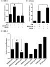
SOCS3 inhibits the activities of C/EBP promoter-luciferase while enhances NF-κB promoter-luciferase activity. MC3T3-E1 cells were transiently transfected with 0.5 μg of DNA comprised of a DEI-4 (A) or a κB (B) luciferase gene, thymidine kinase-luciferase gene, and either SOCS3 expression plasmid or control vector. 24 h after transfection, the cells were incubated with or without 100 ng LPS/ml for 4 h. Cell lysates were used to perform luciferase activity assay. C, MC3T3-E1 cells were transiently transfected with total of 0.5 μg of plasmids as indicated. 24 h later, the cells were treated or left untreated with 100 ng LPS/ml. Luminometer values were normalized for expression from a co-transfected thymidine kinase reporter gene. The data were expressed as means of three experiments ± S.E. *, **, and *** indicate a statistically significant difference, p < 0.05, p < 0.01, and p < 0.001, respectively.
FIGURE 9.
FIGURE 9.
C/EBPβ inhibition by siRNA decreases LPS-induced IL-6 secretion in MC3T3-E1 cells. A, MC3T3-E1 cells were transiently transfected with 40 nm control siRNA or C/EBPβ-specific si RNA. 24 h after transfection, the cells were incubated with 100 ng LPS/ml for 4 h. RNAs were isolated, and RT-PCR was performed by using primers for C/EBPβ and GAPDH, respectively. The level of GAPDH was shown at the bottom as a loading control. B, MC3T3-E1 cells were transiently transfected with 40 nm control siRNA or C/EBPβ si RNA. 24 h later, the cells were incubated with or without 100 ng LPS/ml for 4 h. Supernatants were harvested for ELISA. The data were expressed as means of six experiments ± S.E. *** indicates a statistically significant difference (p < 0.001).
FIGURE 10.
FIGURE 10.
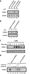
C/EBPβ expression is negatively regulated by SOCS3 at multiple levels. MC3T3-E1 cells were infected with Adeno-X-DsRed2 or pLP-Ad-SOCS3. 48 h after infection, the cells were treated or left untreated with 100 ng LPS/ml for 4 h. A, the total proteins were extracted to conduct Western blot using rabbit anti-C/EBPβ antibody, and rabbit anti-GAPDH antibody, respectively. The level of GAPDH is shown at the bottom as a loading control. B, total cellular RNA was isolated for RT-PCR with primers for C/EBPβ and GAPDH, respectively. The level of GAPDH was shown at the bottom as a loading control. C, MC3T3-E1 cells were incubated with 100 ng LPS/ml for indicated times. Total proteins were extracted to conduct Western blot using rabbit anti-C/EBPβ antibody, and rabbit anti-GAPDH antibody, respectively. The level of GAPDH was shown at the bottom as a loading control. D, MC3T3-E1 cells were infected with Adeno-X-DsRed2 or pLP-Ad-SOCS3. 48 h after infection, cells were cultured in Met/Cys-free medium for 2 h. Cells were then pulsed with Trans35S label in the absence (lanes 1 and 4) or presence (lanes 2–3 and 5–6) of LPS for 2 h, and chased for 5 h with complete medium. Cell lysates were immunoprecipitated by using anti-C/EBPβ antibody and analyzed by SDS-PAGE. Data are representative for two independent experiments.

References

    1. Stoiber D., Kovarik P., Cohney S., Johnston J. A., Steinlein P., Decker T. (1999) J. Immunol. 163, 2640–2647 - PubMed
    1. Cassatella M. A., Gasperini S., Bovolenta C., Calzetti F., Vollebregt M., Scapini P., Marchi M., Suzuki R., Suzuki A., Yoshimura A. (1999) Blood 94, 2880–2889 - PubMed
    1. Bode J. G., Nimmesgern A., Schmitz J., Schaper F., Schmitt M., Frisch W., Häussinger D., Heinrich P. C., Graeve L. (1999) FEBS Lett. 463, 365–370 - PubMed
    1. Yasukawa H., Ohishi M., Mori H., Murakami M., Chinen T., Aki D., Hanada T., Takeda K., Akira S., Hoshijima M., Hirano T., Chien K. R., Yoshimura A. (2003) Nat. Immunol. 4, 551–556 - PubMed
    1. Kubo M., Hanada T., Yoshimura A. (2003) Nat. Immunol. 4, 1169–1176 - PubMed

Publication types

MeSH terms

LinkOut - more resources