. 2010 Nov;137(21):3663-73.
doi: 10.1242/dev.054320. Epub 2010 Sep 28.

The Flamingo ortholog FMI-1 controls pioneer-dependent navigation of follower axons in C. elegans

Affiliations

The Flamingo ortholog FMI-1 controls pioneer-dependent navigation of follower axons in C. elegans

Andreas Steimel et al. Development. 2010 Nov.

Abstract

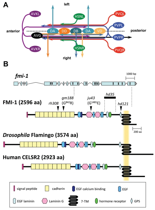
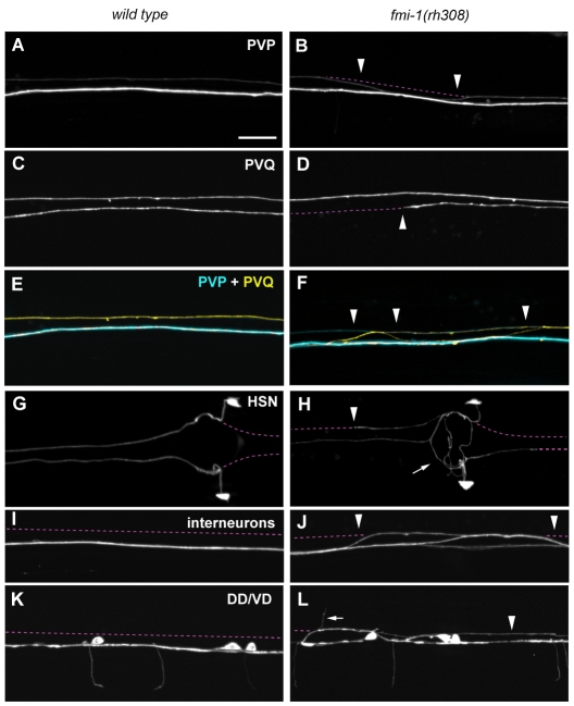
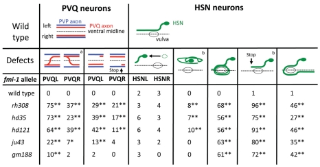
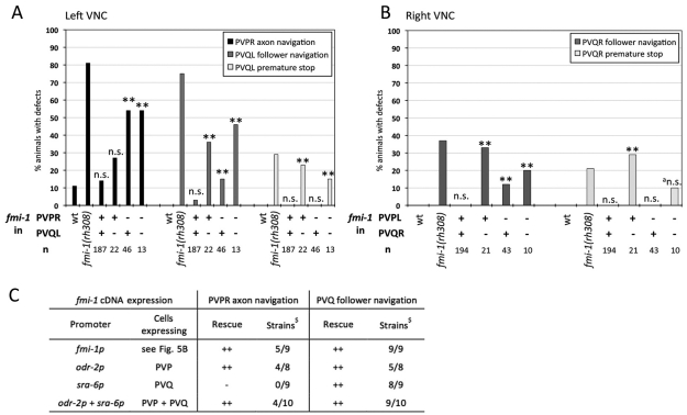
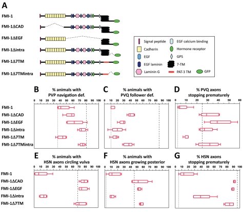
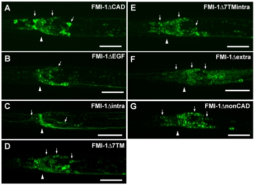
Development of a functional neuronal network during embryogenesis begins with pioneer axons creating a scaffold along which later-outgrowing axons extend. The molecular mechanism used by these follower axons to navigate along pre-existing axons remains poorly understood. We isolated loss-of-function alleles of fmi-1, which caused strong axon navigation defects of pioneer and follower axons in the ventral nerve cord (VNC) of C. elegans. Notably follower axons, which exclusively depend on pioneer axons for correct navigation, frequently separated from the pioneer. fmi-1 is the sole C. elegans ortholog of Drosophila flamingo and vertebrate Celsr genes, and this phenotype defines a new role for this important molecule in follower axon navigation. FMI-1 has a unique and strikingly conserved structure with cadherin and C-terminal G-protein coupled receptor domains and could mediate cell-cell adhesion and signaling functions. We found that follower axon navigation depended on the extracellular but not on the intracellular domain, suggesting that FMI-1 mediates primarily adhesion between pioneer and follower axons. By contrast, pioneer axon navigation required the intracellular domain, suggesting that FMI-1 acts as receptor transducing a signal in this case. Our findings indicate that FMI-1 is a cell-type dependent axon guidance factor with different domain requirements for its different functions in pioneers and followers.


Figures

Fig. 1.
Fig. 1.
Development of the ventral nerve cord (VNC) in C. elegans, and gene model and protein domain organization of FMI-1. (A) The C. elegans VNC consists of two axon tracks with motoneuron cell bodies (light and dark orange, light and dark turquoise) lining the ventral midline (broken line). During embryogenesis, the AVG axon (black) pioneers the right VNC axon track. PVP axons (blue) decussate and extend on the contralateral side of the VNC. The PVPR axon pioneers the left VNC axon track. Both PVP axons are closely followed by PVQ axons (red). DA, DB and DD motoneurons (light and dark turquoise, dark orange, respectively) extend axons into the right axon track and send commissures circumferentially to the DNC. Later, AVK axons (purple) enter the VNC from the anterior. Postembryonically, VD motoneurons (light orange) differentiate and send processes into the VNC and towards the DNC. HSN axons (green) join the VNC at the vulva region. Command interneuron axons, which extend in the right axon track, are omitted for clarity. Ventral view. (B) The fmi-1 gene. The top part illustrates the exon-intron structure of fmi-1. The positions of alleles used in this study are mapped onto the protein structure (arrowheads, bold line). The protein domain organization of Drosophila Flamingo and human CELSR2 are shown for comparison.
Fig. 2.
Fig. 2.
Axonal defects in fmi-1(rh308) animals. Axon guidance of VNC axons in wild-type (A,C,E,G,I,K) and fmi-1(rh308) (B,D,F,H,J,L) animals. (E,F) Overlays of AVG, PVP and PVQ axons. (A) Wild type; (B) PVPR axon crossed into contralateral axon track (arrowheads). (C) Wild type; (D) PVQR axon stopped prematurely (arrowhead). (E) Wild type; (F) PVQL axon (yellow) crossed into contralateral axon track independently of the PVPR axon (cyan) (arrowheads) and extended posteriorly. (G) Wild type; (H) HSN axons circled the vulva (arrow), extended posteriorly along the VNC and stopped prematurely (arrowhead). (I) Wild type; (J) interneurons crossed into the contralateral axon track (arrowheads). (K) Wild type; (L) DD/VD processes extended along the left axon track (arrowhead) and commissures projected along the left side of the animal into the dorsal cord (arrow). The broken line (purple) indicates the position of the left and right VNC axon tracks. Ventral views, anterior towards the left. Markers used: hdIs26 (PVP/PVQ), zdIs13 (HSN), rhIs4 (Interneurons) and hdIs25 (DD/VD). Scale bar: 10 μm.
Fig. 3.
Fig. 3.
Defects of PVQ and HSN follower axons are independent of PVP pioneer axon navigation. aPVQ axons independently crossing the ventral midline and/or leaving the VNC. bPercentage of axons with n=200-220, remaining values are percentage of animals with n=100-110; combined HSNL/R axonal defects are shown; *P<0.05, **P<0.01 (χ2 test) compared with wild type; all graphics are ventral views, anterior towards the left. Marker used are hdIs26 (PVP/PVQ) and zdIs13 (HSN).
Fig. 4.
Fig. 4.
fmi-1 expression and localization. (A) Transcriptional and translational reporter constructs. (B) Cells expressing fmi-1p::GFP. Expression of the fmi-1p construct was detected embryonically (C-H) and postembryonically (I-N). (C,D) Gastrulation-stage embryo. (E,F) Comma-stage embryo. (G,H) 1.5-fold-stage embryo with expression in motoneurons (G, arrows). D, F and H are overlays of Nomarski and GFP channels. (I) L1 larva head region. (J-L) fmi-1p expression (green) in PVP (cyan) and PVQ (red) neurons. (M) Adult animal, vulva region. (N) FMI-1::GFP expression in the adult. Broken white lines indicate the outline of animals where possible. (C-H,N) Lateral views; (I-M) Ventral views. In all pictures, anterior is towards the left. Scale bars: 10 μm.
Fig. 5.
Fig. 5.
Mosaic analysis and targeted expression of fmi-1 in fmi-1(rh308) animals. (A,B) Mosaic analysis; (C) targeted expression. (A,B) Presence (+) or absence (–) of the rescuing array containing fmi-1(+) in PVP and PVQ neurons was determined by presence of nuclear localized GFP. Defects in wild-type (wt) and fmi-1(rh308) animals are shown for comparison. Mosaic analysis results are for (A) left axon track and (B) right axon track neurons. **P<0.01 and not significant (n.s.) with P>0.05 compared with wild type (χ2 test). The marker used was hdIs26. (C) Results of targeted fmi-1 cDNA expression in PVP and PVQ neurons of fmi-1(rh308) animals. Categories: –, no rescue with P>0.05 compared with fmi-1(rh308); ++, rescue with P>0.05 compared with best fmi-1p rescue (χ2 test); $Independent best rescuing strains out of total of analyzed strains; n=27-110. The marker used was hdIs26.
Fig. 6.
Fig. 6.
Domain analysis of FMI-1. (A) For each FMI-1 construct, five transgenic strains were generated and their phenotypes scored. (B-G) Defects in fmi-1(rh308) animals (dashed line) and in best fmi-1 full-length rescue (dotted line) are shown for comparison. Boxes outline 50% of the data set around the median, indicated as a line within the box. The upper and lower ends of the box indicate the 25th and 75th quartiles. Whiskers mark the maximum and minimum values. (B) Rescue of PVP axon guidance. (C) Rescue of PVQ follower axon navigation. (D) Rescue of PVQ premature stop. (E) Rescue of HSN circling phenotype. (F) Rescue of HSN posterior extension. (G) Rescue of HSN premature stop. (B,C) n=39-100; (D) n=92-202; (E,F) n=35-61; (G) n=80-122. Marker used were hdIs26 (PVP,PVQ) and zdIs13 (HSN).
Fig. 7.
Fig. 7.
Localization of FMI-1 domain analysis constructs. (A-G) GFP images of head region with arrows indicating neuronal cell bodies and an arrowhead indicating the position of the nerve ring. Localization of construct (A) FMI-1ΔCAD, (B) FMI-1ΔEGF, (C) FMI-1Δintra, (D) FMI-1Δ7TM, (E) FMI-1Δ7TMintra, (F) FMI-1Δextra and (G) FMI-1ΔnonCAD in head region; lateral views, anterior towards the left. Scale bars: 10 μm.

Similar articles

Cited by

References

    1. Asakura T., Ogura K., Goshima Y. (2007). UNC-6 expression by the vulval precursor cells of Caenorhabditis elegans is required for the complex axon guidance of the HSN neurons. Dev. Biol. 304, 800-810 - PubMed
    1. Bao H., Berlanga M. L., Xue M., Hapip S. M., Daniels R. W., Mendenhall J. M., Alcantara A. A., Zhang B. (2007). The atypical cadherin flamingo regulates synaptogenesis and helps prevent axonal and synaptic degeneration in Drosophila. Mol. Cell. Neurosci. 34, 662-678 - PMC - PubMed
    1. Boulin T., Pocock R., Hobert O. (2006). A novel Eph receptor-interacting IgSF protein provides C. elegans motoneurons with midline guidepost function. Curr. Biol. 16, 1871-1883 - PubMed
    1. Brenner S. (1974). The genetics of Caenorhabditis elegans. Genetics 77, 71-94 - PMC - PubMed
    1. Broadbent I. D., Pettitt J. (2002). The C. elegans hmr-1 gene can encode a neuronal classic cadherin involved in the regulation of axon fasciculation. Curr. Biol. 12, 59-63 - PubMed

Publication types

MeSH terms

LinkOut - more resources