. 2010 Nov;137(21):3603-13.
doi: 10.1242/dev.049189. Epub 2010 Sep 28.

The C. elegans peroxidasin PXN-2 is essential for embryonic morphogenesis and inhibits adult axon regeneration

Affiliations

The C. elegans peroxidasin PXN-2 is essential for embryonic morphogenesis and inhibits adult axon regeneration

Jennifer R Gotenstein et al. Development. 2010 Nov.

Abstract

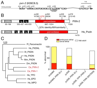
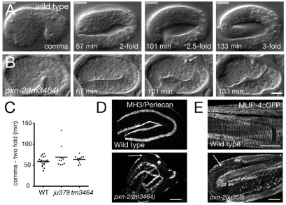
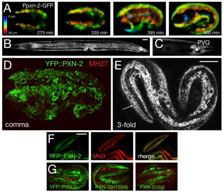
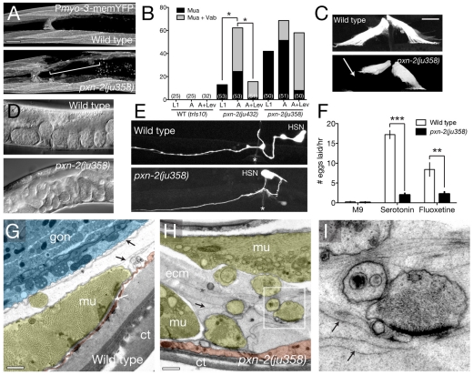
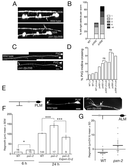
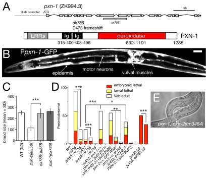
Peroxidasins form a highly conserved family of extracellular peroxidases of unknown cellular function. We identified the C. elegans peroxidasin PXN-2 in screens for mutants defective in embryonic morphogenesis. We find that PXN-2 is essential for specific stages of embryonic morphogenesis and muscle-epidermal attachment, and is also required postembryonically for basement membrane integrity. The peroxidase catalytic activity of PXN-2 is necessary for these developmental roles. pxn-2 mutants display aberrant ultrastructure of the extracellular matrix, suggesting a role in basement membrane consolidation. PXN-2 affects specific axon guidance choice points in the developing nervous system but is dispensable for maintenance of process positions. In adults, loss of pxn-2 function promotes regrowth of axons after injury, providing the first evidence that C. elegans extracellular matrix can play an inhibitory role in axon regeneration. Loss of function in the closely related C. elegans peroxidasin pxn-1 does not cause overt developmental defects. Unexpectedly, pxn-2 mutant phenotypes are suppressed by loss of function in pxn-1 and exacerbated by overexpression of wild-type pxn-1, indicating that PXN-1 and PXN-2 have antagonistic functions. These results demonstrate that peroxidasins play crucial roles in development and reveal a new role for peroxidasins as extracellular inhibitors of axonal regeneration.


Figures

Fig. 1.
Fig. 1.
pxn-2 encodes a C. elegans peroxidasin that is required for body morphogenesis. (A) Genomic structure of pxn-2, showing locations of point mutations and the tm3464 deletion. (B) Domain organization of PXN-2 and human (Hs) peroxidasin. (C) C. elegans PXN-2 and PXN-1 are most closely related to Drosophila and vertebrate peroxidasins, and more distantly related to other myeloperoxidases. Neighbor-joining tree based on ClustalW alignment of peroxidase domains. Ce, C. elegans; Dm, Drosophila melanogaster; Pl, Pacifastacus leniusculus (crayfish); Xt, Xenopus tropicalis; Mm, Mus musculus; Hs, Homo sapiens. Humans have two peroxidasin-encoding genes, PXDN and PXDNL; however, phylogenetic analysis indicates that the two C. elegans peroxidasins diverged separately and are not orthologous to the vertebrate gene pairs. (D) Morphological and lethal phenotypes of pxn-2(tm3464) and pxn-2(ju358) are completely rescued by transgenes containing a pxn-2 cDNA under the control of a 1.8 kb pxn-2 promoter [PXN-2(+), juEx2140] and by the YFP::PXN-2 transgene (juEx2492). Rescue activity of PXN-2(H755A) transgenes (juEx2491) was significantly reduced compared with that of PXN-2(+). Vab, variably abnormal epidermal morphology phenotype.
Fig. 2.
Fig. 2.
pxn-2 mutant embryos are defective in late stages of epidermal elongation, in muscle attachment and in epidermal attachment structures. (A,B) Frames from 4D Nomarski DIC movies of embryogenesis in wild-type N2 (A) and pxn-2(tm3464) (B) C. elegans embryos. Elongation proceeds normally until the twofold stage in pxn-2 mutants. Between 20 and 40 minutes after the twofold stage, visible constrictions appear in the epidermis (arrow), and elongation ceases ∼1 hour later, in most cases not extending to the threefold stage. (C) Early elongation rates are normal in pxn-2 mutants. Dot plot of times from comma stage to twofold as measured in time-lapse movies. Horizontal lines indicate the mean. WT, wild type. (D) In wild-type embryos, perlecan (UNC-52) (MH3 immunostaining) is localized to the basement membrane of muscle quadrants. In threefold pxn-2(tm3464) embryos this localization is retained except where muscles have detached from the epidermis, leaving gaps in the perlecan-staining bands (arrow). (E) In the wild-type larvae, MUP-4::GFP is localized to regularly spaced attachment structures in the epidermis. In pxn-2 mutant larvae, MUP-4::GFP is absent from regions of muscle detachment (arrow). Scale bars: 10 μm.
Fig. 3.
Fig. 3.
pxn-2 is expressed in embryonic and larval epidermis and in selected neurons. (A) The Ppxn-2-GFP transcriptional reporter (juEx1061) is expressed in epidermal cells from late gastrulation (∼210 minutes after the first cleavage) onward. Images are depth-coded projections of confocal z-stacks of a single C. elegans embryo imaged over time. (B) Ppxn-2-GFP expression in epidermis and vulval muscles at the L4 stage. (C) Ppxn-2-GFP is expressed in the PVQ neurons in postembryonic stages. Ppxn-2-GFP is also expressed in the BDU neurons and in four neurons in the anterior ventral ganglion (not shown). (D,E) Expression of functional YFP::PXN-2 (juEx2492) in embryos. Anti-GFP immunostaining (green) and MH27 (anti-AJM-1) staining (red). (D) At comma stage, YFP::PXN-2 is predominantly intracellular in epidermal cells. Maximum transparency projection of confocal sections (7 × 0.3 μm). (E) At the threefold stage, YFP::PXN-2 appears partly extracellular, forming faint longitudinal striations along the quadrants of body wall muscles (arrow). Projection of two confocal sections, 0.3 μm apart. (F) Colocalization of YFP::PXN-2 (green) and perlecan (MH3 immunostaining, red) in the threefold embryo; single confocal section. (G) Equivalent confocal projections of threefold stage embryos transgenic for YFP::PXN-2 (juEx2492), YFP::PXN-2(H755A) (juEx2898) and YFP:: PXN-2(ΔIg) (juEx2899). Images are projections of 6 × 0.5 μm focal planes in the top 3 μm. Scale bars: 10 μm.
Fig. 4.
Fig. 4.
PXN-2 maintains postembryonic muscle-epidermal adhesion and promotes ECM consolidation. (A) Anterior body wall muscle morphology in wild-type (trIs10) and pxn-2(ju358) C. elegans embryos. A region of muscle detachment in pxn-2(ju358) is bracketed. Projections of confocal z-stacks. (B) The muscle attachment (Mua) phenotype in pxn-2(ju432) is progressive in larval development and can be suppressed by paralysis with levamisole. pxn-2(ju358) causes more penetrant Mua phenotypes at the L1 stage; these are slightly progressive and are not significantly reduced by paralysis. The number of animals scored in each column is indicated in parentheses. *, P<0.05; Fisher's exact test. (C) Vulval muscles in the wild type are symmetrically attached, whereas in pxn-2 mutants the muscles become detached (arrow); Pegl-15-GFP (ayIs2). (D) pxn-2 animals exhibit the defective egg laying (Egl) phenotype. (E) In the wild type, the HSN cell body is located posterior to the vulva (asterisk) and extends an axon to the vulval epithelium. In ∼25% of ju358 or ju432 (not shown) adults, the HSN cell body is anterior to its normal position; Ptph-1-GFP (zdIs13). (F) Wild-type animals are inhibited from egg laying in liquid (M9 buffer); this inhibition is overcome by serotonin (35 mM) or the serotonin re-uptake inhibitor fluoxetine (1 mg/ml). Serotonin or fluoxetine only weakly stimulate egg laying in pxn-2(ju358) mutants. **, P<0.01; ***, P<0.001; Student's t-test. (G-I) Ultrastructure of the muscle-epidermal ECM is aberrant in pxn-2(ju358) animals. In the wild type (G), body wall muscles (mu, yellow) are attached to the epidermis (red) and cuticle (ct) via a basement membrane (white arrow); basement membranes covering the internal muscle surface and the gonad (gon, blue) are also visible (black arrows). (H) In pxn-2(ju358) mutants, the body muscles are separated from the epidermis and sometimes surrounded by expanded extracellular material (ecm) containing multiple electron-dense laminae of similar thickness to normal basement membranes (black arrows). (I) Enlargement of boxed region in H, showing muscle finger surrounded by laminae (arrows). Scale bars: 10 μm in A,C; 500 nm in G,H.
Fig. 5.
Fig. 5.
pxn-2 adults display progressive distortion of the pharynx and disorganization of pharyngeal ECM. (A) Deformation of the anterior pharyngeal bulb in pxn-2(ju432) adult C. elegans (DIC images). Vacuoles (white arrows) are frequently seen around the pharyngeal region, possibly resulting from cell necrosis. (B) Progression of pharyngeal distortion in a single pxn-2(ju358) adult. The pharynx morphology defects increase in penetrance during adult life: 19% of day 1 adult ju358 animals displayed overt pharyngeal defects, whereas 69% of day 5 adult ju358 animals displayed defects (n>50 per time point). In the wild type, HIM-4::GFP (rhIs23) is present on the pharyngeal basement membrane and in flexible tracks connecting the anterior pharynx to the epidermis. In pxn-2(ju358), HIM-4::GFP accumulates in extracellular aggregates (arrow). (C) Electron micrographs showing pharyngeal basement membrane (bm) in the wild type. In the enlargement (right), the pharynx (phx) is shaded yellow, surrounding cells in blue. (D) Expanded ECM adjacent to the pharynx of a pxn-2(ju358) mutant. The pharyngeal cytoskeleton is also abnormal. ecm, extracellular material. Scale bars: 10 μm in A,B; 500 nm in C,D.
Fig. 6.
Fig. 6.
PXN-2 functions in selective motoneuron axon guidance choice, prevents midline crossing and inhibits touch neuron axon regrowth after laser axotomy. (A) In wild-type L1 stage C. elegans (Punc-25-GFP, juIs76), commissures of motoneurons DD2-6 extend on the right-hand side. In pxn-2(ju358), commissures frequently extend on the left-hand side (arrow, DD6 commissure). (B) Summary of D neuron commissure handedness defects. The number of commissures that extended incorrectly is shown (n>50 per genotype). (C) Wild-type PVQ axons are fasciculated in the posterior and separate in the anterior. In pxn-2(ju358) animals, PVQ axons inappropriately fasciculate; oyIs14. (D) pxn-2 midline crossing defects do not increase during postembryonic development. Quantitation in L1 and L4 stage animals and in L4 animals reared on levamisole (Lev) plates. n>45 per group; ns, not significant (Fisher's exact test). (E) (Left) Diagram of PLM neuron showing position of axotomy (X). (Right) Images of PLM regrowth 6 hours post-axotomy in wild type and pxn-2(ju358); arrow, site of axotomy. Note the growth-cone-like structure in pxn-2. (F) Quantitation of PLM (zdIs5) regrowth in wild type and pxn-2(ju358) at 6 and 24 hours. Sample size is indicated within the columns. pxn-2 mutants displayed increased regrowth at 6 hours (*, P=0.02; Mann-Whitney test). The increased regrowth of pxn-2 mutants at 24 hours is rescued by the PXN-2(+) transgene juEx2140 (***, P<0.001; ANOVA and Tukey's post-hoc test). (G) By 24 hours post-axotomy, the distal process of the ALM neuron retracts slightly in the wild type, whereas in pxn-2(ju358) mutants the ALM distal process displays significantly increased regrowth. Dot plot; horizontal lines indicate the mean. P=0.003, Mann-Whitney test. Scale bars: 10 μm.
Fig. 7.
Fig. 7.
Antagonistic roles of PXN-1 and PXN-2. (A) Structure of the pxn-1 gene and predicted protein, and location of ok785 deletion. The pxn-1 gene structure has not been completely confirmed by cDNA sequencing, but is strongly supported by alignment with the C. briggsae genome. (B) A Ppxn-1-GFP transcriptional reporter (juEx1101) is expressed in the epidermis, cholinergic motoneurons and vulval muscles. Ventral view of young adult C. elegans hermaphrodite. Scale bar: 20 μm. (C) pxn-1(ok785) suppresses the reduction in brood size of pxn-2 mutants. n=5 broods per genotype; mean ± s.d. ***, P<0.001; Student's t-test. (D) pxn-1(ok785) partially suppresses lethal and morphological defects of pxn-2 mutants, and partly suppresses embryonic lethality of spon-1(ju430). Conversely, transgenes overexpressing wild-type pxn-1(juEx2911) significantly enhance both pxn-2(ju432) and pxn-2(ju358) (not shown). Overexpression of YFP::PXN-2 (juEx2909) results in phenotypes resembling PXN-2 loss of function, which are suppressed by pxn-1 and by RNAi for GFP. Arrays expressing a PXN-2::PXN-1 chimera (juEx2900) slightly enhanced pxn-2(ju432); similar results were obtained for the chimera array juEx2905 (not shown). ***, P<0.001; **, P<0.01; *, P<0.05; Fisher's exact or χ2 test. (E) Approximately 1% of pxn-2(tm3464); pxn-1(ok785) animals develop to sterile adults.

Similar articles

Cited by

References

    1. Alfonso T. B., Jones B. W. (2002). gcm2 promotes glial cell differentiation and is required with glial cells missing for macrophage development in Drosophila. Dev. Biol. 248, 369-383 - PubMed
    1. Andersen S. O. (1964). The cross-links in resilin identified as dityrosine and trityrosine. Biochim. Biophys. Acta 93, 213-215 - PubMed
    1. Aratani Y., Koyama H., Nyui S., Suzuki K., Kura F., Maeda N. (1999). Severe impairment in early host defense against Candida albicans in mice deficient in myeloperoxidase. Infect. Immunol. 67, 1828-1836 - PMC - PubMed
    1. Benard C. Y., Boyanov A., Hall D. H., Hobert O. (2006). DIG-1, a novel giant protein, non-autonomously mediates maintenance of nervous system architecture. Development 133, 3329-3340 - PubMed
    1. Busch S. A., Silver J. (2007). The role of extracellular matrix in CNS regeneration. Curr. Opin. Neurobiol. 17, 120-127 - PubMed

Publication types

MeSH terms

LinkOut - more resources