. 2010 Oct;137(20):3477-87.
doi: 10.1242/dev.052761.

Atoh1a expression must be restricted by Notch signaling for effective morphogenesis of the posterior lateral line primordium in zebrafish

Affiliations

Atoh1a expression must be restricted by Notch signaling for effective morphogenesis of the posterior lateral line primordium in zebrafish

Miho Matsuda et al. Development. 2010 Oct.

Abstract

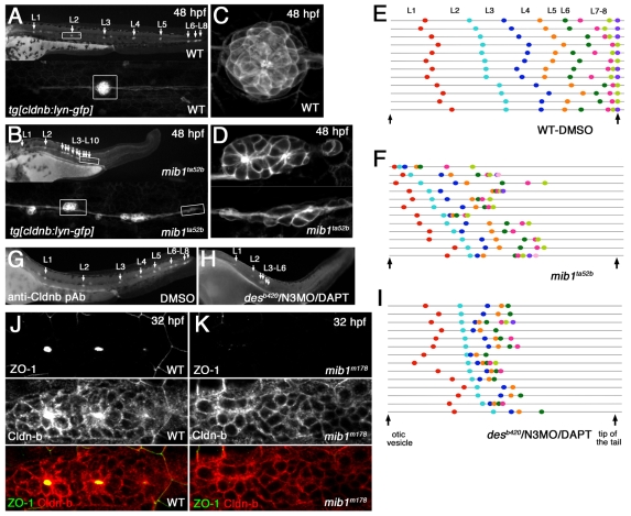
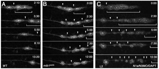
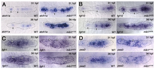
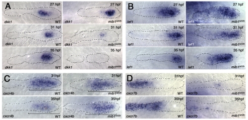
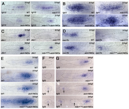
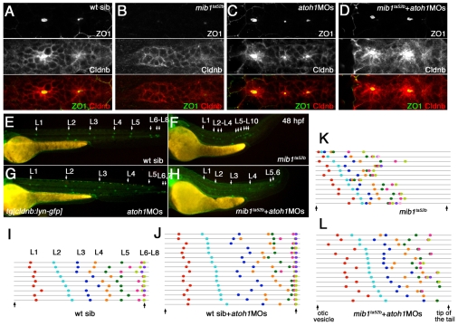
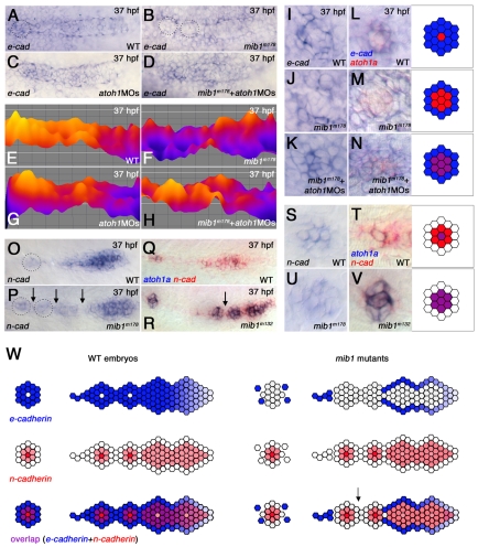
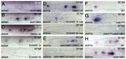
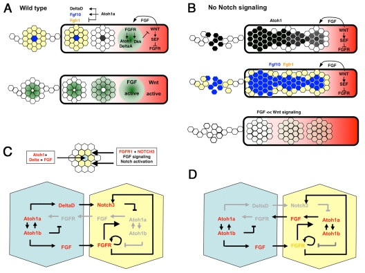
The posterior lateral line primordium (pLLp) migrates caudally, depositing neuromasts to establish the posterior lateral line system in zebrafish. A Wnt-dependent FGF signaling center at the leading end of the pLLp initiates the formation of `proneuromasts' by facilitating the reorganization of cells into epithelial rosettes and by initiating atoh1a expression. Expression of atoh1a gives proneuromast cells the potential to become sensory hair cells, and lateral inhibition mediated by Delta-Notch signaling restricts atoh1a expression to a central cell. We show that as atoh1a expression becomes established in the central cell, it drives expression of fgf10 and of the Notch ligand deltaD, while it inhibits expression of fgfr1. As a source of Fgf10, the central cell activates the FGF pathway in neighboring cells, ensuring that they form stable epithelial rosettes. At the same time, DeltaD activates Notch in neighboring cells, inhibiting atoh1a expression and ensuring that they are specified as supporting cells. When Notch signaling fails, unregulated atoh1a expression reduces Fgfr1 expression, eventually resulting in attenuated FGF signaling, which prevents effective maturation of epithelial rosettes in the pLLp. In addition, atoh1a inhibits e-cadherin expression, which is likely to reduce cohesion and contribute to fragmentation of the pLLp. Together, our observations reveal a genetic regulatory network that explains why atoh1a expression must be restricted by Notch signaling for effective morphogenesis of the pLLp.


Figures

Fig. 1.
Fig. 1.
Loss of Notch signaling causes aberrant neuromast deposition and posterior lateral line primordium (pLLp) migration. (A-D) The distribution and morphology of deposited neuromasts/cell clusters (L1 to L10) in (A,C) wild-type (WT) and (B,D) mib1ta52b tg[cldnb:lynGFP] zebrafish embryos at 48 hpf. (C,D) Epithelial rosettes are disorganized in mib1ta52b mutants. (E,F,I) The pattern of neuromast/cell cluster deposition at 48 hpf in individual wild-type (E), mib1ta52b (F) and desb420/N3MO/DAPT (I) embryos. Colored dots show the position of each deposited neuromast/cell cluster relative to the otic vesicle and tail tip (vertical arrows). Individual embryos are represented on separate lines in an order determined by the relative position of L2. (G,H) The distribution of deposited neuromasts/cell clusters in DMSO-treated (G) and desb420/N3MO/DAPT (H) embryos at 48 hpf. (J,K) There is no central accumulation of ZO1 and Cldnb accumulation is delayed in mib1m178 neuromasts (K) when compared with the wild type (J) at 32 hpf.
Fig. 2.
Fig. 2.
The pLLp eventually undergoes fragmentation when Notch signaling is lost. Images from time-lapse (started at ∼32 hpf) of (A) wild-type, (B) mib1ta52b and (C) N1/N3MO/DAPT tg[cldnb:lynGFP] zebrafish embryos. (A) In wild type, the pLLp migrates as a cohesive structure (bracket); a single break (arrowhead) allows deposition of a neuromast. See Movie 1 in the supplementary material. (B,C) The pLLp fragments as ectopic breaks develop (arrowheads), and proneuromasts are prematurely deposited in both mib1ta52b and N1/N3MO/DAPT embryos. A small portion of the remaining pLLp (bracket) continues to migrate. See Movies 3 and 4 in the supplementary material.
Fig. 3.
Fig. 3.
Notch signaling regulates expression of FGF signaling components. (A,B) atoh1a (A) and fgf10 (B) expression in leading and trailing proneuromasts (arrows) in wild-type and mib1ta52b zebrafish embryos at 31 and 36 hpf. There is 1° expansion of atoh1a and fgf10 expression at 31 hpf and 2° expansion by 36 hpf in mib1ta52b embryos. (C,D) fgfr1 (C) and pea3 (D) are expressed in a pattern complementary to atoh1a and fgf10 in the pLLp. Although subtle, fgfr1 and pea3 expression is reduced in the central cells in maturing proneuromasts (arrowheads). In mib1ta52b embryos, fgfr1 and pea3 expression is reduced in large clusters of cells (1° reduction) at 31 hpf (arrowheads). fgfr1 and pea3 expression is reduced in a broader domain and is restricted to the edges of the pLLp (2° reduction) by 36 hpf.
Fig. 4.
Fig. 4.
Progressive loss of FGF signaling results in loss of FGF-dependent gene expression and in expansion of Wnt-dependent gene expression. (A,B) FGF-dependent dkk1 expression is progressively lost between 27 and 35 hpf in the pLLp of mib1ta52b zebrafish embryos (A), whereas the relative size of the lef1 expression domain progressively expands (B). Note that the lef1 domain becomes progressively smaller in the older wild-type pLLp. (C,D) Whereas cxcr4b expression (C) is not significantly changed in the mib1ta52b pLLp (n=7 at 31 hpf and n=5 at 35 hpf), cxcr7b expression (D) is reduced in mib1ta52b by 31 hpf (n=9 at 31 hpf and n=6 at 35 hpf).
Fig. 5.
Fig. 5.
Knockdown of atoh1 function prevents loss of FGF signaling in the mib1 pLLp. (A,B) atoh1a and atoh1b knockdown prevents unregulated expansion of fgf10 expression in mib1 mutant (mib1ta52b+atoh1MOs) zebrafish embryos (n=10/10) (A). It also prevents loss of fgfr1 expression (B). Statistical analysis is shown in Fig. S10A in the supplementary material. (C) dkk1 expression is recovered in mib1ta52b+atoh1MOs embryos. For statistical analysis, see Fig. S11 in the supplementary material. (D) atoh1MOs allow partial recovery of cxcr7b expression in mib1ta52b embryos (n=5/5). (E) atoh1MOs allow lef1 expression to expand further in mib1m178 mutants. Statistical analysis is shown in Fig. S13 in the supplementary material. (F,G) Whereas there is no fgf3 expression in depositing (F) and deposited (G) neuromasts in wild-type (10/10) and mib1m132 (10/10) embryos, knockdown of atoh1 results in the persistence of fgf3 expression in wild-type (7/10) and mib1m132 (5/10) embryos.
Fig. 6.
Fig. 6.
Knockdown of atoh1 reduces some morphogenesis defects in the mib1 pLLp. (A-D) The central accumulation of ZO1 and Cldnb in maturing proneuromasts (A) is lost in mib1ta52b mutants (B), but is recovered when atoh1 is knocked down (D). (E-L) The relative position of deposited neuromasts (L1 to L10) at 48 hpf (E-H). In mib1ta52b+atoh1MOs zebrafish embryos (H), neuromast distribution and pLLp migration are partially rescued. (I-L) Schematic representation of the pattern of neuromast deposition in individual embryos at 48 hpf. For statistical analysis, see Fig. S3 in the supplementary material.
Fig. 7.
Fig. 7.
Changes in pLLp cohesion correlate with changes in Cadherin gene expression. (A-N) e-cadherin expression in the pLLp (A-H) and deposited proneuromasts (I-N) at ∼37 hpf. (E-H) Color-coded representations of the e-cadherin expression shown in A-D, created using the interactive three-dimensional surface plot plug-in from ImageJ (Abramoff et al., 2004). (A,E) In wild-type, e-cadherin is broadly expressed, being lower at the leading and higher at the trailing proneuromasts prior to deposition. (B,F) e-cadherin expression is suppressed in the pLLp trailing domain of mib1m178 zebrafish embryos (n=8). Dotted circles in B represent domains of suppressed e-cadherin expression. (C,G) atoh1MOs do not substantially alter e-cadherin expression in wild-type embryos (n=7). (D,H) Higher e-cadherin expression in the trailing pLLp is restored in mib1m178+atoh1MOs embryos (n=12). (I,L) e-cadherin expression (blue) is generally more intense, but absent in a central atoh1a-expressing cell (red), in deposited neuromasts (n=6). (J,M) The central atoh1a-expressing domain expands at the cost of e-cadherin in mib1m178 deposited neuromasts (n=6). (K,N) Knockdown of atoh1 in mib1m178 expands e-cadherin expression and allows overlap (purple) with atoh1a (n=13). (O-V) n-cadherin expression in the pLLp (O-R) and deposited proneuromasts (S-V). (O,Q) In wild type, n-cadherin is broadly expressed at the leading end but becomes restricted at the trailing end, prior to neuromast deposition. (P,R) In mib1m178 pLLp, n-cadherin expression is not significantly different from that of wild type. Dotted circles highlight an example of expanded expression of n-cadherin in isolated groups of cells, which are likely to represent prematurely deposited proneuromasts from a recently fragmented pLLp. (Q,R,T,V) Double in situ hybridization showing atoh1a (blue) and n-cadherin (red) expression. (S-V) In deposited neuromasts, n-cadherin expression is similar in wild type (S,T) and mib1 mutants (U,V). In wild type, a central cell expressing atoh1a and n-cadherin (purple in T) is surrounded by cells expressing only n-cadherin (red in T). In mib1 mutants, atoh1a expands and overlaps with n-cadherin (purple in V). (W) The pLLp and neuromasts normally contain cells that express e-cadherin (blue) or n-cadherin (red). Intervening cells expressing both e-cadherin and n-cadherin (purple) provide an adhesive link between cells that express distinct cadherins. When Notch signaling fails, atoh1a expression expands and e-cadherin is lost from intervening cells that would otherwise express both Cadherin genes. As e-cadherin- and n-cadherin-expressing populations are now unable to establish effective adhesive interactions, the pLLp fragments. Arrow indicates predicted point of fragmentation.
Fig. 8.
Fig. 8.
atoh1a and atoh1b cross-activation and establishment of a focal FGF signaling center. (A,B) atoh1MOs eliminate deltaD (A), but not deltaA (B), expression in the pLLp of wild-type zebrafish embryos. (C) SU5402 treatment eliminates deltaA expression after 2 hours. (D) atoh1a expression is retained in the trailing proneuromasts following 15 minutes of SU5402 exposure (middle), but is lost by 30 minutes (bottom). (E) atoh1b knockdown reduces atoh1a expression in the trailing neuromast (top). atoh1b knockdown accompanied by SU5402 exposure for 15 minutes reduces atoh1a expression throughout the entire pLLp (bottom). (F) atoh1b expression is restricted to the trailing neuromast. (G) atoh1b expression expands in mib1ta52b pLLps (top) but is lost when atoh1a MO is injected (bottom). (H) knockdown of atoh1a expands atoh1a expression at the leading end, but expanded expression is not maintained at the trailing end (bottom).
Fig. 9.
Fig. 9.
Establishment of a focal FGF signaling center by atoh1 in maturing proneuromasts. (A) Wnt activation (red) drives FGF and sef expression, prevents local FGF activation and promotes FGF signaling (green) in an adjacent domain. FGF activation drives expression of the Wnt antagonist dkk1 and of atoh1a and deltaA. Delta activates Notch in neighboring cells to restrict atoh1a expression to a central cell (black). atoh1a drives expression of fgf10 (blue) and inhibits fgfr1(yellow), establishing a focal FGF signaling system. (B) In the absence of Notch signaling, additional cells express atoh1a and fgf10, then shut off fgfr1. This results in progressive loss of active FGF signaling, loss of dkk1, and expansion of Wnt activation. (C) Detail of interactions between the central Atoh-expressing cell (blue) and surrounding cells (yellow). Atoh1a-Atoh1b cross-regulation helps maintain atoh1 expression in the central cell. Delta and FGF activate Notch and Fgfr1 in surrounding cells. fgfr1 expression is maintained by autoregulation. FGF activation maintains notch3 expression. Notch inhibits atoh1a expression in surrounding cells. (D) In the absence of Notch activation, surrounding cells (yellow) initiate atoh1a expression; this initiates FGF expression, suppresses Fgfr1 expression, reduces FGF signaling and eventually leads to loss of Notch3 expression.

Similar articles

Cited by

References

    1. Abramoff M. D., Magelhaes P. J., Ram S. J. (2004). Image processing with ImageJ. Biophotonics Int. 11, 36-42
    1. Aman A., Piotrowski T. (2008). Wnt/beta-catenin and Fgf signaling control collective cell migration by restricting chemokine receptor expression. Dev. Cell 15, 749-761 - PubMed
    1. Cavallaro U., Christofori G. (2004). Cell adhesion and signalling by cadherins and Ig-CAMs in cancer. Nat. Rev. Cancer 4, 118-132 - PubMed
    1. Choe E. A., Liao L., Zhou J. Y., Cheng D., Duong D. M., Jin P., Tsai L. H., Peng J. (2007). Neuronal morphogenesis is regulated by the interplay between cyclin-dependent kinase 5 and the ubiquitin ligase mind bomb 1. J. Neurosci. 27, 9503-9512 - PMC - PubMed
    1. D'Souza B., Miyamoto A., Weinmaster G. (2008). The many facets of Notch ligands. Oncogene 27, 5148-5167 - PMC - PubMed

Publication types

MeSH terms