. 2010 Oct 15;123(Pt 20):3587-95.
doi: 10.1242/jcs.073817. Epub 2010 Sep 28.

CMF70 is a subunit of the dynein regulatory complex

Affiliations

CMF70 is a subunit of the dynein regulatory complex

Zakayi P Kabututu et al. J Cell Sci. .

Abstract

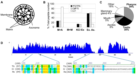
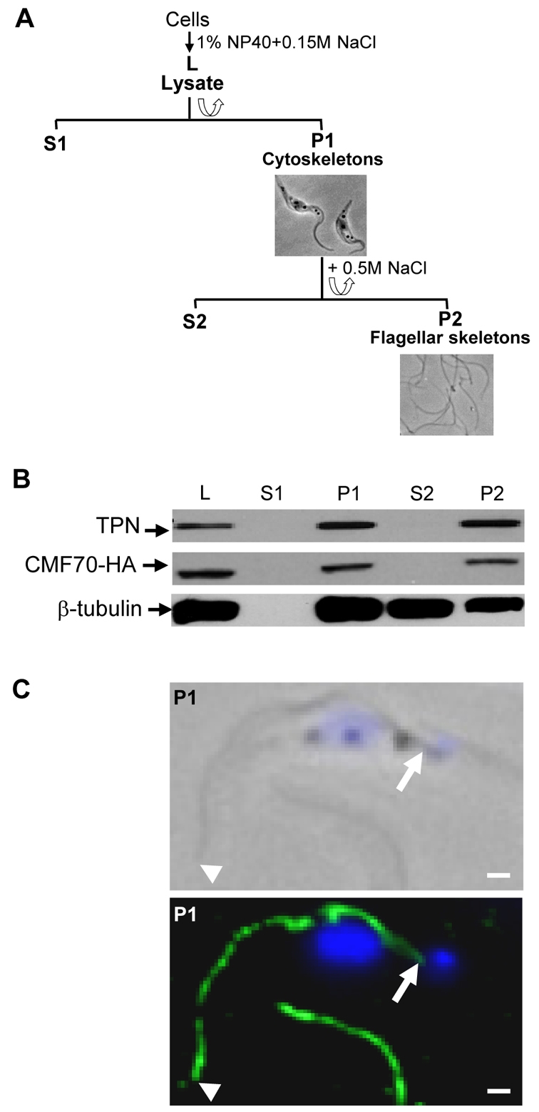
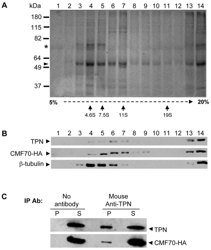
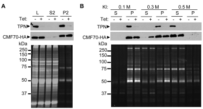
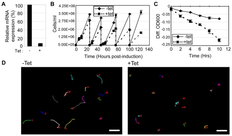
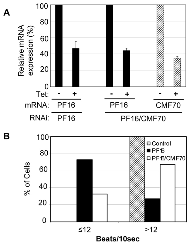
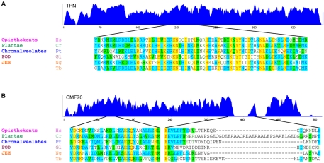
Flagellar motility drives propulsion of several important pathogens and is essential for human development and physiology. Motility of the eukaryotic flagellum requires coordinate regulation of thousands of dynein motors arrayed along the axoneme, but the proteins underlying dynein regulation are largely unknown. The dynein regulatory complex, DRC, is recognized as a focal point of axonemal dynein regulation, but only a single DRC subunit, trypanin/PF2, is currently known. The component of motile flagella 70 protein, CMF70, is broadly and uniquely conserved among organisms with motile flagella, suggesting a role in axonemal motility. Here we demonstrate that CMF70 is part of the DRC from Trypanosoma brucei. CMF70 is located along the flagellum, co-sediments with trypanin in sucrose gradients and co-immunoprecipitates with trypanin. RNAi knockdown of CMF70 causes motility defects in a wild-type background and suppresses flagellar paralysis in cells with central pair defects, thus meeting the functional definition of a DRC subunit. Trypanin and CMF70 are mutually conserved in at least five of six extant eukaryotic clades, indicating that the DRC was probably present in the last common eukaryotic ancestor. We have identified only the second known subunit of this ubiquitous dynein regulatory system, highlighting the utility of combined genomic and functional analyses for identifying novel subunits of axonemal sub-complexes.


Figures

Fig. 1.
Fig. 1.
CMF70 is a conserved component of motile flagella. (A) Cross-section cartoon of a flagellum showing compartments separated by biochemical fractionation. (B) Relative number of peptides identified by Pazour and colleagues (Pazour et al., 2005) in mass spectrometry analyses of C. reinhardtii flagellar fractions corresponding to tergitol-insoluble membrane plus axoneme (M+A), Nonidet-soluble membrane plus matrix (M+M), Nonidet-insoluble axonemes extracted with 0.6 M KCl to yield solubilized extract (KCl Ex) and insoluble extracted axonemes (Ex. Ax.). Data from Pazour and colleagues (Pazour et al., 2005) are tabulated for PF2/trypanin, CMF70 and LC1. The distribution of peptides is similar for PF2/trypanin and CMF70, but different for the outer dynein subunit LC1. (C) Expression profile of the human CMF70 homologue shows enrichment in ciliated tissues (http://www.ncbi.nlm.nih.gov/UniGene/). (D) Amino acid sequence similarity plot (Vector NTI, Invitrogen) of CMF70 homologues from T. brucei (Tb), C. reinhardtii (Cr) and H. sapiens (Hs). A value of +1 is corresponds to a stretch of identical amino acids. Representative regions of high similarity (residues 97–124) and low similarity (residues 366–408) are shown below the chart. Strictly conserved positions are highlighted in yellow, whereas residues identical in two of three sequences are highlighted in blue and conservative substitutions are green.
Fig. 2.
Fig. 2.
CMF70 is tightly associated with the T. brucei flagellar skeleton. (A) Flow chart depicting fractionation procedure to isolate T. brucei salt-extracted flagellar skeletons. Lysates (L) were prepared by extraction with 1% NP-40 and centrifuged for separation of detergent-soluble proteins (S1) from cytoskeletons (P1). The P1 fraction was further extracted with 0.5 M NaCl treatment to solubilize the subpellicular cytoskeleton (S2) and leave the flagellar skeletons (P2) intact. Corresponding phase-contrast images of cell (P1) and flagellar (P2) skeletons are shown. (B) Western blot analysis of whole-cell lysates and subcellular fractions prepared from HA-tagged CMF70 T. brucei cells shows a stable association of CMF70 and trypanin with the flagellar skeleton fraction (P2). The protein preparations were blotted with anti-HA (top panel), anti-trypanin (middle panel) and anti-β-tubulin (bottom panel) monoclonal antibodies. (C) Indirect immunofluorescence analysis. Cytoskeletons (P1) from CMF70-HA T. brucei cells were visualized after labeling with monoclonal anti-HA antibody (green) and DAPI (blue). Phase-contrast (top panel) and merged fluorescence (lower panel) images are shown. Arrowheads indicate the distal tip and arrows show the proximal end of the flagellum, near the kinetoplast. The flagellum of a second cell is visible at the bottom of the image. Scale bars: 1 μm.
Fig. 3.
Fig. 3.
TbCMF70 is in a complex with trypanin. (A,B) Sucrose gradient fractionation of solubilized flagellar skeletons. Flagellar skeletons from CMF70-HA cells were extracted with 0.5 M KI, dialyzed against immunoprecipitation buffer and centrifuged to remove insoluble material. The soluble fraction was subjected to 5–20% sucrose gradient centrifugation and fractions were analyzed by SDS–PAGE and Coomassie Blue staining for total protein (A) or immunoblotting using anti-trypanin (TPN), anti-HA or anti-β-tubulin antibodies as indicated (B). In A, arrows indicate the position of migration in sucrose gradients for calibration standards. Tubulin (arrowhead) and the PFR1 and PFR2 (asterisk) proteins are indicated. (C) Solubilized flagellar skeletons from CMF70-HA T. brucei cells were immunoprecipitated with or without the mouse monoclonal anti-TPN antibody and analyzed by immunoblotting of the supernatants (S) and precipitated beads (P) using rabbit polyclonal anti-trypanin (TPN) or mouse monoclonal anti-HA antibodies (CMF70-HA).
Fig. 4.
Fig. 4.
Loss of trypanin weakens the association of CMF70 with the axoneme. (A) Protein fractions, as defined in Fig. 2, were prepared from CMF70-HA cells harboring a Tet-inducible trypanin RNAi knockdown construct grown with (+) or without (−) 1 μg/ml tetracycline for 72 hours. Samples were analyzed by SDS-PAGE and immunoblotting with antibodies against trypanin (TPN, top panel) or HA (CMF70-HA, middle panel), or stained for total protein with Sypro-ruby (lower panel). (B) P2 flagellar skeletons from uninduced (−) and Tet-induced (+) trypanin RNAi CMF70-HA cells were extracted with the indicated concentration of KI and separated by centrifugation into supernatants (S) and pellets (P). Samples were analyzed as in A. Treatment with 0.3 M KI partially solubilized CMF70-HA from the axoneme when trypanin was ablated by RNAi but not in control cells. Results are representative of three independent experiments.
Fig. 5.
Fig. 5.
CMF70 is required for normal flagellar motility in T. brucei. (A) qRT-PCR analysis shows a dramatic decrease in cmf70 mRNA in CMF70-knockdown cells (+tet) compared with control cells (−tet). (B) Growth curves of uninduced (−Tet) and induced (+Tet) CMF70-knockdown cells show slowed growth within 24 hours of RNAi induction. Each curve represents an average of two independent cell counts and the error bars indicate s.d. (C) Sedimentation assays (Bastin et al., 1999; Ralston et al., 2006) on uninduced (−Tet) versus induced (+Tet) CMF70 RNAi cells show that CMF70-knockdown cells sediment, indicative of a motility defect. The curves represent an average of two independent measurements and the error bars indicate s.d. (D) Motility traces of uninduced (−tet) and induced (+tet) CMF70 RNAi cells. Lines trace the movement of individual cells over a 35 second time interval. Numbers in each panel represent individual cells. Scale bars: 50 μm.
Fig. 6.
Fig. 6.
Loss of CMF70 suppresses flagellar paralysis of central pair knockdowns. (A) qRT-PCR analysis of pf16 and cmf70 transcripts in PF16 single-knockdown or PF16–CMF70 double-knockdown cells. Expression of RNAi-targeted transcripts in tetracycline-induced (+ Tet) cultures is shown relative to uninduced controls (−Tet). Averages of two independent experiments are shown with error bars representing s.d. (B) Analysis of flagellar beating in PF16 single-knockdown (n=55), PF16–CMF70 double-knockdown (n=50) and control cells (n=57). Beats at the tip of the flagellum were quantified over a 10 second interval, as described (Ralston et al., 2006).
Fig. 7.
Fig. 7.
Deep-branching origins of the dynein regulatory complex. Similarity plots (top) with representative sequence alignments (bottom) are shown for trypanin (A) and CMF70 (B) from organisms representing the indicated eukaryotic clades (Fritz-Laylin et al., 2010). JEH encompasses Jakobids, Euglenozoa, Heterolobosea, whereas POD encompasses Parabasalids, Oxymonads and Diplomonads (Fritz-Laylin et al., 2010). Non-trypanosome sequences are from Homo sapiens (Hs), C. reinhardtii (Cr), Paramecium tetraurelia (Pt), Giardia lamblia (Gl) and Naegleria gruberi (Ng). The T. brucei sequence (Tb) is shown for reference. Trypanin proteins show high similarity along their lengths, including several strictly conserved residues. Sequence alignment for the C-terminal portion of the GMAD microtubule-binding domain in the human trypanin homologue (residues 189–260) (Bekker et al., 2007) is shown for reference. CMF70 proteins show high similarity along their lengths. Sequence alignment is shown from conserved and non-conserved regions for reference. Dissimilarity at the N and C-termini of CMF70 derives from unique insertions in the human, Paramecium and trypanosome sequences, suggestive of species-specific functions. Strictly conserved positions are highlighted in yellow, whereas residues identical in three or more sequences are highlighted in blue and conservative substitutions are green.

Similar articles

Cited by

References

    1. Baron D. M., Kabututu Z. P., Hill K. L. (2007a). Stuck in reverse: loss of LC1 in Trypanosoma brucei disrupts outer dynein arms and leads to reverse flagellar beat and backward movement. J. Cell Sci. 120, 1513-1520 - PubMed
    1. Baron D. M., Ralston K. S., Kabututu Z. P., Hill K. L. (2007b). Functional genomics in Trypanosoma brucei identifies evolutionarily conserved components of motile flagella. J. Cell Sci. 120, 478-491 - PubMed
    1. Bastin P., Sherwin T., Gull K. (1998). Paraflagellar rod is vital for trypanosome motility. Nature 391, 548 - PubMed
    1. Bastin P., Pullen T. J., Sherwin T., Gull K. (1999). Protein transport and flagellum assembly dynamics revealed by analysis of the paralysed trypanosome mutant snl-1. J. Cell Sci. 112, 3769-3777 - PubMed
    1. Bekker J. M., Colantonio J. R., Stephens A. D., Clarke W. T., King S. J., Hill K. L., Crosbie R. H. (2007). Direct interaction of Gas11 with microtubules: implications for the dynein regulatory complex. Cell Motil. Cytoskeleton 64, 461-473 - PubMed

Publication types

Substances

LinkOut - more resources