. 2010 Dec;5(12):2180-7.
doi: 10.2215/CJN.02890310. Epub 2010 Sep 28.

Mass spectrometry-based proteomic diagnosis of renal immunoglobulin heavy chain amyloidosis

Affiliations

Mass spectrometry-based proteomic diagnosis of renal immunoglobulin heavy chain amyloidosis

Sanjeev Sethi et al. Clin J Am Soc Nephrol. 2010 Dec.

Abstract

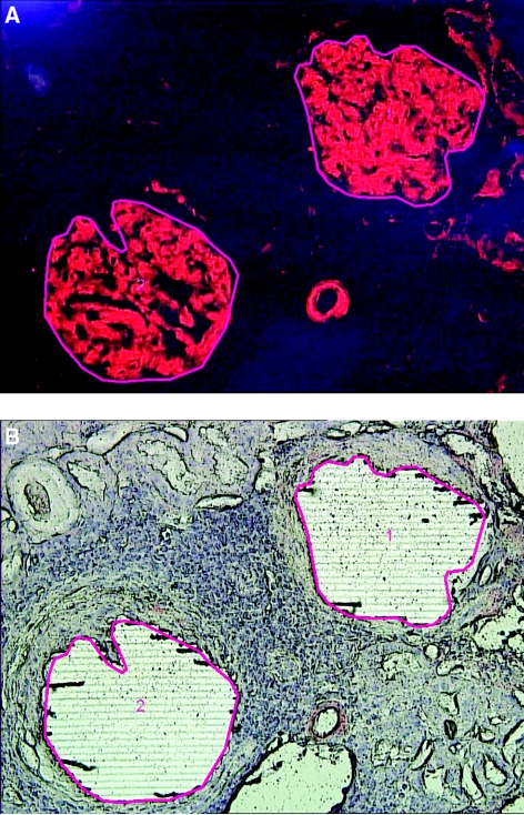
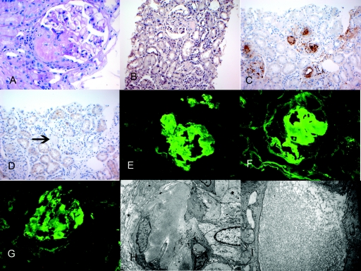
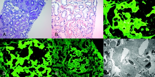
Background and objectives: Amyloidosis is a group of disorders characterized by accumulation of extracellular deposition of proteins as insoluble aggregates. The clinical management of amyloidosis is based on identifying the underlying etiology and accurate typing of the amyloid. Ig heavy chain amyloid involving the kidney is poorly recognized and often poses a diagnostic dilemma. DESIGN, SETTING, PARTICIPANTS, & MEASURES: In this study, we describe the use of laser microdissection (LMD) and mass spectrometry (MS)-based proteomic analysis for the accurate typing of 14 cases of amyloidosis. We also describe the clinicopathologic findings of four problematic cases of renal Ig heavy chain amyloidosis that required LMD/MS proteomic analysis for accurate typing of the amyloid.

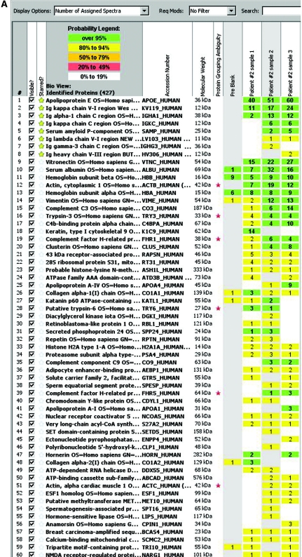
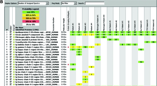
Results: LMD/MS proteomic data of four cases of Ig heavy chain renal amyloidosis showed Ig heavy chains with or without light chains. The break up of the Ig heavy chains was as follows: one case showed Igγ1 chain constant region and λ light chains, one case showed Igα chain constant region and κ light chains variable and constant regions, whereas two cases showed Igγ3 chain constant region and heavy chains variable region I and/or III without light chains. We compare the LMD/MS proteomic data of Ig heavy chain renal amyloid with that of other types of amyloid, including Ig light chains, serum amyloid A, fibrinogen A-α chain renal amyloid, and transthyretin amyloid.

Conclusions: We conclude that LMD/MS is a sensitive and specific tool for diagnosis and accurate typing of renal amyloidosis, including Ig heavy chain amyloid.


Figures

Figure 1.
Figure 1.
Laser microdissection. (A) Representative figure (patient 4) showing Congo red–stained positive glomeruli (1 and 2) to be microdissected. (B) Vacant space on slide after laser microdissection.
Figure 2.
Figure 2.
(A) Scaffold readout of all proteins by spectra of patient 2. MS proteomic data are shown from three samples (1, 2, and 3) prepared from three separate LMDs of Congo red–positive glomeruli. The proteomic data show Igα chain constant region and κ light chains variable and constant region with >95% probability. (B) Representative scaffold readout of proteins of interest for 14 cases of amyloidosis by spectra. The cases are as follows (left to right): four cases of Ig heavy chain amyloid (patients 1 through 4), two cases of fibrogen A-α chain amyloid, two cases of SAA amyloid, four cases of AL amyloid, and two cases of TTR amyloid, respectively. The yellow stars indicate proteins of interest, whereas the red stars indicate protein ambiguity when two proteins share conserved areas.
Figure 2.
Figure 2.
(A) Scaffold readout of all proteins by spectra of patient 2. MS proteomic data are shown from three samples (1, 2, and 3) prepared from three separate LMDs of Congo red–positive glomeruli. The proteomic data show Igα chain constant region and κ light chains variable and constant region with >95% probability. (B) Representative scaffold readout of proteins of interest for 14 cases of amyloidosis by spectra. The cases are as follows (left to right): four cases of Ig heavy chain amyloid (patients 1 through 4), two cases of fibrogen A-α chain amyloid, two cases of SAA amyloid, four cases of AL amyloid, and two cases of TTR amyloid, respectively. The yellow stars indicate proteins of interest, whereas the red stars indicate protein ambiguity when two proteins share conserved areas.
Figure 3.
Figure 3.
Representative biopsy findings of patient 1. (A) Light microscopy showing PAS-negative material at the vascular pole (PAS stain, ×40). (B) Mild positive Congo red stain (×20). (C) Positive SAP stain in the glomeruli and arteriolar walls (×20). (D) Negative SAA stain, arrow points to negative staining (×20); immunofluorescence microscopy shows positive glomerular staining for (E) IgG and (F) λ light chains. (G) κ light chains showed mild (1+) positive staining, and electron microscopy shows randomly oriented amyloid fibrils along arteriolar walls (H) and glomerular capillary walls (I).
Figure 4.
Figure 4.
Representative biopsy findings of patient 2. (A) Light microscopy showing PAS-negative amyloid material in the glomeruli, interstitium, and vessels (PAS stain, ×20). (B) Positive Congo red stain showing positive staining in the glomeruli, interstitium, and vessels (×20); immunofluorescence microscopy shows positive glomerular staining for (C) IgA and (D) κ light chains. (E) λ light chains are essentially negative. (F) Electron microscopy shows randomly oriented amyloid fibrils in the glomerulus.

References

    1. Dember LM: Amyloidosis-associated kidney disease. J Am Soc Nephrol 17: 3458–3471, 2006 - PubMed
    1. Merlini G, Bellotti V: Molecular mechanisms of amyloidosis. N Engl J Med 349: 583–596, 2003 - PubMed
    1. Gillmore JD, Lachmann HJ, Rowczenio D, Gilbertson JA, Zeng C-H, Liu Z-H, Li L-S, Wechalekar A, Hawkins PN: Diagnosis, pathogenesis, treatment, and prognosis of hereditary fibrinogen A{alpha}-chain amyloidosis. J Am Soc Nephrol 20: 444–451, 2009 - PMC - PubMed
    1. Lachmann HJ, Booth DR, Booth SE, Bybee A, Gilbertson JA, Gillmore JD, Pepys MB, Hawkins PN: Misdiagnosis of hereditary amyloidosis as AL (primary) amyloidosis. N Engl J Med 346: 1786–1791, 2002 - PubMed
    1. Miller DV, Dogan A, Sethi S: New-onset proteinuria with massive amorphous glomerular deposits. Am J Kidney Dis 55: 749–754, 2010 - PubMed