. 2011 Jan;39(2):767-80.
doi: 10.1093/nar/gkq812. Epub 2010 Sep 28.

On the molecular basis of uracil recognition in DNA: comparative study of T-A versus U-A structure, dynamics and open base pair kinetics

Affiliations

On the molecular basis of uracil recognition in DNA: comparative study of T-A versus U-A structure, dynamics and open base pair kinetics

Elisa Fadda et al. Nucleic Acids Res. 2011 Jan.

Abstract

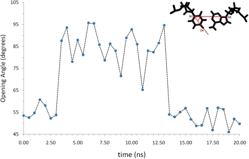
Uracil (U) can be found in DNA as a mismatch paired either to adenine (A) or to guanine (G). Removal of U from DNA is performed by a class of enzymes known as uracil-DNA-glycosylases (UDG). Recent studies suggest that recognition of U-A and U-G mismatches by UDG takes place via an extra-helical mechanism. In this work, we use molecular dynamics simulations to analyze the structure, dynamics and open base pair kinetics of U-A base pairs relative to their natural T-A counterpart in 12 dodecamers. Our results show that the presence of U does not alter the local conformation of B-DNA. Breathing dynamics and base pair closing kinetics are only weakly dependent on the presence of U versus T, with open T-A and U-A pairs lifetimes in the nanosecond timescale. Additionally, we observed spontaneous base flipping in U-A pairs. We analyze the structure and dynamics for this event and compare the results to available crystallographic data of open base pair conformations. Our results are in agreement with both structural and kinetic data derived from NMR imino proton exchange measurements, providing the first detailed description at the molecular level of elusive events such as spontaneous base pair opening and flipping in mismatched U-A sequences in DNA. Based on these results, we propose that base pair flipping can occur spontaneously at room temperature via a 3-step mechanism with an open base pair intermediate. Implications for the molecular basis of U recognition by UDG are discussed.


Figures

Figure 1.
Figure 1.
A T–A base pair in Watson–Crick conformation is shown with green C atoms in panel (a), and a U–A base pair in an open conformation is shown with orange C atoms in panel (b). The open conformation is held together by one hydrogen bond between the O2 of U and the the NH2 of A. Schematic of the proposed 3-steps spontaneous base flipping mechanism is shown in panel (c).
Figure 2.
Figure 2.
Na+ bound in the TAAU minor groove. Only the two base pairs involved in the ion binding are shown. Adenine (A) 7 and thymine (T) 18 are shown with green C atoms, while uracil (U) 8 and A17 are shown with blue C atoms. Na+ is coordinated to the O of U8 and T18.
Figure 3.
Figure 3.
Graphical representation of the four helical parameters monitored throughout the MD simulations. Each rectangle represents a base. The representation is adapted from (51,52). Shear and stretch parameters are measured in Angstrom (Å), while opening and tilt in degrees.
Figure 4.
Figure 4.
Variation of the opening angle for the U7A18 base pair during the first 20 ns of MD simulation of the TAUA dodecamer. The opening angle is defined by the atoms N9-N1-O4, as shown in the panel on the top-right corner. Arrows indicate the timeframe during which the base pair is, respectively, ‘closed’ or in the Watson–Crick configuration or ‘open’. Average opening angle in the ‘closed’ conformation is 53.2°, while in the ‘open’ conformation is 85.2°.
Figure 5.
Figure 5.
Phosphate backbone torsions ε (a) and ζ (b) at the U of the TAUA sequence during the 20-ns MD simulation when the opening takes place. The change in configuration seems to happen as an immediate response to the base opening, which occurs at 3.62 ns. The U–A base pair returns to the Watson–Crick configuration after 13.62 ns. The time interval during which the U–A base pair is in the open conformation is highlighted in pink.
Figure 6.
Figure 6.
Decay kinetics of the open base pair states for all 12 dodecamers. The legend on the right indicates the symbols assigned to each sequence. Trend lines are shown as solid lines for all natural sequences and as dotted lines for sequences containing U. The relative R2-values are shown within brackets for each dodecamer.
Figure 7.
Figure 7.
The distance (dON) between O2 of U6 and NH2 of A19 (see Figure 1 for nomenclature) in one of the AUAT dodecamers used to measure the kinetic decay of open states is monitored along 20 ns trajectory. In the open state, once the H-bond between O2-NH2 breaks, U6 flips out of the stacking. The flipped-out state endures for ∼11 ns before the U6 pairs back with A19, re-establishing the natural Watson–Crick conformation. Snapshots of selected structures are shown above the graph. An image of 2OXM (top-right corner) is also shown for comparison.
Figure 8.
Figure 8.
Values of the ε/ζ torsions for U6 in the AUAT dodecamer >40 ns of MD simulation. The ε is shown in purple, while ζ is shown in green. Ranges defining g+, t and g conformations are highlighted in light green, pink and light blue, respectively, for clarity. The simulation starts with U6-A19 in an open conformation. At 1-ns U6 breaks out of the H bond with A1 and flips-out in an extra-helical position, which holds for 11 ns. During this time interval ε/ζ torsions of the U6 base are in a g/g+ conformation. Closing into a Watson–Crick pairing at 12 ns causes a slight change in conformation. Finally, ε/ζ return in a B-DNA conformation, i.e. t/g, at 22.5 ns.
Figure 9.
Figure 9.
Values of the ε/ζ torsions for for A19, paired to U6, in the AUAT dodecamer >20 ns of MD simulation. The ε is shown in purple, while ζ is shown in green. Ranges defining g+, t and g conformations are highlighted in light green, pink and light blue, respectively, for clarity. Initially ε/ζ of A19 are in a B-DNA t/g conformation. As U6 goes in the flipped-out conformation, ∼1 ns, as for U6 ε/ζ inverts into a g/g+. The latter holds long after U6 closes. The ε/ζ go back into a B-DNA conformation at 30 ns.

References

    1. Sousa MML, Krokan HE, Sluppaugh G. DNA-uracil and human pathology. Mol. Asp. Med. 2007;28:276–306. - PubMed
    1. Krokan HE, Standal R, Sluppaugh G. DNA glycosylases in the base excision repair of DNA. Biochem. J. 1997;325:1–16. - PMC - PubMed
    1. Krokan HE, Drabløs F, Sluppaugh G. Uracil in DNA — occurrence, consequences and repair. Oncogene. 2002;21:8935–8948. - PubMed
    1. Friedberg EC, Walker GC, Siede W, Wood RD, Schultz RA, Ellenberger T. DNA Repair and Mutagenesis. Washington DC: ASM Press; 2006.
    1. Andersen S, Heine T, Sneve R, Konig I, Krokan HE, Epe B, Nilsen H. Incorporation of dUMP into DNA is a major source of spontaneous DNA damage, while excision of uracil is not required for cytotoxicity of fluoropyrimidines in mouse embryonic fibroblasts. Carcinogenesis. 2005;26:547–555. - PubMed

Publication types