

Parathyroid hormone-related protein enhances human ß-cell proliferation and function with associated induction of cyclin-dependent kinase 2 and cyclin E expression
- PMID: 20876711
- PMCID: PMC2992775
- DOI: 10.2337/db09-1796
Parathyroid hormone-related protein enhances human ß-cell proliferation and function with associated induction of cyclin-dependent kinase 2 and cyclin E expression
Abstract
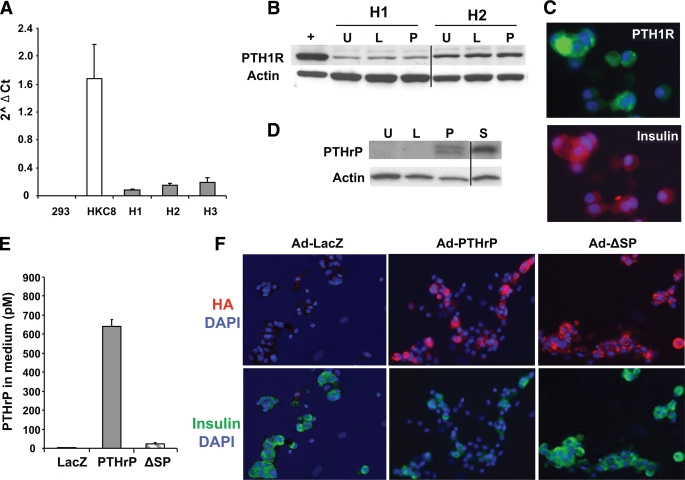
Objective: Inducing human β-cell growth while enhancing function is a major goal in the treatment of diabetes. Parathyroid hormone-related protein (PTHrP) enhances rodent β-cell growth and function through the parathyroid hormone-1 receptor (PTH1R). Based on this, we hypothesized that PTH1R is expressed in human β-cells and that PTHrP has the potential to enhance human β-cell proliferation and/or function.
Research design and methods: PTH1R expression, β-cell proliferation, glucose-stimulated insulin secretion (GSIS), and expression of differentiation and cell-cycle genes were analyzed in human islets transduced with adenoviral PTHrP constructs or treated with PTHrP peptides. The effect of overexpression of late G1/S cell cycle molecules was also assessed on human β-cell proliferation.
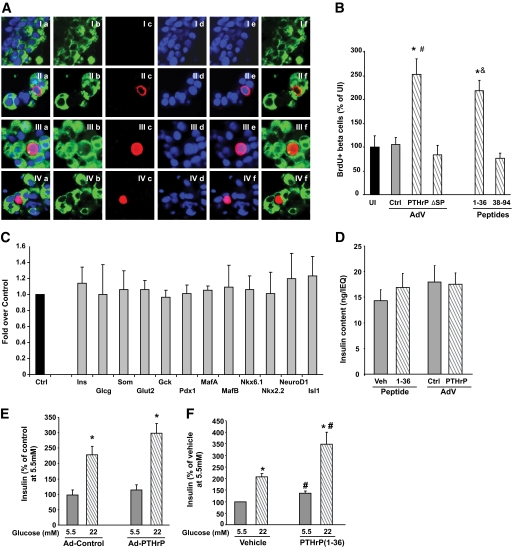
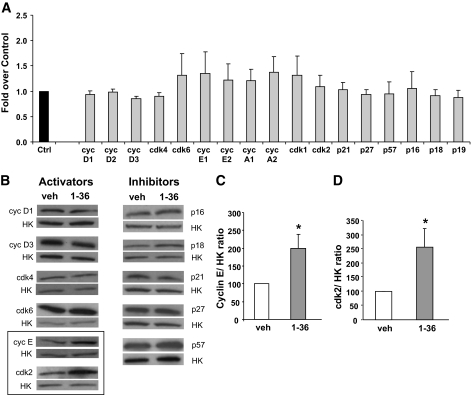
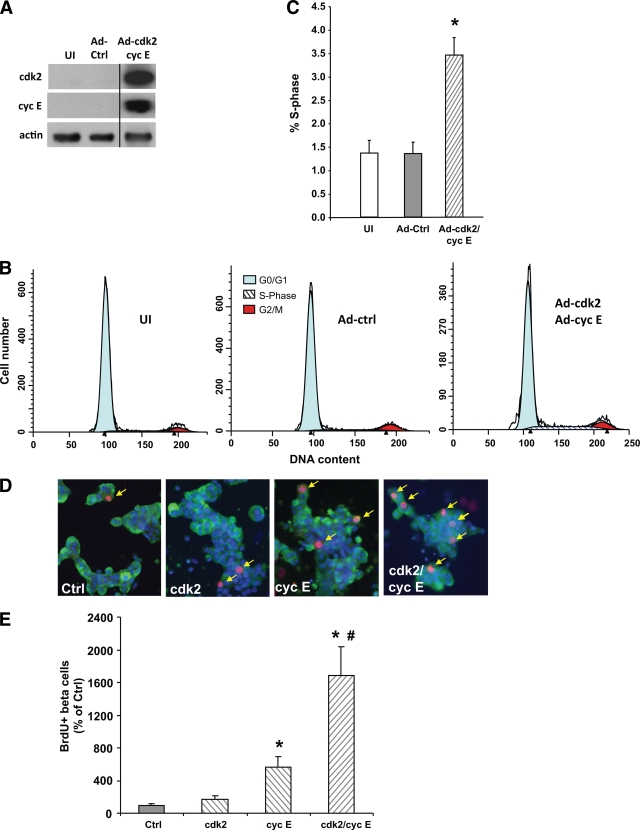
Results: We found that human β-cells express PTH1R. More importantly, overexpression of PTHrP causes a significant approximately threefold increase in human β-cell proliferation. Furthermore, the amino terminus PTHrP(1-36) peptide is sufficient to increase replication as well as expression of the late G1/S cell-cycle proteins cyclin E and cyclin-dependent kinase 2 (cdk2) in human islets. Notably, PTHrP(1-36) also enhances GSIS. Finally, overexpression of cyclin E alone, but not cdk2, augments human β-cell proliferation, and when both molecules are expressed simultaneously there is a further marked synergistic increase in replication.
Conclusions: PTHrP(1-36) peptide enhances human β-cell proliferation as well as function, with associated upregulation of two specific cell-cycle activators that together can induce human β-cell proliferation several fold. The future therapeutic potential of PTHrP(1-36) for the treatment of diabetes is especially relevant given the complementary therapeutic efficacy of PTHrP(1-36) in postmenopausal osteoporosis.
Figures
Similar articles
-
Systemic and acute administration of parathyroid hormone-related peptide(1-36) stimulates endogenous beta cell proliferation while preserving function in adult mice.Diabetologia. 2011 Nov;54(11):2867-77. doi: 10.1007/s00125-011-2260-z. Epub 2011 Jul 29. Diabetologia. 2011. PMID: 21800111
-
Cell cycle actions of parathyroid hormone-related protein in non-small cell lung carcinoma.Am J Physiol Lung Cell Mol Physiol. 2009 Oct;297(4):L578-85. doi: 10.1152/ajplung.90560.2008. Epub 2009 Jul 24. Am J Physiol Lung Cell Mol Physiol. 2009. PMID: 19633068 Free PMC article.
-
Parathyroid Hormone-Related Peptide (1-36) Enhances Beta Cell Regeneration and Increases Beta Cell Mass in a Mouse Model of Partial Pancreatectomy.PLoS One. 2016 Jul 8;11(7):e0158414. doi: 10.1371/journal.pone.0158414. eCollection 2016. PLoS One. 2016. PMID: 27391423 Free PMC article.
-
Abaloparatide, the second generation osteoanabolic drug: Molecular mechanisms underlying its advantages over the first-in-class teriparatide.Biochem Pharmacol. 2019 Aug;166:185-191. doi: 10.1016/j.bcp.2019.05.024. Epub 2019 May 25. Biochem Pharmacol. 2019. PMID: 31136739 Review.
-
PTHrP, PTH, and the PTH/PTHrP receptor in endochondral bone development.Birth Defects Res C Embryo Today. 2003 Nov;69(4):352-62. doi: 10.1002/bdrc.10028. Birth Defects Res C Embryo Today. 2003. PMID: 14745975 Review.
Cited by
-
Human pancreatic β-cell G1/S molecule cell cycle atlas.Diabetes. 2013 Jul;62(7):2450-9. doi: 10.2337/db12-0777. Epub 2013 Mar 14. Diabetes. 2013. PMID: 23493570 Free PMC article.
-
Twenty-five years of PTHrP progress: from cancer hormone to multifunctional cytokine.J Bone Miner Res. 2012 Jun;27(6):1231-9. doi: 10.1002/jbmr.1617. Epub 2012 May 1. J Bone Miner Res. 2012. PMID: 22549910 Free PMC article. Review.
-
Regeneration of Pancreatic β-Cells for Diabetes Therapeutics by Natural DYRK1A Inhibitors.Metabolites. 2022 Dec 29;13(1):51. doi: 10.3390/metabo13010051. Metabolites. 2022. PMID: 36676976 Free PMC article. Review.
-
A Comparison of Vitamin D Levels and Hip Fracture Severity in Elderly Patients With and Without Type 2 Diabetes Mellitus: A Retrospective Clinical Study.Cureus. 2025 Apr 1;17(4):e81574. doi: 10.7759/cureus.81574. eCollection 2025 Apr. Cureus. 2025. PMID: 40313432 Free PMC article.
-
Proximity extension assay inflammatory profiling cannot distinguish the presence of residual C-peptide in patients with long-standing type 1 diabetes.Acta Diabetol. 2025 Jun 5. doi: 10.1007/s00592-025-02537-9. Online ahead of print. Acta Diabetol. 2025. PMID: 40471289
References
-
- Hayek A, Beattie GM, Cirulli V, Lopez AD, Ricordi C, Rubin JS: Growth factor/matrix-induced proliferation of human adult β-cells. Diabetes 1995;44:1458–1460 - PubMed
-
- Vasavada RC, Gonzalez-Pertusa JA, Fujinaka Y, Fiaschi-Taesch N, Cozar-Castellano I, Garcia-Ocaña A: Growth factors and beta cell replication. Int J Biochem Cell Biol 2006;38:931–950 - PubMed
-
- Drucker DJ, Asa SL, Henderson J, Goltzman D: The parathyroid hormone-like peptide gene is expressed in the normal and neoplastic human endocrine pancreas. Mol Endocrinol 1989;3:1589–1595 - PubMed
Publication types
MeSH terms
Substances
Grants and funding
LinkOut - more resources
Full Text Sources
Molecular Biology Databases
Research Materials
