. 2010 Dec;140(6):921-30.
doi: 10.1530/REP-10-0268. Epub 2010 Sep 28.

Cellular stress causes reversible, PRKAA1/2-, and proteasome-dependent ID2 protein loss in trophoblast stem cells

Affiliations

Cellular stress causes reversible, PRKAA1/2-, and proteasome-dependent ID2 protein loss in trophoblast stem cells

W Zhong et al. Reproduction. 2010 Dec.

Abstract

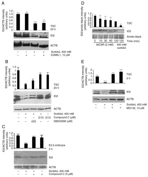
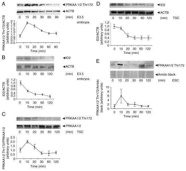
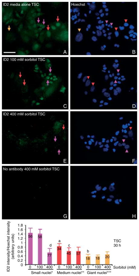
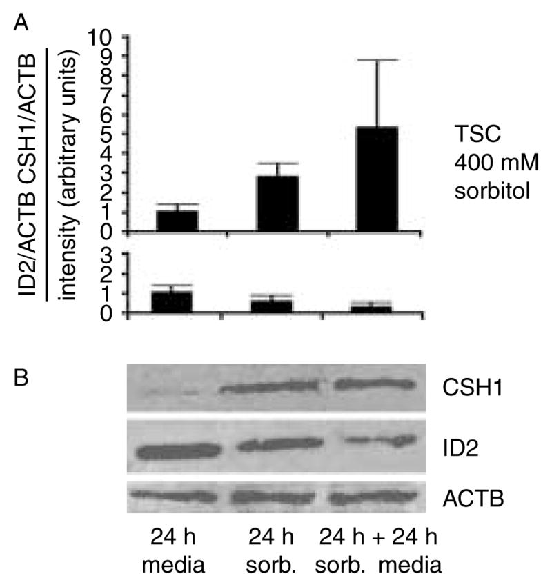
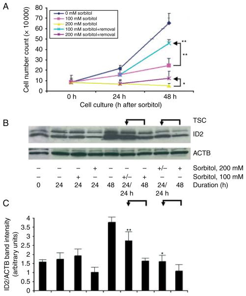
Stress reduces fertility, but the mechanisms mediating this are not understood. For a successful pregnancy, placental trophoblast stem cells (TSCs) in the implanting embryo proliferate and then a subpopulation differentiates to produce hormones. Normally, differentiation occurs when inhibitor of differentiation 2 (ID2) protein is lost in human and mouse placental stem cells. We hypothesize that stress enzyme-dependent differentiation occurs in association with insufficient TSC accumulation. We studied a well-defined model where TSC differentiation requires ID2 loss. The loss of ID2 derepresses the promoter of chorionic somatomammotropin hormone 1 (CSH1), the first hormone after implantation. Csh1 mRNA is known to be induced in stressed TSCs. In this study, we demonstrate that AMP-activated protein kinase (PRKAA1/2, aka AMPK) mediates the stress-induced proteasome-dependent loss of ID2 at high stress levels. At very low stress levels, PRKAA1/2 mediates metabolic adaptation exemplified by the inactivation of acetyl coA carboxylase by phosphorylation without ID2 loss. At the highest stress levels, irreversible TSC differentiation as defined by ID2 loss and slower cell accumulation occurs. However, lower stress levels lead to reversible differentiation accompanied by metabolic adaptation. These data support the hypothesis that PRKAA1/2 mediates preparation for differentiation that is induced by stress at levels where a significant decrease in cell accumulation occurs. This supports the interpretation that enzyme-mediated increases in differentiation may compensate when insufficient numbers of stem cells accumulate.


Conflict of interest statement

Declaration of interest

The authors declare that there is no conflict of interest that could be perceived as prejudicing the impartiality of the research reported.

Figures

Figure 1
Figure 1
Effect of hyperosmolar stress on ID2 protein is independent of MAPK8/9 in TSC, but dependent on PRKAA1/2 in TSC and E3.5 mouse blastocysts. (A) TSCs were cultured in 0 or 400 mM sorbitol with or without the MAPK8/9 inhibitor DJNKI1 (1 μM) for 20 h. Asterisks indicate that the MAPK8/9 inhibitor LJNKL1 has no significant effect on ID2 levels. (B) TSCs were cultured in 0 or 400 mM sorbitol without or with compound C at 1 μM (inhibitor of PRKAA1/2) or SB203580 at 5 μM (inhibitor of MAPK11/14) for 20 h. (C) Embryos were cultured in 0 or 400 mM sorbitol without or with compound C for 2 h. (D) TSCs were incubated with 2 mM AICAR for 0–2 h or with 400 mM for 2 h. (E) TSCs were incubated for 2 h with or without 200 mM sorbitol, with or without 10 μM of the proteasome inhibitor MG132. After treatments, equal amounts of protein were examined with western blots using ID2 and ACTB antibody or amido black. Histograms show the ratio of ID2/ACTB or ID2/amido black band intensity. Error flags are S.D.s for triplicate experiments for TSC or duplicate experiments for embryos.
Figure 2
Figure 2
Rapid transient activation of phosphorylated PRKAA1/2 Thr172 in TSC, ESC, and embryos occurs simultaneously with rapid loss of ID2 protein in TSCs and embryos. Embryos (A and B), TSCs (C and D), and ESC (E) were cultured under optimal conditions, sorbitol was added for 0–90/120 min, and cells and embryos were lysed, proteins fractionated by SDS–PAGE and immunoblotted for PRKAA1/2 Thr172 or ID2 and reprobed for actin (ACTB) or amido black as a loading control. Histograms below each immunoblot show the PRKAA1/2 Thr172 normalized to the loading control and this ratio is set to 1 for unstressed cells/embryos. Y error bars show S.D. of triplicate experiments in TSC and ESC or duplicate experiments in embryos.
Figure 3
Figure 3
Hyperosmolar stress activates PRKAA1/2 over a wide dose range, but ACACA Ser79 is phosphorylated at low doses and ID2 loss and decrease in TSC accumulation occur at high doses. (A) TSCs were plated and cultured for 24 h in media without sorbitol and with 12.5, 25, 50, and 100 mM sorbitol and then counted. (B) TSCs were cultured with the indicated doses of sorbitol for 2 h, fractionated by SDS–PAGE, and immunoblotted and stained for antigens using polyclonal antibodies. All experiments were repeated three times and histograms show X±S.E.M. Superscript ‘a’ indicates significant changes for 0 mM sorbitol stress for cell counts and proteins, and superscript ‘b’ indicates no significant difference for the same unstressed TSCs. Significance was determined by ANOVA followed by Duncan’s post hoc tests. For proteins, the earliest unstressed time point shown was set to 1 and served as a denominator to normalize the experimental values from stressed TSCs.
Figure 4
Figure 4
Hyperosmolar stress leads to destruction of ID2 protein in nearly all cells at high doses (400 mM), but lower doses (100 mM) spared ID2 in TSCs with small nuclei. TSCs were cultured in 0 mM (A and B), 100 mM (C and D), or 400 mM (E and F) sorbitol for 30 h and then fixed and stained for ID2 using indirect immunocytochemistry. In (G and H), 400 mM sorbitol-treated TSCs were probed without primary antibody; (B, D, F, and H) are the Hoechst-stained nuclei corresponding to cells in (A, C, E, and G) respectively. The orange arrow and arrowhead show the position of the protein corresponding to its nucleus of a giant cell (>2000 arbitrary units), red arrows and arrowheads show the protein and medium-sized nuclei (1000–2000 arbitrary units), and purple arrows and arrowheads show the protein and small TSC nuclei (100–999 arbitrary units). Histogram shows the mean intensity of ID2/intensity of Hoechst to normalize depth of nuclei, Y error flags are S.D., and numbers in bars show number of nuclei counted. Statistical comparisons show, in unstressed TSCs, the decrease in ID2 intensity from small to medium (a) and medium to giant nuclei (b) are each significant (P<0.005). The decreases in ID2 intensity in medium-sized nuclei from unstressed to 100 mM (c) and in small nuclei from unstressed or 100–400 mM (d) are significant (P<0.001). This is a representative experiment from three similar experiments.
Figure 5
Figure 5
CSH1 increases and ID2 decreases in 24 h after 400 mM sorbitol is removed from cultured TSCs. (A) TSCs were cultured for 24 h without or with 400 mM sorbitol, or cultured with sorbitol for 24 h followed by 24 h without sorbitol, cells were lysed, proteins were fractionated by SDS–PAGE and immunoblotted for CSH1, ID2, and reprobed for actin (ACTB) as a loading control. (B) Histograms below each immunoblot show either CSH1 or ID2 normalized to the loading control and this ratio is set to 1 for unstressed cells/embryos. Y error bars show S.D. of triplicate experiments.
Figure 6
Figure 6
Cell accumulation rates and ID2 increase 24 h after 100 or 200 mM sorbitol is removed from cultured TSCs. (A) TSCs were cultured without sorbitol or with 100 or 200 mM sorbitol for 24 h, and then sorbitol was removed or continued. TSCs were counted using Trypan blue at 0, 24, and 48 h. The constant stress groups were designated by unchanged color for 0–48 h at 100 mM (yellow) and 200 mM (light purple). The blue line represents a removal of 200 mM sorbitol in 24–48 h and the dark purple line represents a removal of 100 mM sorbitol in 24–48 h. (B) TSCs were cultured with or without 100 or 200 mM sorbitol for 24 h, then sorbitol was removed or continued, and cell lysates were fractionated by SDS–PAGE and probed for ID2 and reprobed for ACTB. (C) Histogram shows the ratio of ID2/ACTB band intensity. Error flags are S.D.s for triplicate experiments. In (A) and (C), arrows show the comparison of 48 h with sorbitol and 24 h of sorbitol followed by 24 h without it. In (A), * and ** show the significant (P<0.01 and P<0.001 respectively) increase in cell number when 200 and 100 mM sorbitol were removed for the second 24 h. The top ** indicate a significant decrease in cell number from 0 to 100 mM sorbitol. In (C), * and ** show the significance (P<0.01 and P<0.001 respectively) when 200 or 100 mM sorbitol was removed for the second 24 h.

References

    1. Abell AN, Granger DA, Johnson NL, Vincent-Jordan N, Dibble CF, Johnson GL. Trophoblast stem cell maintenance by fibroblast growth factor 4 requires MEKK4 activation of Jun N-terminal kinase. Molecular and Cellular Biology. 2009;29:2748–2761. 10.1128/MCB.01391-08. - PMC - PubMed
    1. Bell CE, Lariviere NM, Watson PH, Watson AJ. Mitogen-activated protein kinase (MAPK) pathways mediate embryonic responses to culture medium osmolarity by regulating Aquaporin 3 and 9 expression and localization, as well as embryonic apoptosis. Human Reproduction. 2009;24:1373–1386. 10.1093/humrep/dep010. - PubMed
    1. Caenepeel S, Charydczak G, Sudarsanam S, Hunter T, Manning G. The mouse kinome: discovery and comparative genomics of all mouse protein kinases. PNAS. 2004;101:11707–11712. 10.1073/pnas.0306880101. - PMC - PubMed
    1. Chai N, Patel Y, Jacobson K, McMahon J, McMahon A, Rappolee DA. FGF is an essential regulator of the fifth cell division in preimplantation mouse embryos. Developmental Biology. 1998;198:105–115. 10.1016/S0012-1606(98)80031-6. - PubMed
    1. Chen J, Downs SM. AMP-activated protein kinase is involved in hormone-induced mouse oocyte meiotic maturation in vitro. Developmental Biology. 2008;313:47–57. 10.1016/j.ydbio.2007.09.043. - PMC - PubMed

Publication types

Substances