. 2010 Sep;22(9):3020-33.
doi: 10.1105/tpc.110.078527. Epub 2010 Sep 28.

Distinct roles of the ATR kinase and the Mre11-Rad50-Nbs1 complex in the maintenance of chromosomal stability in Arabidopsis

Affiliations

Distinct roles of the ATR kinase and the Mre11-Rad50-Nbs1 complex in the maintenance of chromosomal stability in Arabidopsis

Simon Amiard et al. Plant Cell. 2010 Sep.

Abstract

Signaling of chromosomal DNA breaks is of primary importance for initiation of repair and, thus, for global genomic stability. Although the Mre11-Rad50-Nbs1 (MRN) complex is the first sensor of double-strand breaks, its role in double-strand break (DSB) signaling is not fully understood. We report the absence of γ-ray-induced, ATM/ATR-dependent histone H2AX phosphorylation in Arabidopsis thaliana rad50 and mre11 mutants, confirming that the MRN complex is required for H2AX phosphorylation by the ATM and ATR kinases in response to irradiation-induced DSB in Arabidopsis. rad50 and mre11 mutants spontaneously activate a DNA damage response, as shown by the presence of γ-H2AX foci and activation of cell cycle arrest in nonirradiated plants. This response is ATR dependent as shown both by the absence of these spontaneous foci and by the wild-type mitotic indices of double rad50 atr and mre11 atr plants. EdU S-phase labeling and fluorescence in situ hybridization analysis using specific subtelomeric probes point to a replicative S-phase origin of this chromosome damage in the double mutants and not to telomere destabilization. Thus, the data presented here show the exclusive involvement of ATR in DNA damage signaling in MRN mutants and provide evidence for a role for ATR in the avoidance of S-phase DNA damage.


Figures

Figure 1.
Figure 1.
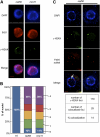
The MRN Complex Is Essential for Activation of the ATM and ATR Kinases in Response to Irradiation. (A) Detection of γ-H2AX immunofluorescence in mitotic root tip nuclei. In nonirradiated plants, no foci were detected in wild-type (left panel) plants, and a small number are seen in rad50 mutants (right panel). 25 Gy γ-irradiation induces γ-H2AX foci in wild-type plants, but there is no change in the number of foci in rad50 mutants. DNA is stained with DAPI (blue), γ-H2AX foci are colored in green, and merged images overlay γ-H2AX foci onto chromosomes. A 2-μm scale bar is shown at the bottom left. (B) Graphic representation of the number of foci detected in nonirradiated and irradiated wild-type, rad50, and mre11 mutant plants. The numbers of foci per nucleus corresponding to the differently colored blocks are given to the right, except for the irradiated wild-type bar, for which these are given in the bar. The table underneath gives the percentage of nuclei containing at least one focus (% of nuclei +) and the mean number of foci per mitotic nucleus (n = 25; numbers in brackets are standard deviations).
Figure 2.
Figure 2.
H2AX Phosphorylation in rad50 and mre11 Mutant Plants Is Increased in S- and Early G2-Phase Nuclei. (A) EdU labeling shows enrichment of γ-H2AX foci in S- and early G2-phase nuclei of rad50 and mre11 mutants. DNA is stained with DAPI (blue), EdU incorporation is shown in red, and γ-H2AX foci are colored in green. Bar = 2 μm. (B) Graphic representation of numbers of γ-H2AX foci in EdU+ nuclei. The numbers of foci per nucleus corresponding to the differently colored blocks are given to the right. The percentages of nuclei without foci (blue) or with more than four foci (purple) are indicated. In each case, 120 EdU-positive nuclei were counted. (C) Immunostaining and subtelomeric FISH labeling of root tip nuclei of rad50 mutants reveal that γ-H2AX signal is partially located at telomeres. Nuclei were stained with DAPI (blue), γ-H2AX foci are colored in green, and FISH signals are red. Images are a single focal plane from a deconvolved three-dimensional image. Colocalized foci are indicated with white arrows. A 2-μm scale bar is shown at the bottom left.
Figure 3.
Figure 3.
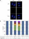
Plant Cells Mutated in the MRN Complex Activate the ATR Kinase. (A) Immunofluorescence of root tip mitotic nuclei of rad50 single (left panel) and rad50 atr double (right panel) mutants shows the absence of γ-H2AX foci in the rad50 atr mutant. DNA is stained with DAPI (blue), γ-H2AX foci are colored in green, and merged images overlay γ-H2AX foci onto chromosomes. Bar = 2 μm. (B) Graphic representation of the number of foci detected in wild-type, atr, rad50, mre11, rad50 atr, and mre11 atr mutant plants. The numbers of foci per nucleus corresponding to the differently colored blocks are given in the bars. The table underneath gives the percentage of nuclei containing at least one focus (% of nuclei +) and the mean number of foci per mitotic nucleus (n = 25; numbers in brackets are standard deviations).
Figure 4.
Figure 4.
ATM Independence of Spontaneous γ-H2AX Foci in rad50. Immunofluorescence of mitotic root tip nuclei indicates that γ-H2AX foci formation 20 min after irradiation is almost abolished in the presence of IATM (ku55933) (A) and that foci detected in rad50 mutants are not ATM dependent (B). DNA was stained with DAPI (blue), γ-H2AX foci are colored in green, and merged images overlay γ-H2AX foci onto chromosomes. The percentage of nuclei containing at least one focus (% of nuclei +) and the mean number of foci per nucleus from counting 25 nuclei are indicated. Bars = 2 μm.
Figure 5.
Figure 5.
rad50 atr or mre11 atr Mutants Present Severe Morphological Defects. Phenotypic appearance of the wild type, rad50, mre11, atr, rad50 atr, and mre11 atr mutants 5 d (top photograph; bar = 5 mm) or 6 weeks after germination. Phenotypes of double rad50 atr and mre11 atr mutants are also presented 10 weeks after germination.
Figure 6.
Figure 6.
Cell Cycle Regulation and Cell Death Profile in Root Tips of rad50 atr and mre11 atr Mutants. (A) The reduction in the number of mitotic nuclei per root tip observed in rad50 and mre11 mutants depends on ATR. Numbers of mitotic nuclei per root tip are shown for 5-d-old wild-type and rad50, mre11, atr, atr rad50, and atr mre11 seedlings. Error bars indicate standard errors (n = 5) and an asterisk significant differences (t test; P < 0.05) between the wild type and rad50 or mre11 mutants. (B) Abundant cell death in double rad50 atr or mre11 atr mutants. Representative images of root tips stained with propidium iodide, which stains dead cells (images are representative of 10 root tips). No cell death is observed in wild-type plants, limited cell death is observed in the region around the quiescent center in rad50 or mre11 mutants, and abundant cell death is observed all along the root tip in rad50 atr and mre11 atr. Cell death in the atr rad50 and atr mre11 mutants is not affected by inhibition of ATM kinase with IATM. Bar = 50 μm.
Figure 7.
Figure 7.
Increased Cytological Abnormalities and Chromosome Fragmentation in the rad50 atr Mutant. (A) Cytogenetic analysis of flower bud nuclei of rad50 atr mutant plants. Mitotic anaphase bridges are classified in two distinct categories: classical (thin and complete bridges) or fragmented (thick and fragmented bridges). DNA stained with DAPI is white. (B) FISH analysis using a pool of nine subtelomeric (subtel) BAC fluorescent probes (red). DAPI (blue), FISH signal (red), and merged images are shown for two anaphase nuclei: one with subtelomeric signals in the bridge (right) and a second without subtelomeric signals (left). Bar = 2 μm. (C) Numeric results recapitulating the percentage of anaphases with chromosomal bridges and the percentage of subtelomeric signal in bridges in flower buds of rad50 and rad50 atr mutants (nb, number of nuclei).
Figure 8.
Figure 8.
Model Explaining the Role for MRN and ATR in Maintaining Proper Replication. Absence of functional MRN complex induces stalled replication forks (represented by a red cross). In presence of ATR (1), the kinase is activated through the presence of ssDNA generated at the stalled fork. ATR kinase will then phosphorylate H2AX (visible in S-phase), stabilize blocked replication forks, and facilitate completion of S-phase synthesis (major pathway symbolized by a thick arrow). Failure to rescue replication forks results in fork collapse and chromatid breakage, giving rise to activation of ATR and appearance of γ-H2AX foci either in S-phase or in mitosis (minor pathway symbolized by a thin arrow). In absence of ATR in rad50 atr or mre11 atr double mutants (2), fork stabilization is less efficient, leading to increased DSB and genomic instability.

Similar articles

Cited by

References

    1. Adams K.E., Medhurst A.L., Dart D.A., Lakin N.D. (2006). Recruitment of ATR to sites of ionising radiation-induced DNA damage requires ATM and components of the MRN protein complex. Oncogene 25: 3894–3904 - PMC - PubMed
    1. Bi X., Srikanta D., Fanti L., Pimpinelli S., Badugu R., Kellum R., Rong Y.S. (2005). Drosophila ATM and ATR checkpoint kinases control partially redundant pathways for telomere maintenance. Proc. Natl. Acad. Sci. USA 102: 15167–15172 - PMC - PubMed
    1. Bleuyard J.-Y., Gallego M.E., White C.I. (2004). Meiotic defects in the Arabidopsis rad50 mutant point to conservation of the MRX complex function in early stages of meiotic recombination. Chromosoma 113: 197–203 - PubMed
    1. Brown E.J., Baltimore D. (2000). ATR disruption leads to chromosomal fragmentation and early embryonic lethality. Genes Dev. 14: 397–402 - PMC - PubMed
    1. Bundock P., Hooykaas P. (2002). Severe developmental defects, hypersensitivity to DNA-damaging agents, and lengthened telomeres in Arabidopsis MRE11 mutants. Plant Cell 14: 2451–2462 - PMC - PubMed

Publication types