. 2010 Nov;16(11):2277-90.
doi: 10.1261/rna.2091710. Epub 2010 Sep 28.

A novel mRNA affinity purification technique for the identification of interacting proteins and transcripts in ribonucleoprotein complexes

Affiliations

A novel mRNA affinity purification technique for the identification of interacting proteins and transcripts in ribonucleoprotein complexes

Boris Slobodin et al. RNA. 2010 Nov.

Abstract

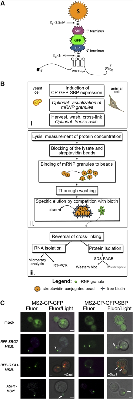
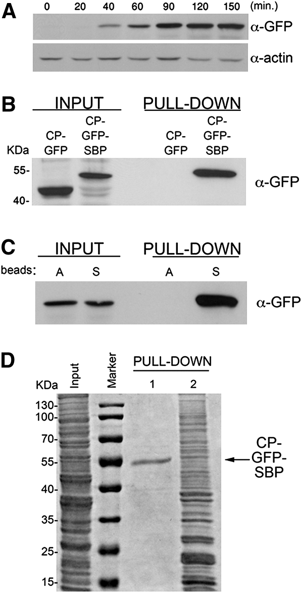
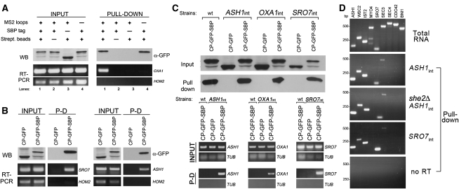
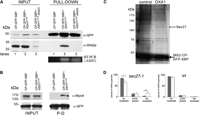
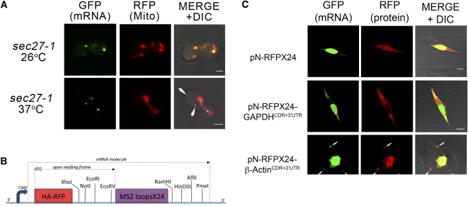
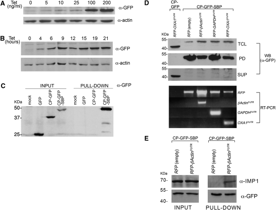
Intracellular mRNA targeting and localized translation are potential determinants for protein localization. To facilitate targeting, mRNAs possess specific cis-acting sequence motifs that are recognized by trans-acting RNA-binding proteins (RBPs). While many mRNAs are trafficked, our knowledge of the RBPs involved and presence of additional transcripts within these ribonucleoprotein (RNP) complexes is limited. To facilitate the identification of RBPs and transcripts that bind to specific mRNAs, we developed RNA-binding protein purification and identification (RaPID), a novel technique that allows for the affinity purification of MS2 aptamer-tagged mRNAs and subsequent detection of bound RBPs and transcripts using mass-spectometry and RT-PCR, respectively. RaPID effectively isolated specific mRNAs from both yeast and mammalian cells, and identified known mRNA-RBP interactions (e.g., ASH1-She2; β-Actin-IMP1). By isolating tagged OXA1 mRNA using RaPID, we could identify a yeast COPI subunit (i.e., Sec27) as a candidate interacting protein. This finding was strengthened by the observation that a portion of OXA1 mRNA was delocalized in a sec27-1 temperature-sensitive mutant at restrictive temperatures. Finally, RaPID could also be used to show biochemically the coexistence of different RNA species within the same RNP complex (e.g., coprecipitation of the yeast SRO7, WSC2, SEC3, and IST2 mRNAs with ASH1 mRNA) for the first time.


Figures

FIGURE 1.
FIGURE 1.
RaPID and its components. (A) Functional moieties of MS2-CP-GFP-SBP. MS2-CP-GFP-SBP binds to MS2 aptamer-tagged mRNAs via the MS2-CP moiety located at the N-terminal and interacts with immobilized streptavidin via the SBP moiety at the C-terminal. The affinities of both interactions are indicated. The GFP moiety located in the central region allows for mRNA visualization via fluorescence microscopy and is recognized by anti-GFP antibodies in Westerns. (B) A flowchart of the RaPID procedure. The schematic is divided into three main steps; the first, (i) involves cell growth, induction of the MS2-CP-GFP-SBP expression, and cross-linking after harvesting. The second step (ii) includes cell lysis, the pull-down reaction, and gentle elution of the bound material with biotin. In the last step (iii), the cross-links are reversed and the RNA and protein fractions are isolated and subjected to further analysis. (C) Addition of the SBP tag does not alter mRNA visualization using the MS2 system. Yeast expressing either MS2-CP-GFP (CP-GFP) or MS2-CP-GFP-SBP (CP-GFP-SBP) were transformed with plasmids expressing MS2L-tagged RFP-SRO7 and RFP-OXA1, or ASH13′UTR. Yeast were grown to mid-log phase, transferred to medium lacking methionine for 1 h, fixed, and visualized by confocal microscopy. In cells expressing RFP-OXA1-MS2L, the RFP-tagged Oxa1 protein acts as a mitochondrial marker. White arrows indicate localization of the mRNA granules, i.e., bud-tip in the case of ASH1 and SRO7 mRNAs, and mitochondria in the case of OXA1 mRNA. (Fluor) The localization of GFP-labeled mRNA or GFP-labeled mRNA and RFP-labeled mitochondria; (Fluor/Light) the merge between the fluorescence and light microscopy (differential interference contrast) windows. Size bars, 1 μm.
FIGURE 2.
FIGURE 2.
Characterization of the MS2-CP-GFP-SBP fusion protein. (A) Starvation-induced expression of MS2-CP-GFP-SBP. Yeast transformed with pMS2-CP-GFP-SBP plasmid were grown to mid-log phase (O.D.600 ≅ 1) in synthetic medium, shifted to medium lacking methionine, grown for the indicated times (minutes), and then collected. Cells were lysed and 50 μg of protein samples were analyzed by Western blotting using anti-GFP antibodies to detect MS2-CP-GFP-SBP or anti-actin antibodies to detect actin, as a loading control. (B) MS2-CP-GFP-SBP, but not MS2-CP-GFP, is efficiently pulled down with streptavidin-conjugated beads. Yeast transformed with plasmids expressing MS2-CP-GFP or MS2-CP-GFP-SBP were grown in 200-mL cultures to mid-log phase, starved for methionine for 75 min, and harvested. Following lysis, 5 mg of total protein extract derived from each transformant was incubated with streptavidin beads, washed with lysis buffer, and eluted using free biotin. Eluates (Pull-down) were resolved by SDS-PAGE along with 50-μg samples of total protein (Input) and probed with anti-GFP antibodies. Molecular mass is given in kilodaltons (kDa). (C) MS2-CP-GFP-SBP does not bind to immobilized avidin. Yeast transformed with pMS2-CP-GFP-SBP were grown to mid-log phase in 400-mL cultures, incubated in medium lacking methionine for 75 min, and harvested. After lysis, separate aliquots of 8 mg of total protein were incubated overnight with beads conjugated to either avidin (A beads) or streptavidin (S beads). Following washing, proteins were eluted with biotin and both the eluates (Pull-down) and 50 μg of samples of total protein (Input) were analyzed by Western blotting using anti-GFP antibodies. (D) Avidin blocking and biotin-mediated elution greatly improve the signal-to-noise ratio. Yeast transformed with pMS2-CP-GFP-SBP were grown in 400-mL cultures, starved for methionine for 75 min, lysed, and two aliquots each of 2 mg of total protein were incubated overnight with streptavidin-conjugated beads. One aliquot was blocked for 1 h with free avidin prior to the pull-down, and the bound material was eluted by competition with free biotin (PULL-DOWN 1), while the second aliquot (PULL-DOWN 2) was not avidin blocked and was eluted by boiling in sample buffer containing 0.1 M dithiothreitol for 5 min. Both eluates were resolved by SDS-PAGE along with a 50-μg aliquot of total protein (Input) and were stained with a general protein stain (Imperial; Sigma).
FIGURE 3.
FIGURE 3.
RaPID allows for the specific isolation of MS2L-tagged mRNAs. (A) MS2 aptamer-tagged OXA1 mRNA is specifically precipitated using RaPID. Wild-type yeast cultures (300 mL each) expressing RFP-OXA1-MS2L mRNA and either pMS2-CP-GFP or pMS2-CP-GFP-SBP, and yeast expressing RFP-OXA1 mRNA and pMS2-CP-GFP-SBP were grown to mid-log phase and processed as described in the Materials and Methods. Following lysis, 10 mg of total protein from each cell type was subjected to RaPID to yield the respective eluates. For Western analysis (WB), 30% of each eluate (PULL-DOWN) and 40 μg from samples of total protein (INPUT) were resolved by SDS-PAGE and detected using anti-GFP antibodies. The remaining 70% of each eluate and 40-μg samples of total extract from each cell type were taken for RNA isolation and subsequent RT–PCR analysis (RT–PCR) with OXA1 or HOM2 primers. (Lane 1) Input from cells expressing RFP-OXA1-MS2L mRNA (+ MS2 loops) and MS2-CP-GFP-SBP (+ SBP tag) pulled down with immobilized streptavidin (+ Strept. beads). For controls, we performed the same experiment using avidin beads (lane 2), MS2-CP-GFP protein lacking the SBP tag (lane 3), or the RFP-OXA1 message lacking the MS2 loops (lane 4). HOM2 primers were used to detect HOM2 as a control mRNA. (B) MS2 aptamer-tagged SRO7 and ASH1 mRNAs can be specifically precipitated using RaPID. Wild-type yeast expressing MS2L-tagged RFP-SRO7 mRNA (left) or MS2L-tagged ASH13′UTR (right), along with either MS2-CP-GFP-SBP or MS2-CP-GFP were grown, lysed, subjected to RaPID, and analyzed as in A. (C) Endogenous MS2L-tagged ASH1, OXA1, and SRO7 transcripts can be specifically isolated using RaPID. Wild-type (wt) cells and yeast strains bearing MS2L-tagged ASH1, OXA1, and SRO7 loci (i.e., ASH1int, OXA1int, and SRO7int) were transformed with pMS2-CP-GFP-SBP or pMS2-CP-GFP plasmids and were grown in 400-mL cultures, treated, and collected. After lysis and RaPID, 25% of the eluate (Pull-down) and 40 μg of the total protein (INPUT) were resolved by SDS-PAGE and detected using anti-GFP antibodies (top). For RNA analysis (bottom), RNA was isolated from the remaining 75% of the eluate (P-D) and from 25 μL of the total extract (INPUT), and analyzed by RT–PCR using the indicated primer pairs (bottom). TUB1 primers (TUB) were used to detect TUB1 mRNA as control. (D) Additional mRNAs coprecipitate with endogenous MS2L-tagged ASH1 mRNA. Yeast (i.e., ASH1int, she2Δ ASH1int, and SRO7int) expressing the indicated endogenous MS2L-tagged mRNAs and pMS2-CP-GFP-SBP were grown in 400-mL cultures and 20 mg of total extract from each lysate was subjected to RaPID. RNAs derived from the total extract (Total RNA) or the eluates (Pull-down) were analyzed by RT–PCR using primer pairs corresponding to known or suspected bud-localized messages (e.g., ASH1 [control], WSC2, IST2, MYO4, SRO7, SEC3, SEC4, CDC42, and BNI1). mRNA coprecipitation was examined in precipitates derived from ASH1int cells (ASH1int), she2Δ ASH1int cells (she2Δ ASH1int), and SRO7int cells (SRO7int). RNA samples derived from the eluate of ASH1int cells were also subjected to PCR without reverse transcription, as controls (no RT). PCR products were resolved on 1.5% agarose gels.
FIGURE 4.
FIGURE 4.
Identification of RNA-binding proteins using RaPID. (A) Endogenous She2 interacts with the 3′UTR of ASH1 mRNA. Wild-type and she2Δ yeast expressing pMS2-CP-GFP-SBP and MS2L-tagged ASH13UTR mRNA, or wild-type yeast expressing pMS2-CP-GFP-SBP alone were grown in 400-mL cultures, treated, lysed, and 25 mg of the total protein extract from each sample was subjected to RaPID. For Western analysis, 85% of the eluates (PULL-DOWN) and 40-μg samples of the total protein (INPUT) were resolved by SDS-PAGE and analyzed using anti-She2 and anti-GFP antibodies. Note that the faint band detected with anti-She2 antibodies in input lane 3 is probably due to recognition of a nonspecific protein. For RT–PCR analysis, RNA was isolated from the remaining 15% of the eluate and analyzed by RT–PCR with a primer pair that recognizes the ASH13UTR. (B) Endogenous Myo4 is identified in the precipitated ASH1 mRNP complex. Yeast expressing pMS2-CP-GFP-SBP and MS2L-tagged ASH13UTR mRNA or cells expressing pMS2-CP-GFP-SBP alone were grown in 400-mL cultures, treated, lysed, and 30 mg of the total protein extract from each sample was subjected to the RaPID procedure. The eluates (PULL-DOWN) and 40-μg samples of the total protein (INPUT) were resolved by SDS-PAGE and analyzed with anti-Myo4 and anti-GFP antibodies. (C) Identification of Sec27 as a candidate OXA1 mRNA interacting protein. Wild-type yeast expressing RFP-OXA1-MS2L (OXA1) or RFP-MS2L (control) were grown in cultures of 800 mL, treated, lysed and aliquots of 50 mg of total protein extract were processed using RaPID. Following the reversal of cross-linking, the eluate was resolved by SDS-PAGE using a 20 x15 cm 9% polyacrylamide gel, silver stained, and select bands were analyzed by mass spectrometry. The thin arrow marks Sec27, while the thick arrow marks the precipitated MS2-CP-GFP-SBP protein. (D) Inactivation of Sec27 increases the proportion of delocalized OXA1 mRNA granules. Wild-type or sec27-1 yeast strains expressing RFP-OXA1-MS2L mRNA and MS2-CP-GFP(x3) were grown to mid-log phase (O.D.600 ≅ 1) at 26°C. Cells were shifted for 1 h to medium lacking methionine at the permissive temperature (26°C) and then either shifted to the restrictive temperature (37°C) for 1 h or maintained at 26°C. The cells were fixed and the localization of OXA1 mRNA granules relative to mitochondrial structures (as labeled with Oxa1-RFP) was analyzed using confocal microscopy. The gray-filled and white (unfilled) columns of the histogram indicate the distribution of granules in cells incubated at 26oC and 37oC, respectively. Statistics show the average percentage (±SE) of colocalized, juxtaposed (within ≤0.2 μm), or delocalized granules relative to the closest mitochondrial structure. (*) P = 0.035; (**) P = 0.006.
FIGURE 5.
FIGURE 5.
Effect of SEC27 inactivation upon the localization of OXA1 mRNA in yeast and establishment of a dual mRNA and protein detection system in animal cells. (A) SEC27 inactivation enhances the delocalization of OXA1 mRNA. sec27-1 cells expressing RFP-OXA1-MS2L mRNA and MS2-CP-GFP(x3) were shifted to either restrictive temperatures for 1 h or maintained at permissive temperatures (as detailed in the legend to Fig. 4D) and visualized. White arrows indicate delocalized OXA1 mRNA granules. Size bars, 2 μm. (B) A schematic representation of the pN-RFPX24 expression vector, which is based upon pcDNA3.1(-) (Invitrogen). Downstream of the constitutive CMV promoter is an open reading frame that begins with an encoded HA epitope-tagged RFP gene that lacks a stop codon (HA-RFP), followed by a multiple cloning site (MCS; see indicated sites) for insertion of a gene of interest. Downstream of the MCS are 24 repeats of the MS2 aptamer (MS2 loops x24) and a second MCS (see indicated sites) that serves for the optional insertion of a 3′UTR sequence. (C) Visualization of GAPDH and β-Actin mRNAs and their respective translation products in animal cells. NIH3T3 fibroblasts were seeded onto round glass coverslips (13-mm diameter; in 24-well plates), grown for 24 h, and transfected with a plasmid expressing MS2-CP-GFP bearing nuclear localization sequence (100 ng DNA/well), as well as either the pN-RFPX24 plasmid alone or pN-RFPX24 plasmids that express the GADPH or β-Actin coding regions (CDRs) and corresponding 3′UTRs (500 ng DNA/well), as indicated. Fresh medium was added after 6 h and the cells were grown for an additional 12 h prior to fixation in a 4% formaldehyde solution, mounting on slides, and visualization by confocal microscopy. No asymmetric distribution of either RFP (top) or GAPDH (middle) mRNA and protein was observed. RFP protein was found in the nucleus and cytoplasm, while GAPDH (a cytosolic protein) was restricted to the cytoplasm, as expected. White arrows indicate the colocalization of β-Actin mRNA and protein at polarized extensions of the cell, as seen by others. Size bars, 10 μm.
FIGURE 6.
FIGURE 6.
Use of RAPID in animal cells. (A) Dose-dependent induction of MS2-CP-GFP-SBP expression. 293TRex cells were transiently transfected with pcDNA4-MS2-CP-GFP-SBP (6 μg of DNA/10-cm dish), harvested after 8 h, and reseeded into 6-well plates. After an additional 12 h, tetracycline was added at the indicated concentration and the cells were grown for an additional 20 h. After harvesting, the cells were lysed and 10% of the total protein extract was resolved by SDS-PAGE and analyzed in Western blots using anti-GFP to detect MS2-CP-GFP-SBP and anti-actin antibodies to detect actin, as a loading control. (B) Time-dependent induction of MS2-CP-GFP-SBP expression. Stable 293TRex-MS2-CP-GFP-SBP cells were grown to ∼50% confluency, and then tetracycline was added to the medium (100 ng/mL) for the indicated times. After harvesting, the cells were lysed and 30 μg of the total protein extract from each time point was resolved by SDS-PAGE and analyzed in blots, as described in A. (C) Mammalian expressed MS2-CP-GFP-SBP precipitates with immobilized streptavidin. HEK293 cells were transiently transfected with pcDNA4/TO plasmids coding for GFP, MS2-CP-GFP, or MS2-CP-GFP-SBP (8 μg of DNA/100-mm dish). Nontransfected HEK293 cells were also included (mock). After 24 h, the cells were harvested, total protein was extracted, and 3.5 mg from each sample was taken for pull-down with streptavidin-conjugated beads. Both the eluates (PULL-DOWN) and 40 μL of the total protein extract (INPUT) samples were resolved by SDS-PAGE and analyzed by Western blotting using anti-GFP antibodies. (D) Isolation of mammalian MS2L-tagged mRNAs using RaPID. 293TRex cells stably expressing MS2-CP-GFP-SBP were transfected (6 μg of DNA/100-mm dish) with pN-RFPX24 plasmids expressing HA-RFP alone or as a fusion with the 3′UTRs of the β-Actin, GAPDH, or OXA1 mRNAs, as indicated. In addition, 293TRex cells stably expressing MS2-CP-GFP were transfected (6 μg of DNA/100-mm dish) with a pN-RFPX24 plasmid expressing HA-RFP fused to the 3′UTR of OXA1 mRNA. The cells were treated with tetracycline (100 ng/mL, for 12 h), collected, cross-linked with 0.1% formaldehyde, lysed, and 5 mg of total cellular extract from each sample was taken for RaPID. For Western analysis, 25% of the eluate (PD), 30 μg of total protein (TCL), and 30 μg of the supernatant remaining after incubation with the streptavidin beads (SUP) were resolved by SDS-PAGE and detected in blots using anti-GFP antibodies. For RT–PCR analysis (RT–PCR), RNA was isolated from the remaining 75% of the eluate, subjected to reverse transcription, and analyzed by PCR using the indicated primers. (E) Precipitation of endogenous IMP1 with human β-Actin mRNA. 293TRex cells stably expressing MS2-CP-GFP-SBP were transfected (10 μg of DNA/10-cm dish) with either pN-RFPX24 (empty) or pN-RFPX24 containing the 3′UTR of β-Actin. Cells were grown in the presence of tetracycline (100 ng/mL) for 12 h, collected, cross-linked using 0.01% formaldehyde, and 18 mg of each total extract was processed by RaPID. The eluates and 50 μg of the total input were analyzed by Western blots using the indicated antibodies.

References

    1. Aronov S, Gerst JE 2004. Involvement of the late secretory pathway in actin regulation and mRNA transport in yeast. J Biol Chem 279: 36962–36971 - PubMed
    1. Aronov S, Gelin-Licht R, Zipor G, Haim L, Safran E, Gerst JE 2007. mRNAs encoding polarity and exocytosis factors are cotransported with the cortical endoplasmic reticulum to the incipient bud in Saccharomyces cerevisiae. Mol Cell Biol 27: 3441–3455 - PMC - PubMed
    1. Bachler M, Schroeder R, von Ahsen U 1999. StreptoTag: A novel method for the isolation of RNA-binding proteins. RNA 5: 1509–1516 - PMC - PubMed
    1. Beach DL, Keene JD 2008. Ribotrap : Targeted purification of RNA-specific RNPs from cell lysates through immunoaffinity precipitation to identify regulatory proteins and RNAs. Methods Mol Biol 419: 69–91 - PubMed
    1. Beach DL, Salmon ED, Bloom K 1999. Localization and anchoring of mRNA in budding yeast. Curr Biol 9: 569–578 - PubMed

Publication types

MeSH terms