. 2010 Oct 21;55(20):6141-55.
doi: 10.1088/0031-9155/55/20/007. Epub 2010 Sep 29.

In vivo transcranial cavitation threshold detection during ultrasound-induced blood-brain barrier opening in mice

Affiliations

In vivo transcranial cavitation threshold detection during ultrasound-induced blood-brain barrier opening in mice

Yao-Sheng Tung et al. Phys Med Biol. .

Abstract

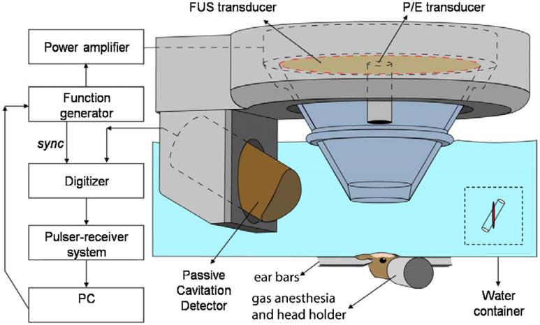
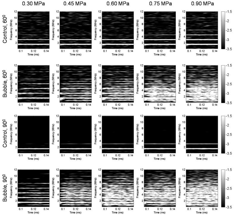
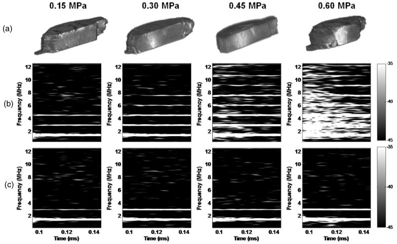
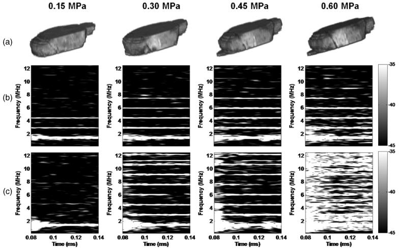
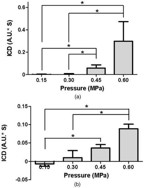
The in vivo cavitation response associated with blood-brain barrier (BBB) opening as induced by transcranial focused ultrasound (FUS) in conjunction with microbubbles was studied in order to better identify the underlying mechanism in its noninvasive application. A cylindrically focused hydrophone, confocal with the FUS transducer, was used as a passive cavitation detector (PCD) to identify the threshold of inertial cavitation (IC) in the presence of Definity® microbubbles (mean diameter range: 1.1-3.3 µm, Lantheus Medical Imaging, MA, USA). A vessel phantom was first used to determine the reliability of the PCD prior to in vivo use. A cerebral blood vessel was simulated by generating a cylindrical channel of 610 µm in diameter inside a polyacrylamide gel and by saturating its volume with microbubbles. The microbubbles were sonicated through an excised mouse skull. Second, the same PCD setup was employed for in vivo noninvasive (i.e. transdermal and transcranial) cavitation detection during BBB opening. After the intravenous administration of Definity® microbubbles, pulsed FUS was applied (frequency: 1.525 or 1.5 MHz, peak-rarefactional pressure: 0.15-0.60 MPa, duty cycle: 20%, PRF: 10 Hz, duration: 1 min with a 30 s interval) to the right hippocampus of twenty-six (n = 26) mice in vivo through intact scalp and skull. T1 and T2-weighted MR images were used to verify the BBB opening. A spectrogram was generated at each pressure in order to detect the IC onset and duration. The threshold of BBB opening was found to be at a 0.30 MPa peak-rarefactional pressure in vivo. Both the phantom and in vivo studies indicated that the IC pressure threshold had a peak-rarefactional amplitude of 0.45 MPa. This indicated that BBB opening may not require IC at or near the threshold. Histological analysis showed that BBB opening could be induced without any cellular damage at 0.30 and 0.45 MPa. In conclusion, the cavitation response could be detected without craniotomy in mice and IC may not be required for BBB opening at relatively low pressures.


Figures

Figure 1
Figure 1
Block diagram of the experimental setup. The PCD was positioned at 60° relative to the longitudinal axis of the FUS beam. The overlap between the focal regions of PCD (blue) and FUS (red) occurring inside the murine brain is illustrated in the inset.
Figure 2
Figure 2
Illustration of the ICD calculation of the in vivo experiments. (a) The spectrogram of the first pulse from 0.02 to 0.15 ms after a 4 MHz high-pass filter (Chebshev type 1) at 0.60 MPa. The harmonics and the broadband response could be observed in this figure. The corresponding VRMS is depicted in (b) and the ICD was obtained using the integral of the VRMS curve between two dashed lines.
Figure 3
Figure 3
Spectrogram of the first pulse in the phantom study without and with the microbubbles at five distinct acoustic pressures. Acoustic emissions were acquired with the hydrophone positioned at 60° and 90° from the longitudinal axis of the FUS beam. No broadband acoustic emissions were detected without the microbubbles while, with the microbubbles, the broadband acoustic emissions were detected at pressure equal to or higher than 0.45 MPa. At 0.30 MPa, the ultra-harmonics were detected at the 90° PCD but not at the 60° PCD.
Figure 4
Figure 4
BBB opening using transducer-A and microbubbles as confirmed by (a) 3D sagittal MRI and (b) the corresponding spectrogram (on the signal acquired by the hydrophone) of the first pulse from 0.095 to 0.145 ms showing that the broadband acoustic emissions were detected at 0.45 MPa and 0.60 MPa but not at 0.15 MPa and 0.30 MPa. 3D MR confirmed that BBB could open at 0.30 MPa, i.e. without inertial cavitation. (c) Spectrogram of the first pulse from 0.095 to 0.145 ms in the absence of microbubbles showing that only the first and second harmonics could be detected without microbubbles.
Figure 5
Figure 5
BBB opening using transducer-B and microbubbles as confirmed by (a) 3D-MR images, and (b) the corresponding spectrogram (on the signal acquired by the hydrophone) of the first pulse from 0.095 to 0.145 ms showing that broadband acoustic emissions were detected at 0.45 MPa and 0.6 MPa but not at 0.15 MPa and 0.3 MPa. The 3D-MRI images confirmed that the BBB could open at 0.3 MPa, i.e. without inertial cavitation. (c) Spectrograms (on the signal acquired by the 10 MHz P/E transducer) also showed that the threshold of inertial cavitation was 0.45 MPa (see quantitative results in figure 6).
Figure 6
Figure 6
ICD at four distinct acoustic pressures using (a) transducer-A or (b) transducer-B. ICD was quantified as the area under the VRMS curve from 0.095 ms to 0.145 ms, at each pressure. The signal used for quantification was from a focused hydrophone. For transducer-A, the ICD at 0.45 MPa and 0.60 MPa was significantly higher than at 0.15 MPa and 0.30 MPa (*P < 0.05). For transducer-B, the ICD at 0.60 MPa was significantly higher than at 0.30 MPa and 0.15 MPa. The ICD at 0.45 MPa was also significantly higher than 0.15 MPa (* denotes P < 0.05).
Figure 7
Figure 7
2D-MRI images and H&E-stained horizontal sections of the BBB-opened hippocampi at (a-c) 0.15 MPa, (d-f) 0.30 MPa, (g-i) 0.45 MPa and (j-l) 0.60 MPa using transducer-A and microbubbles. Sonicated brains at 0.15 MPa, 0.30 MPa and 0.45 MPa showed no histological damage. Brain samples sonicated at 0.60 MPa (j)-(l) showed higher incidence of microscopic damage at multiple distinct damaged sites. The black arrows point to the RBC extravasations. The black boxes in the left and middle columns indicate the enlarged regions shown in the middle column and right column, respectively. Magnifications and scale bars in (a), (d), (g) and (j) are 40× and 200 μm, in (b), (e), (h) and (k) 100× and 100 μm and in (c), (f), (i) and (l) 200× and 50 μm, respectively.
Figure 8
Figure 8
2D-MR images and H&E-stained horizontal sections of the BBB-opened hippocampi at (a-c) 0.15 MPa, (d-f) 0.30 MPa, (g-i) 0.45 MPa and (j-l) 0.60 MPa using transducer-B and microbubbles. Sonicated brains at 0.15 MPa, 0.30 MPa and 0.45 MPa showed no histological damage. Minor microscopic damage was noticeable in one location of the right hippocampus sonicated at 0.60 MPa, constituting one distinct damaged site (g)-(i). Black arrows point to RBC extravasations. The black boxes inside the left and middle column show enlarged regions in the middle and right columns, respectively. Magnifications and scale bars in (a), (d), (g) and (j) are 40× and 200 μm, in (b), (e), (h) and (k) 100× and 100 μm and in (c), (f), (i) and (l) 200× and 50 μm, respectively.

Similar articles

Cited by

References

    1. Baseri B, Choi JJ, Tung YS, Konofagou EE. Multi-modality safety assessment of blood–brain barrier opening using focused ultrasound and Definity microbubbles: a short-term study. Ultrasound Med. Biol. 2010;36:1445–59. - PMC - PubMed
    1. Caskey CF, Stieger SM, Qin S, Dayton PA, Ferrara KW. Direct observations of ultrasound microbubble contrast agent interaction with the microvessel wall. J. Acoust. Soc. Am. 2007;122:1191–200. - PubMed
    1. Chen B, Friedman B, Cheng Q, Tsai P, Schim E, Kleinfeld D, Lyden PD. Severe blood–brain barrier disruption and surrounding tissue injury. Stroke. 2009;40:e666–74. - PMC - PubMed
    1. Choi JJ, Feshitan JA, Baseri B, Wang S, Tung YS, Borden MA, Konofagou EE. Microbubble-size dependence of focused ultrasound-induced blood–brain barrier opening in mice in vivo. IEEE Trans. Biomed. Eng. 2010a;57:145–54. - PMC - PubMed
    1. Choi JJ, Pernot M, Brown TR, Small SA, Konofagou EE. Spatio-temporal analysis of molecular delivery through the blood–brain barrier using focused ultrasound. Phys. Med. Biol. 2007a;52:5509–30. - PubMed

Publication types