. 2010 Oct;120(10):3508-19.
doi: 10.1172/JCI43621. Epub 2010 Sep 27.

A β(IV)-spectrin/CaMKII signaling complex is essential for membrane excitability in mice

Affiliations

A β(IV)-spectrin/CaMKII signaling complex is essential for membrane excitability in mice

Thomas J Hund et al. J Clin Invest. 2010 Oct.

Abstract

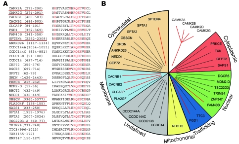
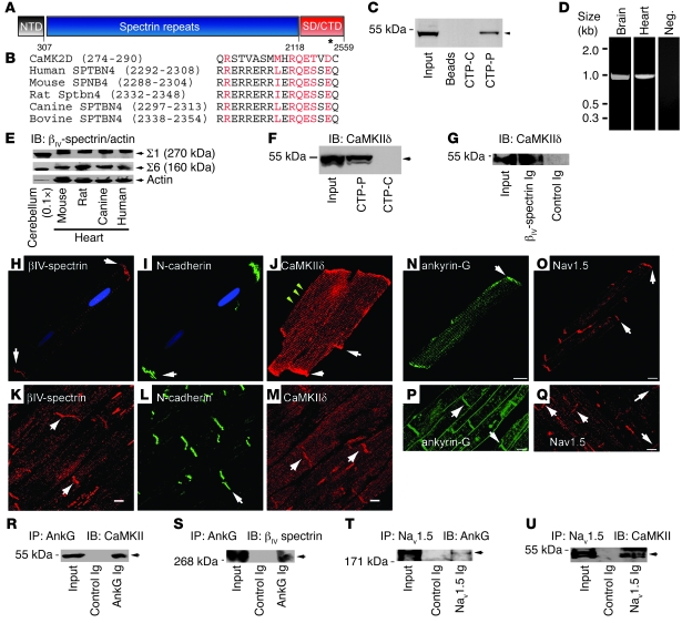
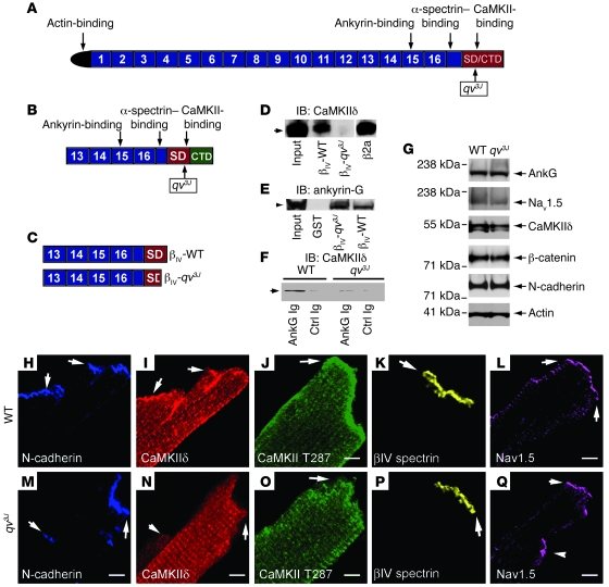
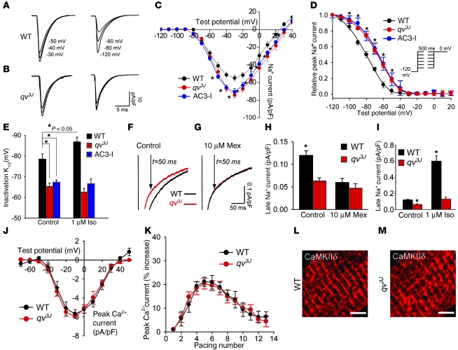
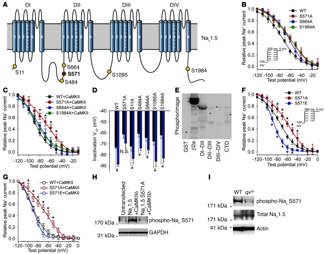
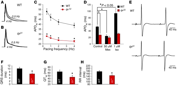
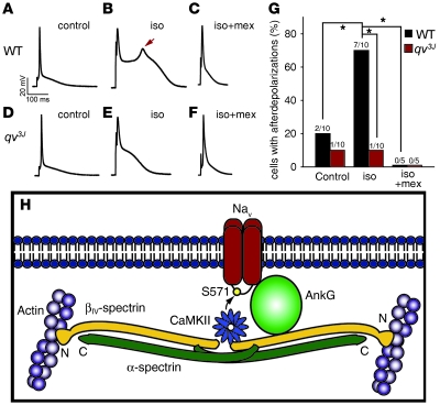
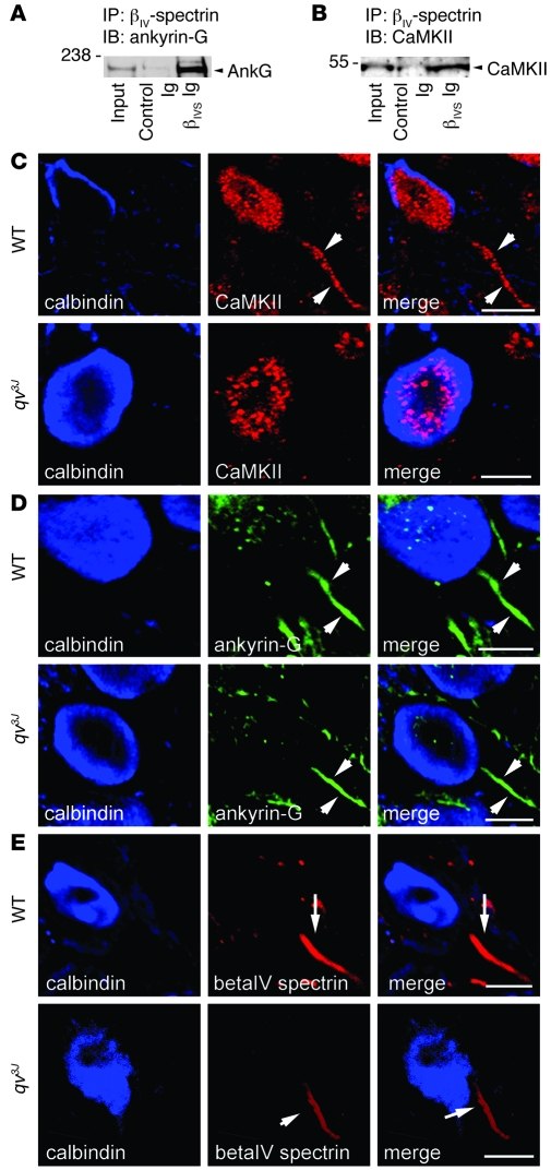
Ion channel function is fundamental to the existence of life. In metazoans, the coordinate activities of voltage-gated Na(+) channels underlie cellular excitability and control neuronal communication, cardiac excitation-contraction coupling, and skeletal muscle function. However, despite decades of research and linkage of Na(+) channel dysfunction with arrhythmia, epilepsy, and myotonia, little progress has been made toward understanding the fundamental processes that regulate this family of proteins. Here, we have identified β(IV)-spectrin as a multifunctional regulatory platform for Na(+) channels in mice. We found that β(IV)-spectrin targeted critical structural and regulatory proteins to excitable membranes in the heart and brain. Animal models harboring mutant β(IV)-spectrin alleles displayed aberrant cellular excitability and whole animal physiology. Moreover, we identified a regulatory mechanism for Na(+) channels, via direct phosphorylation by β(IV)-spectrin-targeted calcium/calmodulin-dependent kinase II (CaMKII). Collectively, our data define an unexpected but indispensable molecular platform that determines membrane excitability in the mouse heart and brain.


Figures

Figure 1
Figure 1. Identification of putative new CaMKII-binding proteins.
(A) Putative CaMKII-targeting proteins identified through a screen of the human genome using a sequence from the CaMKII autoregulatory domain as bait. Candidates with the highest homology to CaMKII autoregulatory domain are listed first. (B) Candidate targeting molecules included cytoskeletal, nuclear, cytoplasmic, and mitochondrial proteins with identified roles in cell metabolism, cytoskeletal dynamics, and signaling. All CaMKII gene products (α, β, γ, and δ) were recognized by the screen; notably, only 1 known CaMKII-binding partner was identified (β2a). Candidates were cloned from human cDNA, and CaMKII-binding activity was assessed by in vitro binding assays using radiolabeled target proteins and activated CaMKII (CaMKII T287D). Notably, only clones underlined in red in A showed robust binding for CaMKII.
Figure 2
Figure 2. βIV-spectrin is a CaMKII-binding protein in heart.
(A) βIV-spectrin contains an N-terminal actin-binding domain (NTD), 17 spectrin repeats, and specific and C-terminal domains (SD/CTD). The putative CaMKII-binding site is denoted by an asterisk. (B) The putative CaMKII-binding domain in βIV-spectrin was homologous to a CaMKIIδ autoregulatory domain motif and conserved across orthologs. (C) CTP-P bound radiolabeled CaMKIIδ; CTP-C and GST beads alone lacked binding. (D) βIV-spectrin RNA levels in adult rat brain and heart. (E) βIV-spectrin (Σ1 and Σ6) in ventricular lysates from multiple species. βIV-spectrin was expressed approximately 8–10 fold higher in cerebellum than in heart. Cardiac βIV-spectrin migrated approximately 4 kDa larger than did cerebellar βIV-spectrin. (F) CTP-P, but not CTP-C, associated with CaMKIIδ from rat heart. (G) Endogenous CaMKIIδ and βIV-spectrin coimmunoprecipitated from adult heart lysate. (H and I) βIV-spectrin and N-cadherin in rat cardiomyocytes. Nuclei are shown by Topro-3 dye (blue). (J) CaMKIIδ localization in adult rat myocytes. CaMKIIδ localized to the intercalated disc (white arrows) here and to a second population at transverse-tubules (yellow arrows). Ventricular sections stained for (K) βIV-spectrin, (L) N-cadherin, and (M) CaMKIIδ showed coexpression of these proteins at the intercalated disc (white arrows). (N and P) Ankyrin-G and (O and Q) Nav1.5 were also found at the intercalated disc (white arrows) in rat myocytes and tissue sections. (RU) Coimmunoprecipitation studies demonstrate cardiac complex of βIV-spectrin, CaMKIIδ, ankyrin-G (AnkG), and Nav1.5. Scale bars: 10 μm (HQ).
Figure 3
Figure 3. βIV-spectrin is required for CaMKIIδ targeting.
(A) βIV-spectrin organization in WT and qv3J animals. qv3J animals lacked a CaMKII-binding domain, but retained actin-, ankyrin-, and α-spectrin–binding domains. (B and C) Schematic of control GST–βIV-spectrin fusion protein encompassing spectrin repeats 13–17 and the CaMKII-binding domain (βIV-WT) and truncated mutant lacking CaMKII-binding domain (βIV-qv3J). (D) βIV-spectrin WT and qv3J GST fusion proteins were incubated with detergent-soluble rat heart lysate and analyzed by immunoblot (CaMKIIδ). L-type Ca2+ channel β2a subunit was also expressed as a GST fusion protein and used as positive control. CaMKIIδ bound to the β2a and WT GST fusion proteins, but not to the qv3J GST fusion protein. Lanes were run on the same gel but were noncontiguous (white line). (E) βIV-spectrin WT and qv3J GST fusion proteins retained binding activity for ankyrin-G. (F) Coimmunoprecipitation studies showing association of ankyrin-G and CaMKIIδ in WT, but not qv3J, hearts. (G) Expression of ankyrin-G, Nav1.5, CaMKIIδ, β-catenin, and N-cadherin in heart lysates from WT and qv3J animals. Actin is shown as loading control. (HQ) Permeabilized adult rat cardiomyocytes from (HL) WT and (MQ) qv3J hearts were immunostained for (H and M) N-cadherin, (I and N) total CaMKIIδ, (J and O) CaMKII-phospho-T287, (K and P) βIV-spectrin, and (L and Q) Nav1.5. Localization of CaMKIIδ (total and phospho-T287) to the intercalated disc (white arrows) was disrupted in qv3J cardiomyocytes. Scale bars: 10 μm (HQ).
Figure 4
Figure 4. βIV-spectrin/CaMKII complex is required for myocyte Na+ channel function.
(A and B) Whole-cell patch clamp INa traces from WT and qv3J cardiomyocytes. Test pulse potential is listed next to each trace. (C) Current-voltage relationship for cardiomyocytes from WT, qv3J, and AC3-I mice (n = 8 per group). *P < 0.05 versus WT. (D and E) Voltage-gated Na+ channel steady-state inactivation measured from WT, qv3J, and AC3-I cardiomyocytes (n = 8 per group). *P < 0.05 versus WT. Pulse protocol is shown in the inset of D. (F and G) INa,p from WT and qv3J mice with or without 10 μM mexiletine. Persistent current was determined as amplitude 50 ms after time of peak. (H and I) INa,p in WT (n = 10) and qv3J (n = 17) cardiomyocytes at baseline and in the presence of 10 μm mexiletine or isoproterenol. *P < 0.05. (J) Whole-cell patch clamp Ca2+ current-voltage relationship for WT (n = 9) and qv3J (n = 8) cardiomyocytes. (K) Ca2+ current facilitation (peak current in response to a train of depolarizing voltage pulses to 0 mV) in WT (n = 9) and qv3J (n = 7) cardiomyocytes. Peak current is expressed as percent increase from first pulse. (L and M) Transverse-tubule CaMKII labeling was preserved in qv3J mouse myocytes. Scale bars: 5 μm (L and M).
Figure 5
Figure 5. βIV-spectrin/CaMKII regulates Nav1.5 phosphorylation.
(A) CaMKII consensus phosphorylation sites in intracellular domains of Nav1.5. (B and C) Na+ channel steady-state inactivation measured from WT and Nav1.5 mutant channels expressed in HEK cells. *P < 0.05 versus WT. (D) Summary data showing depolarizing shift in steady-state inactivation V1/2 following CaMKII activation for WT and all mutants except S571A. Black, before CaMKII activation; blue, after CaMKII activation. (E) CaMKII phosphorylation assay on intracellular domains of Nav1.5. β2a was used as positive control. Asterisks denote location of purified proteins. (F and G) Na+ channel steady-state inactivation measured from WT, S571A, and S571E (phosphomimetic) channels expressed in HEK cells. *P < 0.05 versus WT. (H) Nav1.5 S571 antibody recognized WT, but not Nav1.5 S571A mutant, channels. WT and mutant channels were expressed at equivalent levels in HEK293 cells overexpressing active CaMKIIδ. (I) Immunoblots showing reduced levels of phospho–Nav1.5 S571, but unchanged total Nav1.5 levels, in qv3J versus WT heart lysates. Actin is shown as loading control.
Figure 6
Figure 6. βIV spectrin/CaMKII signaling complex is critical for normal cardiac cell membrane excitability.
(A and B) Representative APs and (C) APD90 in WT (n = 14) and qv3J (n = 16) cardiomyocytes paced at 4, 2, 1, or 0.5 Hz. *P < 0.05. (D) Differences in APD90 were eliminated by application of 50 μM mexiletine, but exaggerated by 1 μM isoproterenol (pacing frequency, 0.5 Hz). n = 14 (WT); 16 (qv3J). *P < 0.05. (E) Representative electrocardiograms recorded from Langendorff-perfused WT and qv3J hearts and summary data for (F) QRS duration (G), QT interval at 90% repolarization (QT90), and (H) RR interval for qv3J versus WT. n = 10 (WT); 10 (qv3J). *P < 0.05.
Figure 7
Figure 7. βIV-spectrin/CaMKII complex regulates afterdepolarization formation in response to isoproterenol treatment.
Representative APs recorded from WT and qv3J cardiomyocytes at baseline (A and D) and following application of 1 μM isoproterenol (B and E) or isoproterenol applied with 10 μM mexiletine (C and F). (G) Summary data showing percent cells displaying afterdepolarizations in WT and qv3J at baseline (n = 10 per genotype) and following application of isoproterenol (n = 10 per genotype) or isoproterenol applied with mexiletine (n = 5 per genotype). *P < 0.05. (H) βIV-spectrin–based complex targets CaMKII to effector proteins in excitable cells.
Figure 8
Figure 8. βIV-spectrin targets CaMKII in neurons.
(A and B) βIV-spectrin associated with both ankyrin-G and CaMKII from mouse cerebellar lysates. (C) CaMKII (red) was observed at AISs (white arrows) in WT, but not qv3J, cerebellum. (D) AIS integrity (shown by positive ankyrin-G staining) was not markedly affected in qv3J mice. (E) βIV-spectrin (red) was present at qv3J mouse AISs, but at reduced levels compared with WT cerebellum. Calbindin (blue) was used to label the Purkinje cell body in CE. Scale bars: 10 μm (CE).

Comment in

Similar articles

Cited by

References

    1. Frankland PW, O’Brien C, Ohno M, Kirkwood A, Silva AJ. Alpha-CaMKII-dependent plasticity in the cortex is required for permanent memory. Nature. 2001;411(6835):309–313. doi: 10.1038/35077089. - DOI - PubMed
    1. Karcher RL, et al. Cell cycle regulation of myosin-V by calcium/calmodulin-dependent protein kinase II. Science. 2001;293(5533):1317–1320. doi: 10.1126/science.1061086. - DOI - PubMed
    1. Lisman J, Schulman H, Cline H. The molecular basis of CaMKII function in synaptic and behavioural memory. Nat Rev Neurosci. 2002;3(3):175–190. doi: 10.1038/nrn753. - DOI - PubMed
    1. Nutt LK, et al. Metabolic regulation of oocyte cell death through the CaMKII-mediated phosphorylation of caspase-2. Cell. 2005;123(1):89–103. doi: 10.1016/j.cell.2005.07.032. - DOI - PMC - PubMed
    1. Zhang R, et al. Calmodulin kinase II inhibition protects against structural heart disease. Nat Med. 2005;11(4):409–417. - PubMed

Publication types