. 2011 Apr;21(4):588-99.
doi: 10.1038/cr.2010.137. Epub 2010 Sep 28.

Identification of a small molecule activator of novel PKCs for promoting glucose-dependent insulin secretion

Affiliations

Identification of a small molecule activator of novel PKCs for promoting glucose-dependent insulin secretion

Shuai Han et al. Cell Res. 2011 Apr.

Abstract

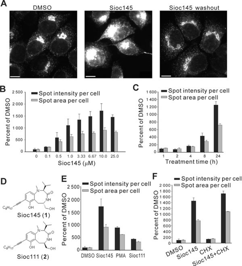
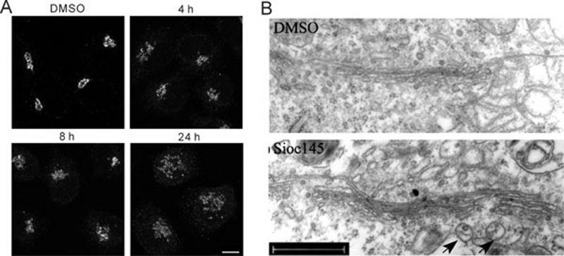
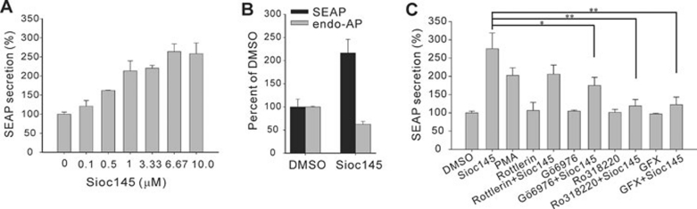
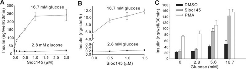
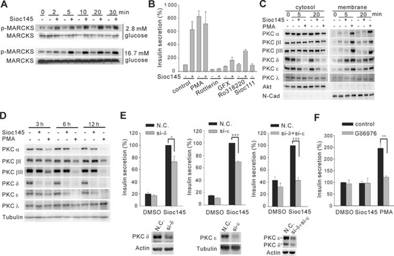
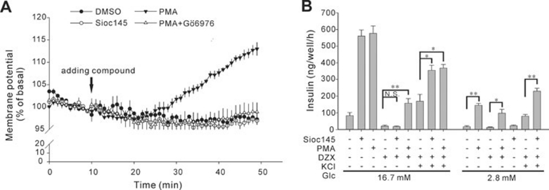
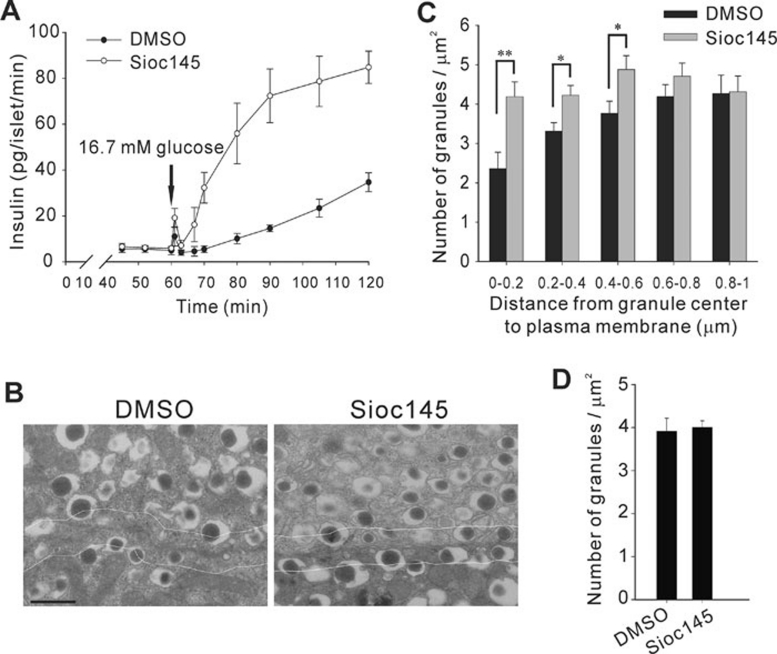
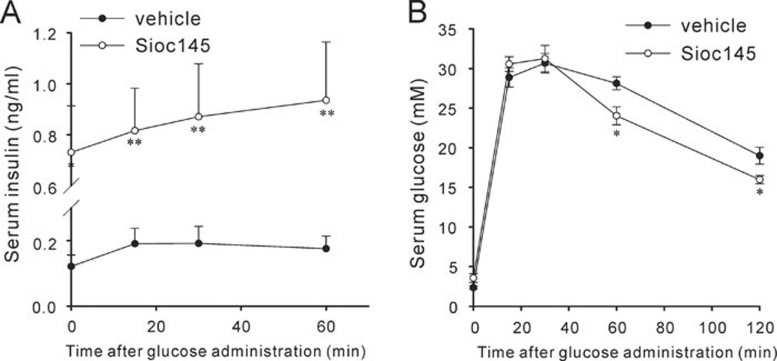
Using an image-based screen for small molecules that can affect Golgi morphology, we identify a small molecule, Sioc145, which can enlarge the Golgi compartments and promote protein secretion. More importantly, Sioc145 potentiates insulin secretion in a glucose-dependent manner. We show that Sioc145 selectively activates novel protein kinase Cs (nPKCs; δ and ɛ) but not conventional PKCs (cPKCs; α, βI and βII) in INS-1E insulinoma cells. In contrast, PMA, a non-selective activator of cPKCs and nPKCs, promotes insulin secretion independent of glucose concentrations. Furthermore, we demonstrate that Sioc145 and PMA show differential abilities in depolarizing the cell membrane, and suggest that Sioc145 promotes insulin secretion in the amplifying pathway downstream of K(ATP) channels. In pancreatic islets, the treatment with Sioc145 enhances the second phase of insulin secretion. Increased insulin granules close to the plasma membrane are observed after Sioc145 treatment. Finally, the administration of Sioc145 to diabetic GK rats increases their serum insulin levels and improves glucose tolerance. Collectively, our studies identify Sioc145 as a novel glucose-dependent insulinotropic compound via selectively activating nPKCs.


Figures

Figure 1
Figure 1
Sioc145 enhances the signal of p58-YFP in H4 cells. (A) H4-p58-YFP cells were treated with DMSO or 5 μM Sioc145 for 24 h or then exchanged with fresh medium without Sioc145 for an additional 24 h. Images were analyzed for spots' intensity and area per cell. Scale bar, 10 μm. (B) Dose-response of Sioc145 in H4-p58-YFP cells treated for 24 h. (C) Time course response of 5 μM Sioc145 in H4-p58-YFP cells. (D) Chemical structures of Sioc145 and Sioc111. (E) p58-YFP signal in cells treated with DMSO, 5 μM Sioc145, 5 μM Sioc111 or 100 ng/ml PMA. (F) p58-YFP signal in cells treated with DMSO, 5 μM Sioc145, 10 μg/ml CHX alone or CHX together with 5 μM Sioc145 for 24 h. All graphics represent means ± SD obtained from three independent experiments.
Figure 2
Figure 2
Effect of Sioc145 on the morphology and structure of the Golgi. (A) Immunofluorescence images of H4 cells treated with DMSO, 5 μM Sioc145 for 4 h, 8 h or 24 h using GM130 antibody. Scale bar, 10 μm. (B) Representative electron microscopy images of the Golgi in H4 cells treated with DMSO or 5 μM Sioc145 for 24 h. Arrows indicate the representatives of big secretory vesicles. Scale bar, 500 nm.
Figure 3
Figure 3
Sioc145 promotes SEAP secretion via a PKC-dependent pathway. (A) Dose-response of Sioc145 in promoting SEAP secretion in H4-SEAP cells treated for 24 h. (B) H4-SEAP cells were treated with DMSO or 5 μM Sioc145 for 24 h. The levels of alkaline phosphatase enzyme in culture medium (SEAP) or total cell lysate (endo-AP) were analyzed. (C) H4-SEAP cells were treated with DMSO, 5 μM Sioc145 or 100 ng/ml PMA in the presence or absence of 10 μM Rottlerin, 2 μM Gö6976, 2 μM Ro318220 or 2.5 μM GFX for 24 h as indicated. Secreted SEAP was measured. Data are normalized to that of DMSO-treated cells, which is set as 100% and error bars represent SD from three independent experiments. *P < 0.05; **P < 0.01.
Figure 4
Figure 4
Sioc145 potentiates insulin secretion in a glucose-dependent manner. (A) Insulin secretion from INS-1E cells stimulated with either 2.8 mM or 16.7 mM glucose for 30 min in the presence of Sioc145 at indicated concentrations. (B) Dose-response of Sioc145 in rat pancreatic islets stimulated with 2.8 mM or 16.7 mM glucose concentration (see Materials and Methods). (C) Insulin secretion in response to various concentrations of glucose was examined in the presence of 1 μM Sioc145 or 0.05 μM PMA in INS-1E cells. Error bars represent SEM from three independent experiments.
Figure 5
Figure 5
Selective activation of nPKCs is required for insulinotropic action of Sioc145. (A) MARCKS phosphorylation in 2.8 mM or 16.7 mM glucose was analyzed in INS-1E cells treated with 1 μM Sioc145 for indicated time points. (B) 16.7 mM glucose-stimulated insulin secretion from INS-1E cells treated with Sioc145 (1 μM) alone or together with indicated compounds: PMA, 0.05 μM; Rottlerin, 5 μM; GFX, 2.5 μM; Ro318220, 2 μM. Data are normalized to that of DMSO-treated cells which is set as 100%. (C) The translocation of PKC isoforms in INS-1E cells treated with Sioc145 (1 μM) or PMA (0.05 μM). N-cadherin (N-cad) and Akt were used as membrane and cytosol markers, respectively. (D) Effect of prolonged treatment with Sioc145 (1 μM) or PMA (0.05 μM) on protein levels of PKC isoforms. (E) Insulin secretion in PKC δ and/or ɛ downregulated cells. Cells transfected with indicated siRNA(s) were stimulated with DMSO or 1 μM Sioc145 at 16.7 mM glucose concentration. Insulin secretion from Sioc145-treated control cells (N.C.) were set as 100%. (F) Gö6976 abolished PMA-induced secretion. INS-1E cells treated with Sioc145 (1 μM) or PMA (0.05 μM) were co-treated with Gö6976 (1 μM) for 30 min in 2.8 mM glucose. Error bars represent SEM from at least three independent experiments. *P < 0.05; **P < 0.01; ***P < 0.001.
Figure 6
Figure 6
Cell membrane depolarization is a prerequisite for insulinotropic effect of Sioc145. (A) Cell membrane potential was monitored as bisoxonol fluorescence in INS-1E cells stimulated with Sioc145 (1 μM), PMA (0.05 μM) or PMA plus Gö6976 (1 μM), in the presence of 2.8 mM glucose. Arrow indicates the time point when indicated compounds were added. (B) INS-1E cells were treated with indicated combinations of Sioc145 (1 μM), PMA (0.05 μM), DZX (250 μM) and KCl (30 mM), in the presence of 2.8 mM or 16.7 mM glucose for 1 h. Data are means ± SEM from three independent experiments. N.S., not significant; *P < 0.05; **P < 0.01.
Figure 7
Figure 7
Characterization of secretion kinetics and insulin granule distribution in Sioc145-treated rat pancreatic islets. (A) Rat pancreatic islets pretreated with 5 μM Sioc145 or DMSO for 30 min were perifused with 16.7 mM glucose buffer for 1 h, and insulin secretion was measured at indicated time points (see Materials and Methods). (B) Islets were treated with DMSO or 5 μM Sioc145 for 30 min, followed by a 60 min equilibration in 2.8 mM glucose buffer. Representative electron micrographs of islet β-cell sections are shown. Scale bar, 0.5 μm. White lines indicate a distance of 0.2 μm from the plasma membrane. (C) Density of insulin granules located in 0.2 μm concentric shells within the first 1 μm area from the plasma membrane. Granules located in the single section were categorized according to their distance from the granule center to the plasma membrane. (D) The average density of granules in cytoplasm. The area of cytoplasm is calculated as the cell area minus the nuclear area. Error bars represent SEM from three independent experiments (A) or 10 individual β-cells derived from four rats (C, D). *P < 0.05; **P < 0.01.
Figure 8
Figure 8
Effect of Sioc145 in diabetic GK rats. Fasted GK rats were administered intraperitoneally with Sioc145 (0.414 mg/kg) or vehicle 2 h before glucose loading (2 g/kg, i.p). Sera were collected before or at indicated time points after glucose administration to measure insulin levels (A) and glucose levels (B). n ≥ 5. Data shown are means ± SEM (*P < 0.05; **P < 0.01 vs vehicle; Student's t-test).

References

    1. Straub SG, Sharp GW. Glucose-stimulated signaling pathways in biphasic insulin secretion. Diabetes Metab Res Rev. 2002;18:451–463. - PubMed
    1. Henquin JC. Triggering and amplifying pathways of regulation of insulin secretion by glucose. Diabetes. 2000;49:1751–1760. - PubMed
    1. Henquin JC. Pathways in β-cell stimulus-secretion coupling as targets for therapeutic insulin secretagogues. Diabetes. 2004;53 Suppl 3:S48–58. - PubMed
    1. Gembal M, Detimary P, Gilon P, Gao ZY, Henquin JC. Mechanisms by which glucose can control insulin release independently from its action on adenosine triphosphate-sensitive K+ channels in mouse B cells. J Clin Invest. 1993;91:871–880. - PMC - PubMed
    1. Knutson KL, Hoenig M. Identification and subcellular characterization of protein kinase-C isoforms in insulinoma β-cells and whole islets. Endocrinology. 1994;135:881–886. - PubMed

Publication types

MeSH terms