. 2011 Feb;19(2):408-16.
doi: 10.1038/mt.2010.201. Epub 2010 Sep 28.

Ex-vivo gene therapy restores LEKTI activity and corrects the architecture of Netherton syndrome-derived skin grafts

Affiliations

Ex-vivo gene therapy restores LEKTI activity and corrects the architecture of Netherton syndrome-derived skin grafts

Wei-Li Di et al. Mol Ther. 2011 Feb.

Abstract

Netherton syndrome (NS) is a debilitating congenital skin disorder caused by mutations in the SPINK5 gene encoding the lymphoepithelial Kazal-type-related inhibitor (LEKTI). It is characterized by defective keratinization, recurrent infections, and hypernatraemic dehydration with a mortality rate of about 10% in the first year of life. Currently, there are no curative treatments for NS. We have developed a HIV-1 based, self-inactivating lentiviral vector to express SPINK5 in keratinocytes as part of an ex-vivo gene therapy strategy for NS. High transduction efficiency was achieved in NS keratinocytes and reconstitution of LEKTI expression was confirmed in previously deficient cells. These genetically corrected keratinocytes were further tested in an in vitro organotypic culture (OTC) system and in vivo mouse/human skin engraftment model. Results showed correction of epidermal architecture in both OTCs and regenerated skin grafts. Importantly, the results from corrected skin grafts indicated that even where detectable LEKTI expression was restored to a limited numbers of cells, a wider bystander benefit occurred around these small populations. As LEKTI is a secreted protein, the genetically modified graft may provide not only an immediate local protective barrier, but also act as a source of secreted LEKTI providing a generalized benefit following ex-vivo gene therapy.


Figures

Figure 1
Figure 1
Morphology and LEKTI expression in the skin with Neterton syndrome (NS). (a,b) H&E staining and (c,d) immunofluorescence staining in healthy donor skin and NS skin. In NS, the acanthotic, psoriasiform epidermis was covered by parakeratotic stratum corneum (b) and LEKTI expression was absent in the uppermost layers of the skin (d), compared to healthy donor skin, where intense membranous staining (green color) was detectable in the granular and cornified layers (c). (a,b) Bar = 100 µm; (c,d) = 20 µm. LEKTI, lymphoepithelial Kazal-type-related inhibitor.
Figure 2
Figure 2
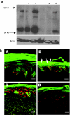
SPINK5/eGFP lentiviral vector mediate LEKTI expression in keratinocytes. (a) Schematic representation of lentiviral vectors used in this study. The vectors used were self-inactivating HIV-1-based viruses, encoding the spleen focus-forming virus (SFFV) LTR promoter, the HIV-1 cPPT element, and a mutated WPRE. The accessory plasmids mediated vector packaging and pseudotyping with the vesicular stomatitis virus (vsv) envelope. (b) Western blotting of N-TERT cells following transduction with increasing MOIs of SPINK5/eGFP lentivirus. Lane 1 shows nontransduced N-TERT cells in which full length, unprocessed LEKTI is not detectable. Lanes 2–6 show cells transduced with an increasing MOI of vector-encoding SPINK5/eGFP (0.1, 0.6, 3.2, 16, and 25). Lane 7 was loaded 1 µg of recombinant LEKTI and lane 8 was loaded tissue lysate from human skin. Increasing levels of full length (~145 kd) and cleaved LEKTI bands (~68) were detected in transduced cells. Lane 7 and 8 are used as positive controls showing the size of full-length LEKTI and processed LEKTI. (c) Western blotting of culture media to detect secreted LEKTI fragments. Lane 1 was N-TERT cell lysate; lane 2 was health donor skin lysate; lane 3 was culture medium of nontransduced N-TERT cells; lane 4 was culture medium of cells transduced with eGFP alone; and lane 5 was culture medium of cells transduced with SPINK5/eGFP. Results showed an intensive band at size ~68 kd, representing secreted LEKTI in lane 5, but not lane 3 or 4 which were derived from media of nontransduced cells and cells transduced with eGFP alone. Thus, gene-modified cells expressed, processed, and secreted LEKTI. CMV, cytomegalovirus; cPPT, central polypurine tract; eGFP, enhanced green fluorescent protein; LEKTI, lymphoepithelial Kazal-type-related inhibitor; LTR, long-terminal repeat; MOI, multiplicity of infection; SIN, self-inactivating; WPRE, Woodchuck postregulatory element.
Figure 3
Figure 3
Flow cytometry analysis of cell proliferation and death in transduced N-TERT cells. (a) Cells transduced with eGFP or SPINK5/eGFP expressing viral vector were treated with EdU. Examples of the distribution of cell populations in different stages of the cell cycles are shown. There were no significant differences between groups, suggesting that lentiviral transduction and LEKTI expression do not affect cell turnover. (b) Cell viability of transduced N-TERT cells was analyzed using propidium iodide staining and flow cytometry. The distribution of live and dead cells in the eGFP+, transduced cell populations is shown. There were no significant differences in cell death or survival between groups indicating that transduction with vectors encoding SPINK5/eGFP does not influence cell survival. EdU, 5-ethynyl-2′-deoxyuridine; eGFP, enhanced green fluorescent protein; LEKTI, lymphoepithelial Kazal-type-related inhibitor.
Figure 4
Figure 4
Lentiviral vector delivery of SPINK5 transgene into primary keratinocytes. (a) Transduced healthy donor or NS primary keratinocytes were harvested and the LEKTI expression was assessed using anti-LEKTI antibody by western blot analysis. Lane 1 (healthy donor) and lane 4 (NS) were nontransduced cells. Endogenous full length (145 kd) and cleaved (~68 kd) LEKTI could be detected in normal keratinocytes nontransduced or transduced with eGFP alone expressing vector (lane 1 and 2), but not in NS cells (lanes 4 and 5). In contrast, in cells transduced with SPINK5/eGFP vector, increased expression of LEKTI was observed in both healthy donor (lane 3) and NS cells (lane 6) compared to the cells transduced with eGFP alone (lanes 2 and 5). These results demonstrate successful reconstitution of LEKTI expression in NS keratinocytes following lentiviral transduction. (b) Primary keratinocytes from health donors or NS were transduced with lentiviral vector expressing SPINK5/eGFP or eGFP alone. GFP+ cells were sorted by flow cytometry to enrich eGFP+ cells and then deployed in organotypic cultures (OTC). eGFP in frozen sections from (OTC) was directly detected under fluorescence microscopy. (A,C) Cultures derived from healthy donor keratinocytes, and (B,D) cultures derived from NS keratinocytes. Cells transduced with eGFP alone (A,B) or SPINK5/eGFP (C,D) all expressed high levels of eGFP (green) with an intense staining pattern especially in the uppermost layers of the epidermis. NS keratinocytes transduced with SPINK5/eGFP showed reduced numbers of nuclei in the cornified layer of the epidermis (D) compared to the culture generated by NS cells transduced with eGFP alone (B, arrowed). Red shows nuclei staining by propidium iodide. Bar = 40 µm. eGFP, enhanced green fluorescent protein; LEKTI, lymphoepithelial Kazal-type-related inhibitor; NS, Netherton syndrome.
Figure 5
Figure 5
LEKTI expression in OTCs transduced with SPINK5/eGFP. (a) OTC generated from healthy donor keratinocytes and (b) is the OTC generated from NS keratinocytes. Cells were transduced with (A–C, G–I) vector-encoding eGFP vector or (D–F, J–L) SPINK5/eGFP vector and used in OTCs. Sections from OTCs were stained with anti-LEKTI antibody and the expression of LEKTI (red) and eGFP (green) was verified by Confocal microscopy. Endogenous LEKTI expression could be detected in OTCs derived from normal cells (B) but not in OTC generated from NS cells (H). In contrast, both eGFP and LEKTI were detected in OTCs derived from healthy donor (E) or NS cells (K) transduced with SPINK5/eGFP expressing vectors. Merged images showed the overlapped expression of SPINK5 and eGFP in (C), (F), and (L) but not where NS cells had been transduced with vector expressing eGFP alone (I). These results demonstrate that LEKTI expression can be restored in NS using lentiviral vector technology and that the corrected cells can grow and differentiate as seen in healthy donor cells. Bar = 40 µm. (c) Nuclei numbers in the stratum corneum of OCTs in optical sections were analyzed by Image-Pro and results showed a significant reduction in the OTC using NS keratinocytes transduced with SPINK5/eGFP compared to nontransduced or eGFP transduced cells (*single factor ANOVA, P < 0.01). eGFP, enhanced green fluorescent protein; LEKTI, lymphoepithelial Kazal-type-related inhibitor; NS, Netherton syndrome; OTC, organotypic culture.
Figure 6
Figure 6
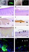
In vivo assessment of NS keratinocyte correction in a humanized mouse model: macroscopic and histological examination following lentiviral transduction. NS keratinocytes were transduced with (a–e,k) eGFP or (f–j,m) SPINK5/eGFP and grafted onto nude mice in two independent series of experiments. Regenerated skin grafts were examined 8 weeks after grafting. (a,f) macroscopic appearance of the graft under transmit light and (b,g) a real-time eGFP expression under 488 nm light. (c,h) Histological appearance of grafts. (d,i) Human involucrin expression using antihuman involucrin antibody to indicate mouse (Mo)—human (Hu) skin boundary. (e) eGFP expression using GFP antibody. (j) The LEKTI expression using antihuman LEKTI antibody and arrows indicate isolated or clustered LEKTI+ cells within a wider area of skin. (k,m) Both frozen sections represent the overlapping between real-time eGFP (green) expression and LEKTI expression (red) stained with LEKTI antibody. These images showed that there was reversal of papillary changes in the graft transduced with SPINK5/eGFP (h) compared to the graft transduced with control vector eGFP (c). Low numbers of LEKTI expression cells in the grafts transduced with SPINK5/eGFP vector in both paraffin and frozen sections (j and m, arrowed areas) suggest small population of corrected cells sufficient to mediate wider correction of the epidermal architecture. bars: (a,b,f,g) = 5 µm; (c,d,h,i) = 200 µm; (e,j) = 100 µm; (k,m) = 40 µm. eGFP, enhanced green fluorescent protein; NS, Netherton syndrome.

References

    1. Bitoun E, Micheloni A, Lamant L, Bonnart C, Tartaglia-Polcini A, Cobbold C, et al. LEKTI proteolytic processing in human primary keratinocytes, tissue distribution and defective expression in Netherton syndrome. Hum Mol Genet. 2003;12:2417–2430. - PubMed
    1. Chavanas S, Bodemer C, Rochat A, Hamel-Teillac D, Ali M, Irvine AD, et al. Mutations in SPINK5, encoding a serine protease inhibitor, cause Netherton syndrome. Nat Genet. 2000;25:141–142. - PubMed
    1. Borgoño CA, Michael IP, Komatsu N, Jayakumar A, Kapadia R, Clayman GL, et al. A potential role for multiple tissue kallikrein serine proteases in epidermal desquamation. J Biol Chem. 2007;282:3640–3652. - PubMed
    1. Descargues P, Deraison C, Prost C, Fraitag S, Mazereeuw-Hautier J, D'Alessio M, et al. Corneodesmosomal cadherins are preferential targets of stratum corneum trypsin- and chymotrypsin-like hyperactivity in Netherton syndrome. J Invest Dermatol. 2006;126:1622–1632. - PubMed
    1. Ishida-Yamamoto A, Deraison C, Bonnart C, Bitoun E, Robinson R, O'Brien TJ, et al. LEKTI is localized in lamellar granules, separated from KLK5 and KLK7, and is secreted in the extracellular spaces of the superficial stratum granulosum. J Invest Dermatol. 2005;124:360–366. - PubMed

Publication types

Substances