. 2010 Aug 30:4:163.
doi: 10.3389/fnins.2010.00163. eCollection 2010.

Enhanced post-ischemic neurogenesis in aging rats

Affiliations

Enhanced post-ischemic neurogenesis in aging rats

Yao-Fang Tan et al. Front Neurosci. .

Abstract

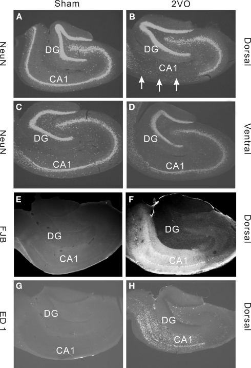
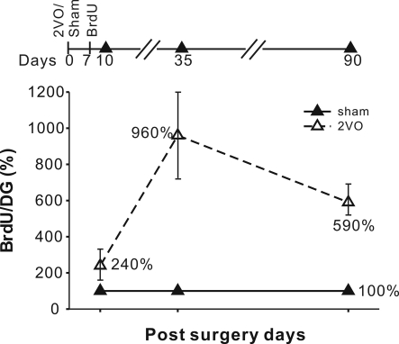
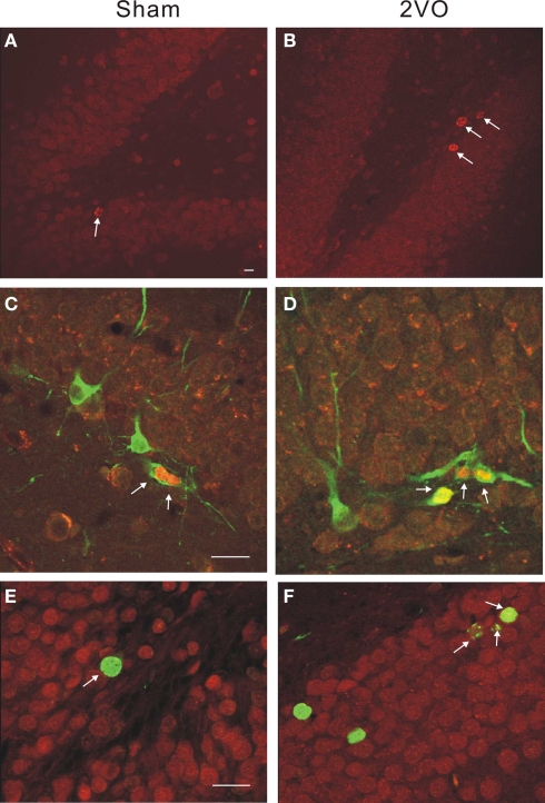
Hippocampal neurogenesis persists in adult mammals, but its rate declines dramatically with age. Evidence indicates that experimentally-reduced levels of neurogenesis (e.g., by irradiation) in young rats has profound influence on cognition as determined by learning and memory tests. In the present study we asked whether in middle-aged, 10- to 13-months-old rats, cell production can be restored toward the level present in young rats. To manipulate neurogenesis we induced bilateral carotid occlusion with hypotension. This procedure is known to increase neurogenesis in young rats, presumably in a compensatory manner, but until now, has never been tested in aging rats. Cell production was measured at 10, 35, and 90 days after ischemia. The results indicate that neuronal proliferation and differentiation can be transiently restored in middle-aged rats. Furthermore, the effects are more pronounced in the dorsal as opposed to ventral hippocampus thus restoring the dorso-ventral gradient seen in younger rats. Our results support previous findings showing that some of the essential features of the age-dependent decline in neurogenesis are reversible. Thus, it may be possible to manipulate neurogenesis and improve learning and memory in old age.

Keywords: adult neurogenesis; aging; dentate gyrus; hippocampus; ischemia; stroke.


Figures

Figure 1
Figure 1
Immunohistochemical staining demonstrating effects of sham (control) and 2VO (ischemia) procedure in dorsal and ventral regions of the hippocampus. (A–D) Show sections stained with a neuronal marker NeuN. Note, a severe loss of cells in dorsal CA1 field (arrows) but no obvious cell loss in DG (A,B). Much lesser effect is seen in ventral CA1 (C,D). (E,F) Staining with a neuronal degeneration marker Fluorojade B (FJB) shows clear degeneration in CA1 field in dorsal region. (G,H) Staining with a microglia marker ED1 shows strong inflammatory response in CA1 and Hilus but not in DG. Sections in (A–D) are from representative animals within the 35 day group. Sections in (E–H) are from animals within the 10 day group.
Figure 2
Figure 2
Experimental time line and the increases in the BrdU+ cell numbers at 10, 35 and 90 days. The increases were expressed in terms of % of the sham control values with standard errors indicated. Increases in the number of BrdU+ cells were significant at all three time points. The absolute values are given in Table 2.
Figure 3
Figure 3
Immunohistochemical staining illustrating neurogenesis in control and ischemic rats. (A,B) Single-labeled BrdU+ cells are more numerous (arrows) within a DG in 2VO, ischemic animal at 10 days. (C,D) Double-labeled neuroblasts show BrdU+ (red) and DCX+ (green) neuroblasts at 10 days. (E,F) Double-labeled cells show BrdU+ (green) and CaBP+ (red) neurons at 35 days. Calibration bar = 40 microns in all cases.

Similar articles

Cited by

References

    1. Bannerman D. M., Rawlins J. N., McHugh S. B., Deacon R. M., Yee B. K., Bast T., Zhang W. N., Pothuizen H. H., Feldon J. (2004). Regional dissociations within the hippocampus--memory and anxiety. Neurosci. Biobehav. Rev. 28, 273–28310.1016/j.neubiorev.2004.03.004 - DOI - PubMed
    1. Bizon J. L., Gallagher M. (2003). Production of new cells in the rat dentate gyrus over the lifespan: relation to cognitive decline. Eur. J. Neurosci. 18, 215–21910.1046/j.1460-9568.2003.02733.x - DOI - PubMed
    1. Burke S. N., Barnes C. A. (2006). Neural plasticity in the ageing brain. Nat. Rev. Neurosci. 7, 30–4010.1038/nrn1809 - DOI - PubMed
    1. Cameron H. A., McKay R. D. G. (1999). Restoring production of hippocampal neurons in old age. Nat. Neurosci. 2, 894–89710.1038/13197 - DOI - PubMed
    1. Darsalia V., Heldmann U., Lindvall O., Kokaia Z. (2005). Stroke-induced neurogenesis in aged brain. Stroke 36, 1790–179510.1161/01.STR.0000173151.36031.be - DOI - PubMed

LinkOut - more resources