. 2010:67:1-94.
doi: 10.3114/sim.2010.67.01.

Species and ecological diversity within the Cladosporium cladosporioides complex (Davidiellaceae, Capnodiales)

Affiliations

Species and ecological diversity within the Cladosporium cladosporioides complex (Davidiellaceae, Capnodiales)

K Bensch et al. Stud Mycol. 2010.

Abstract

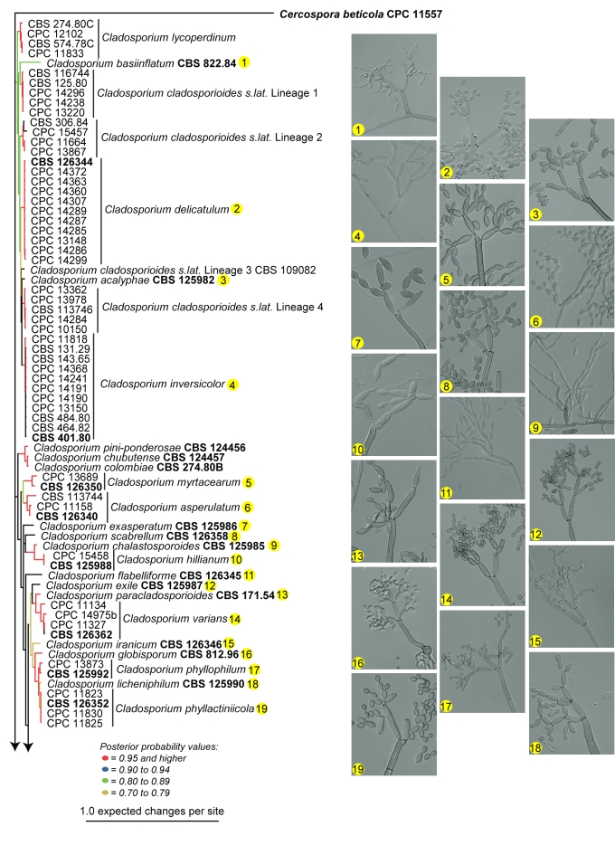
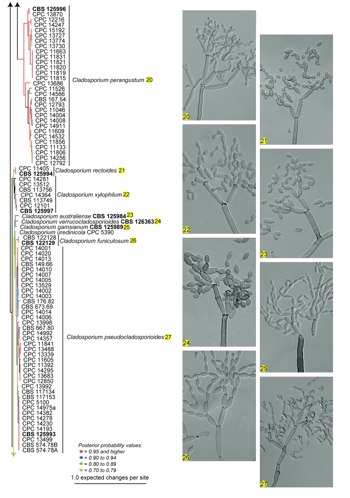
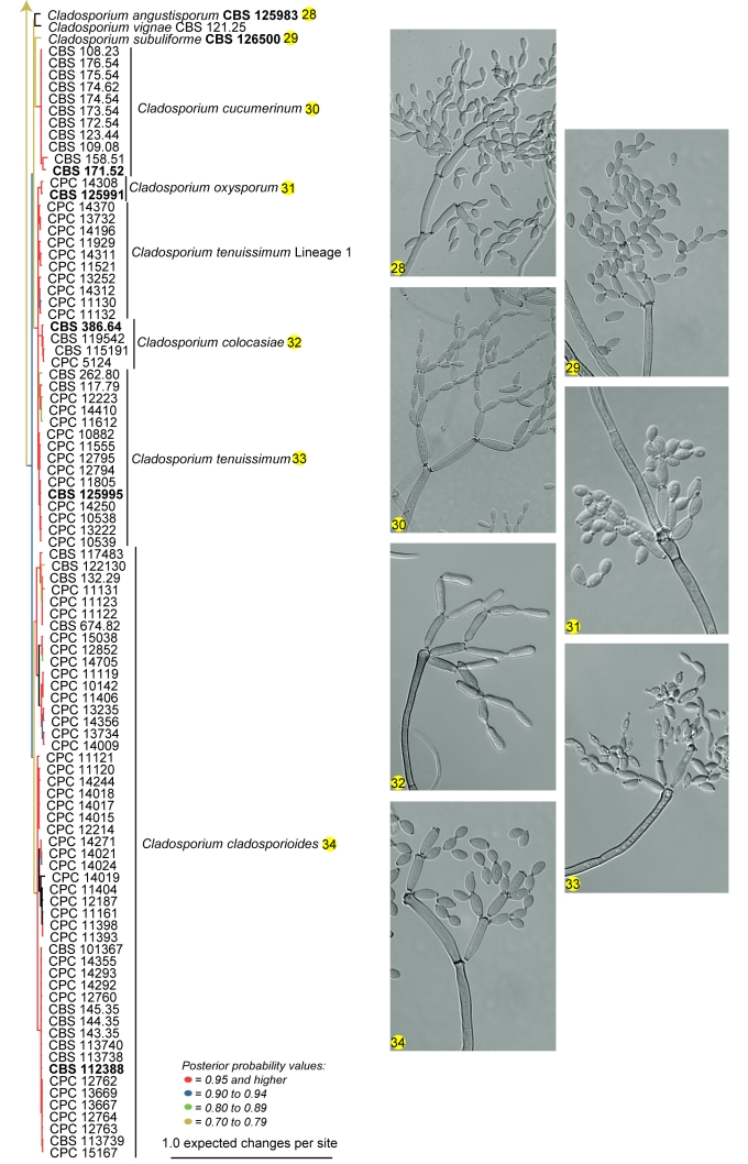
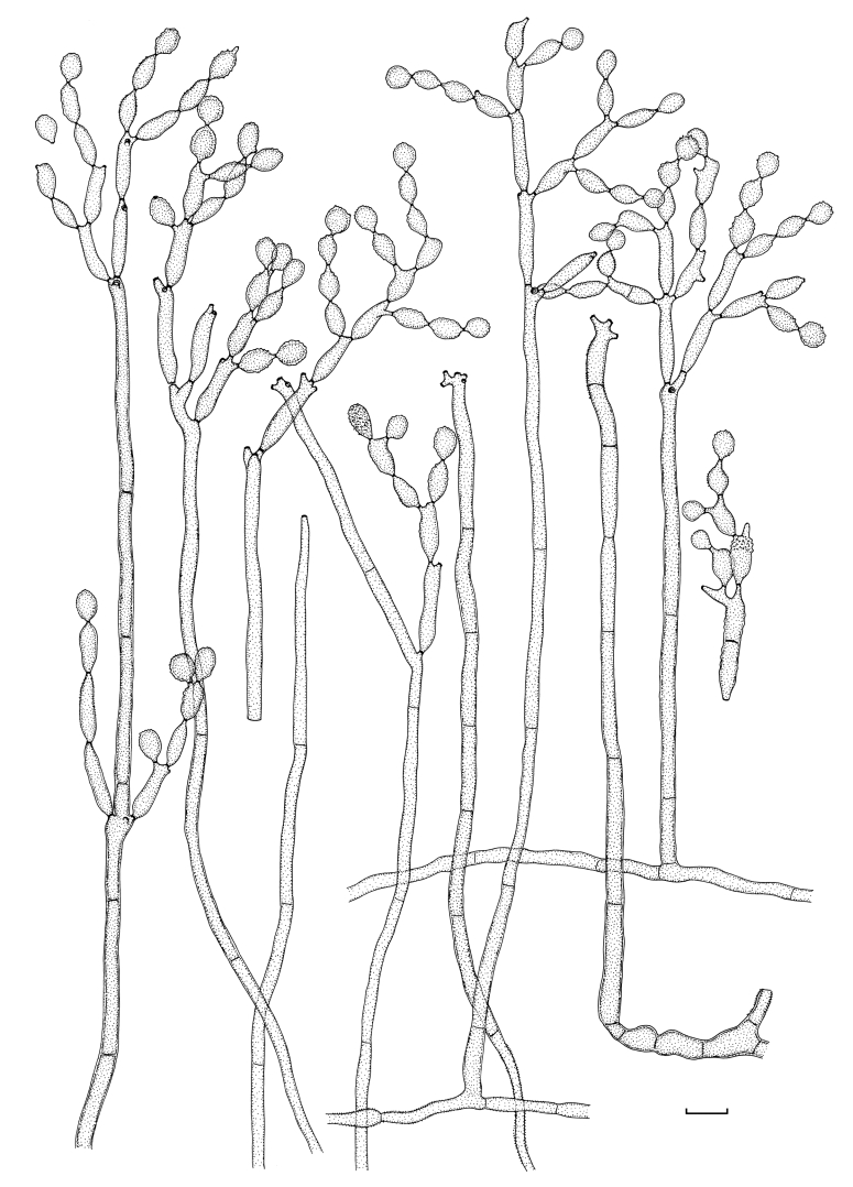
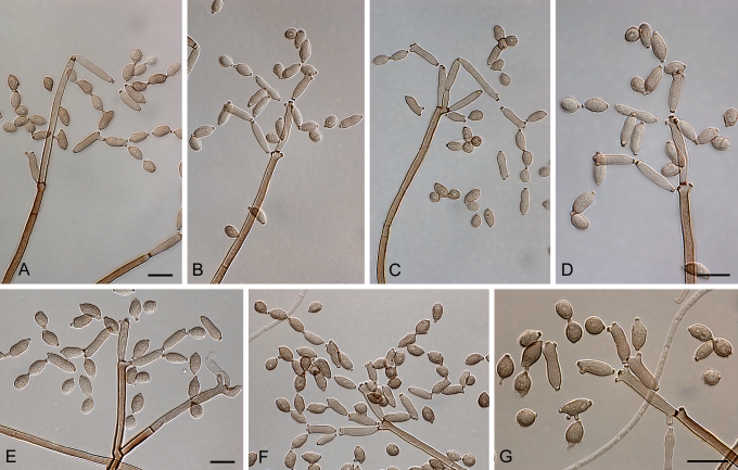
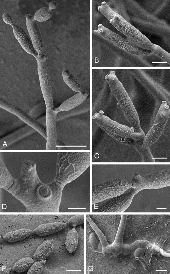
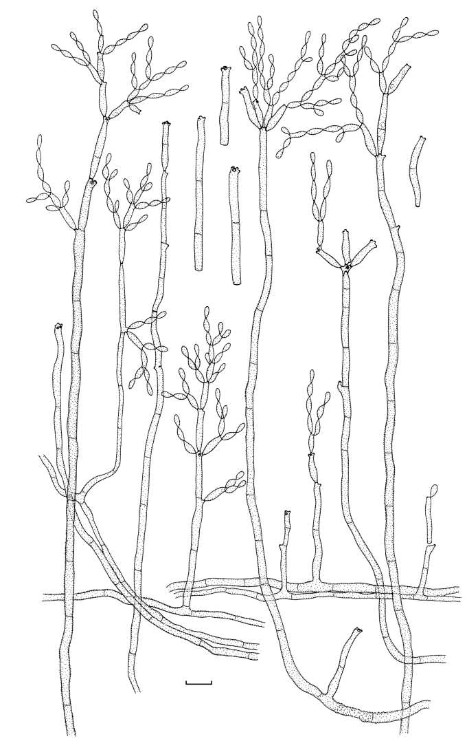
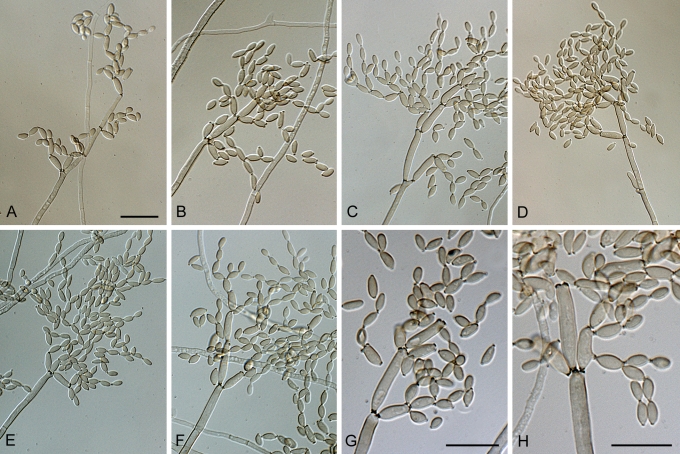
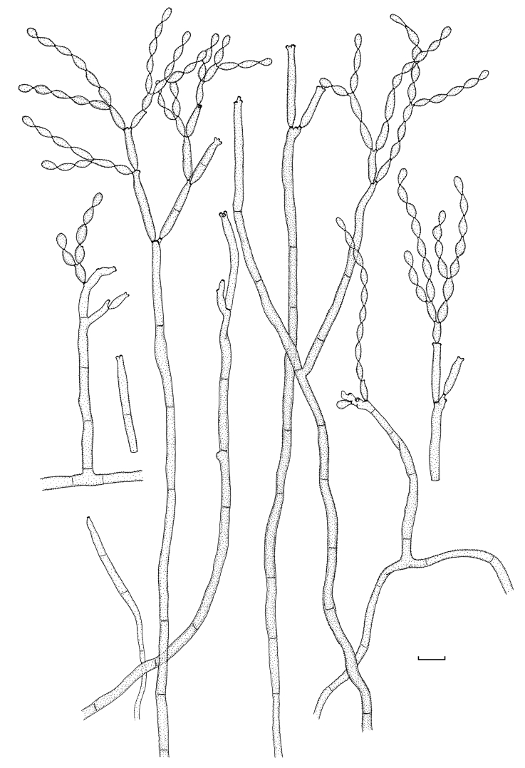
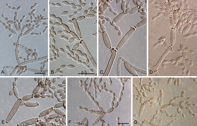
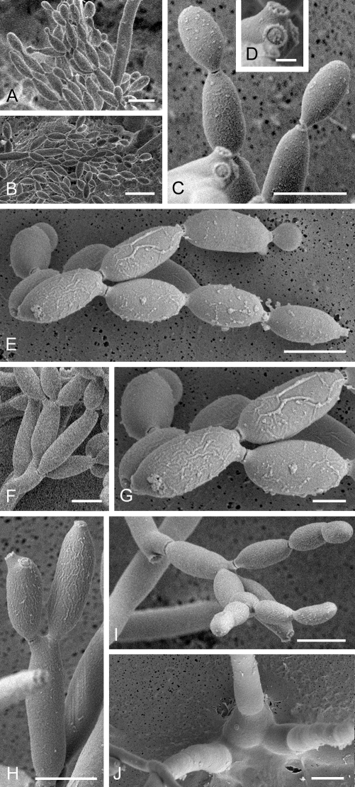
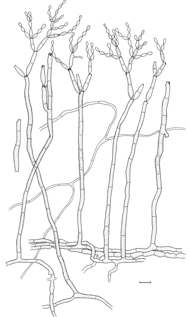
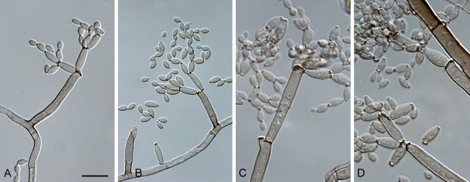
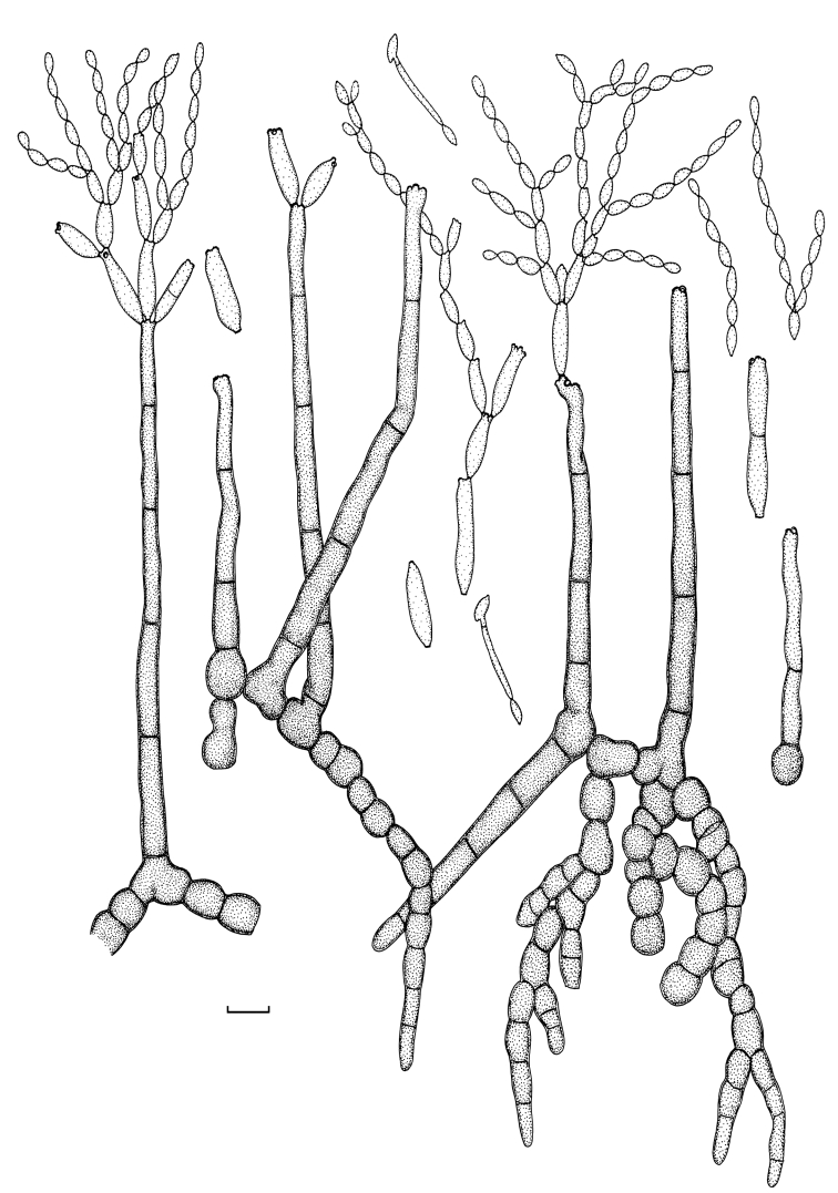
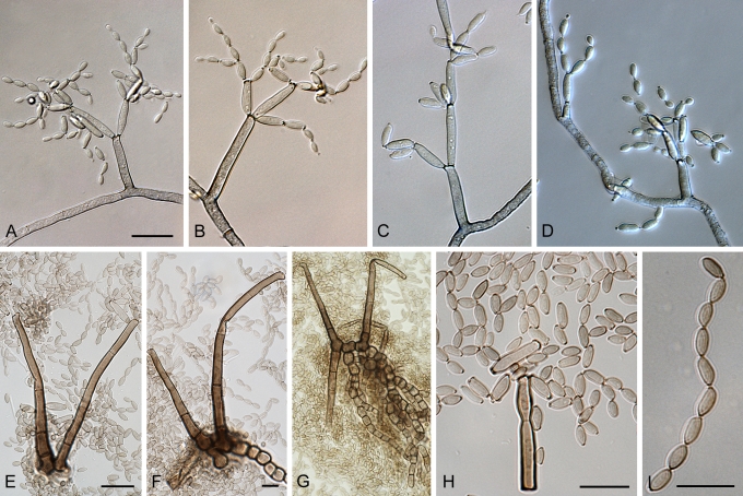
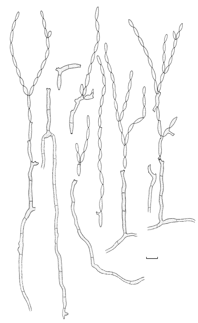
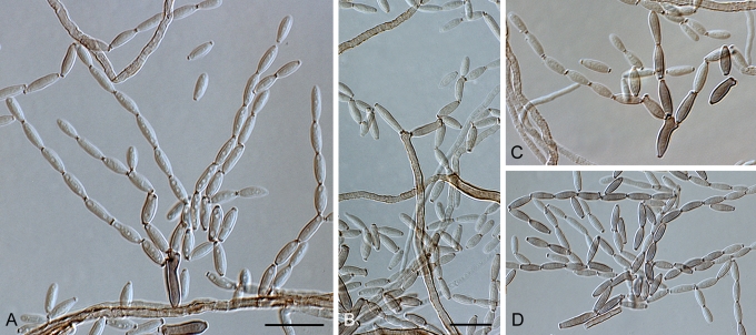
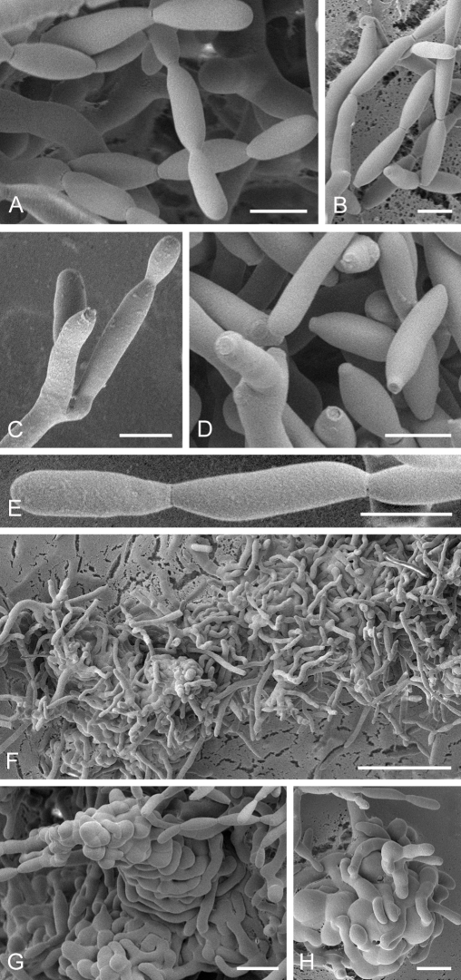
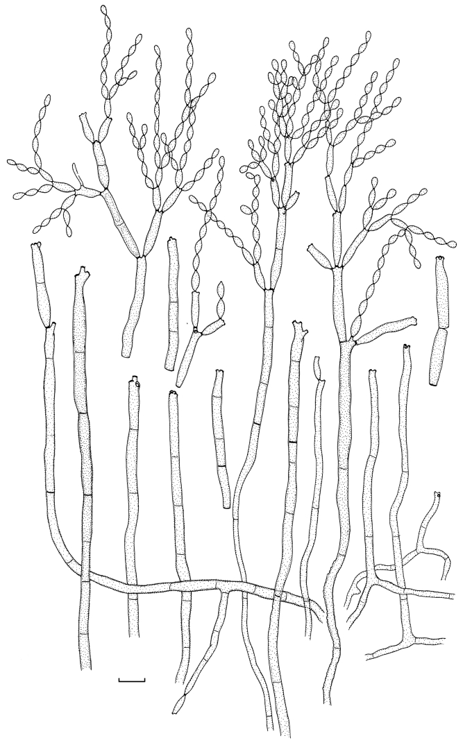
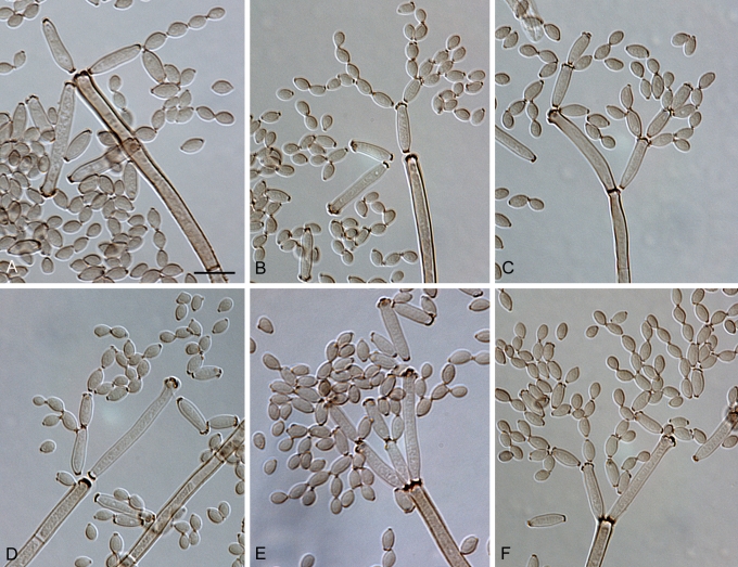
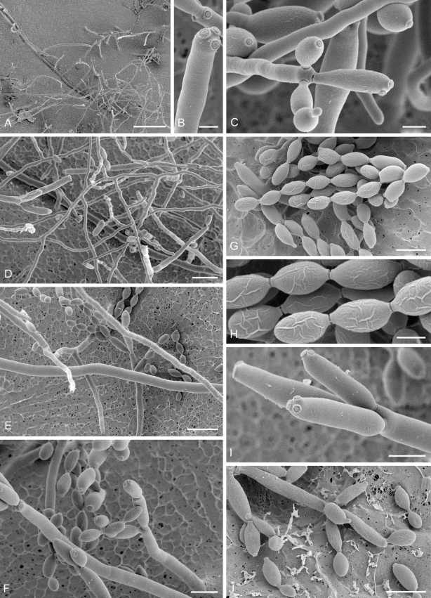
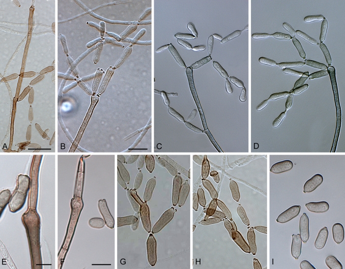
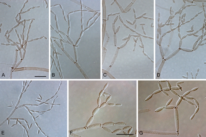
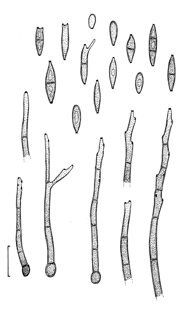
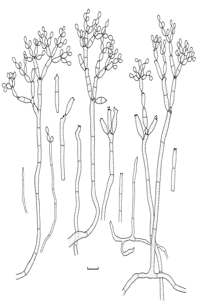
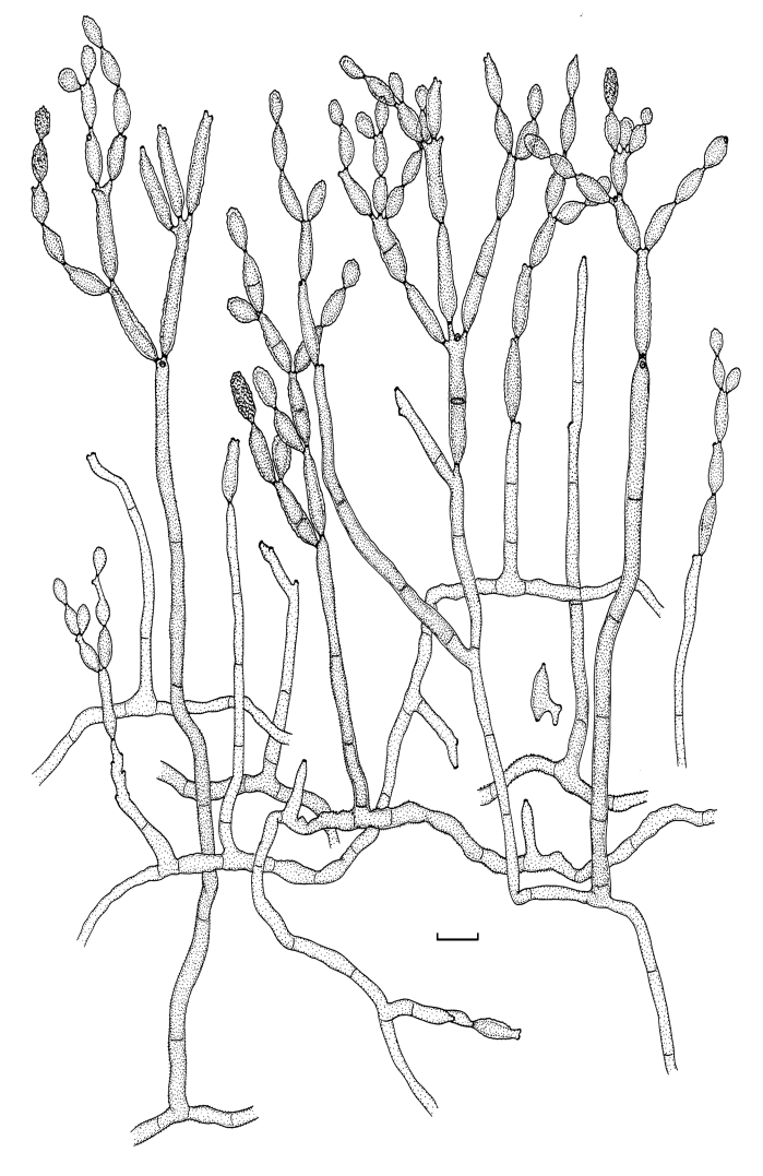
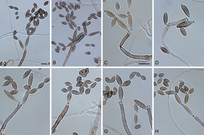
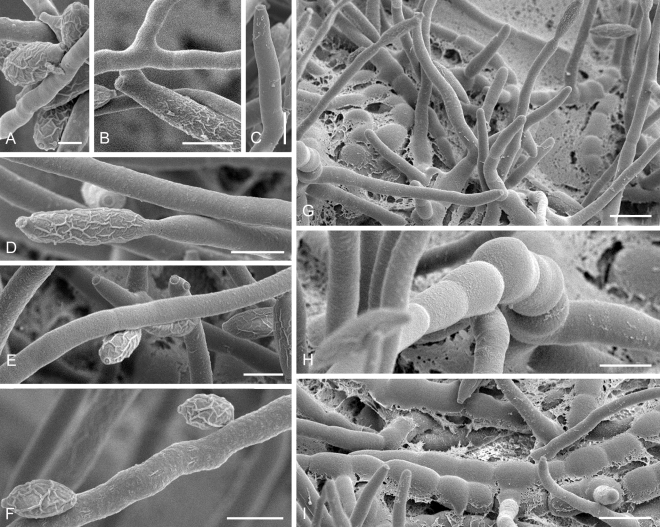
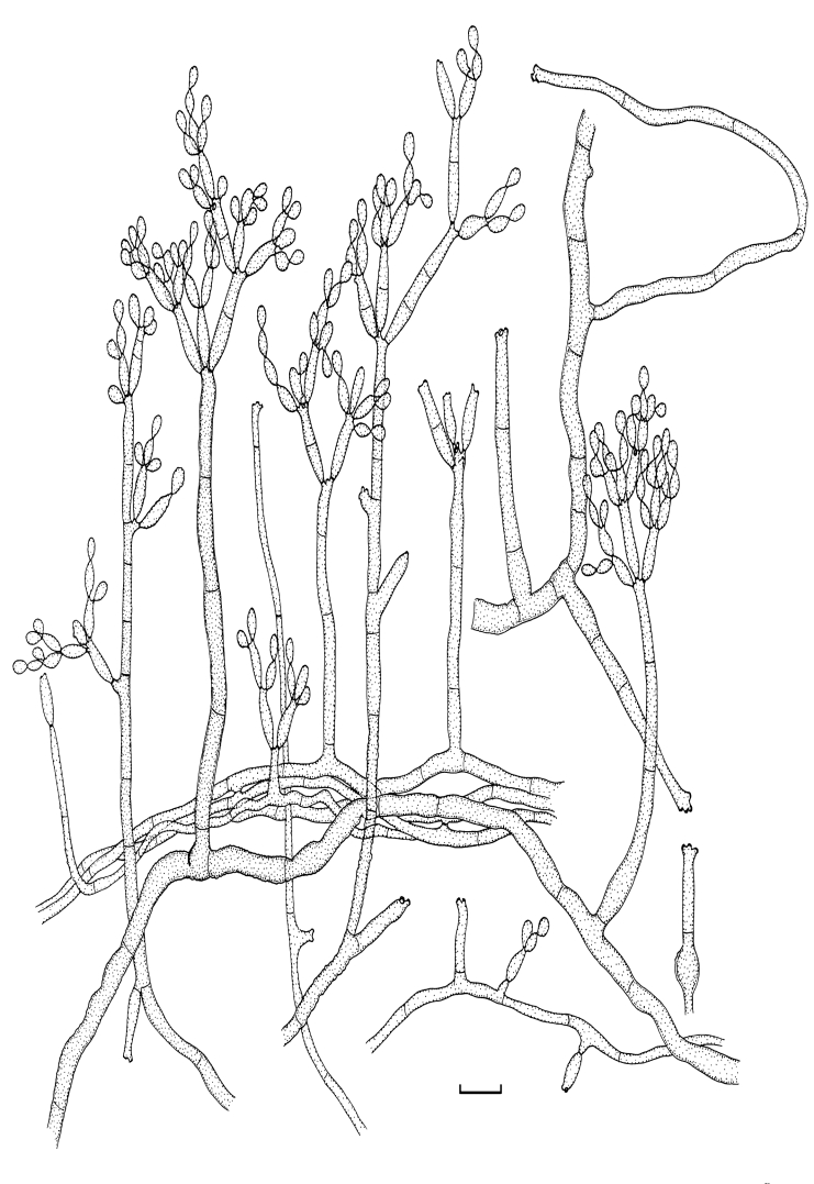
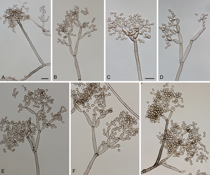
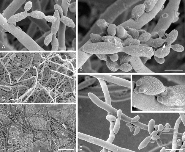
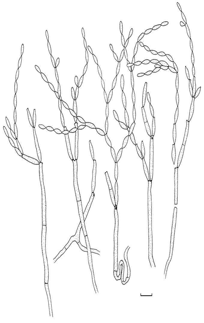
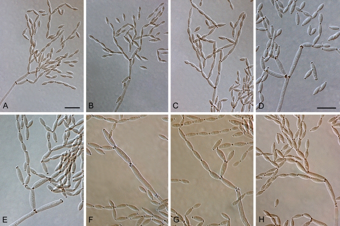
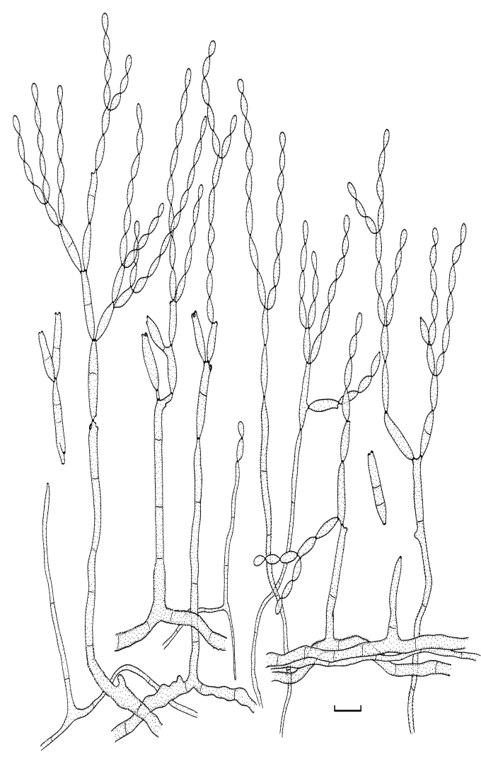
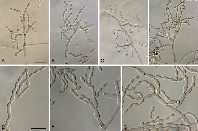
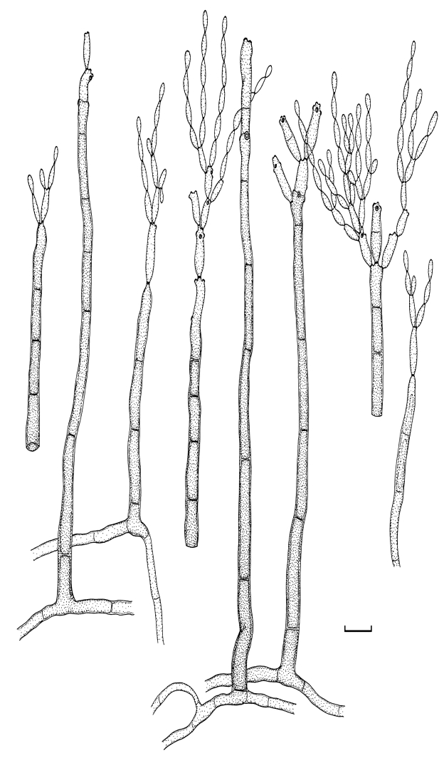
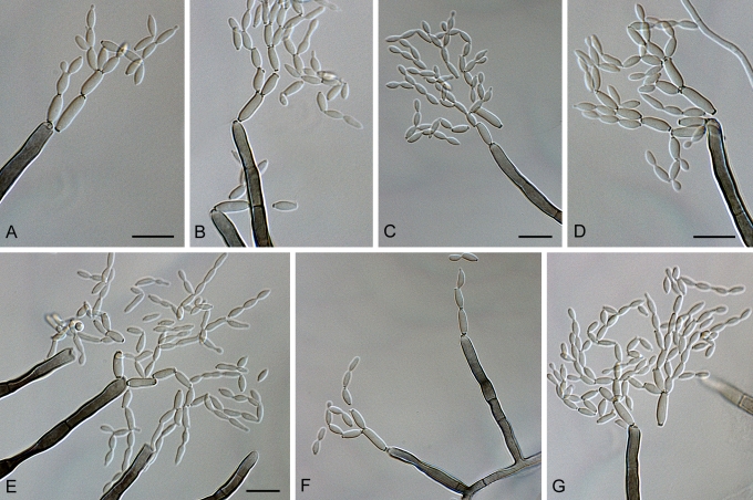
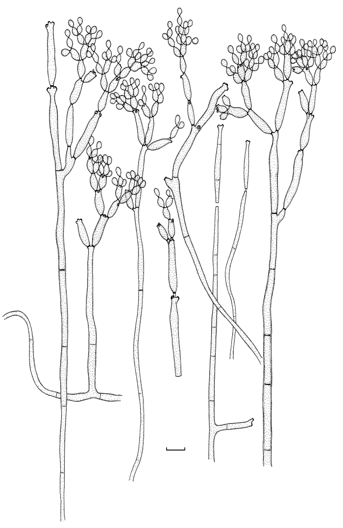
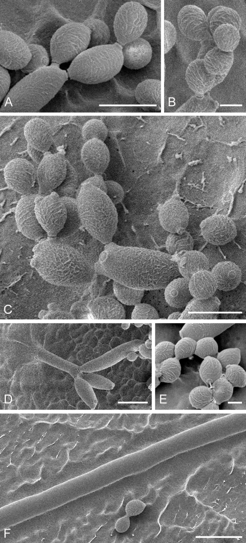
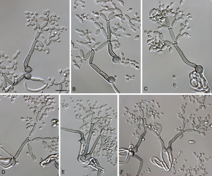
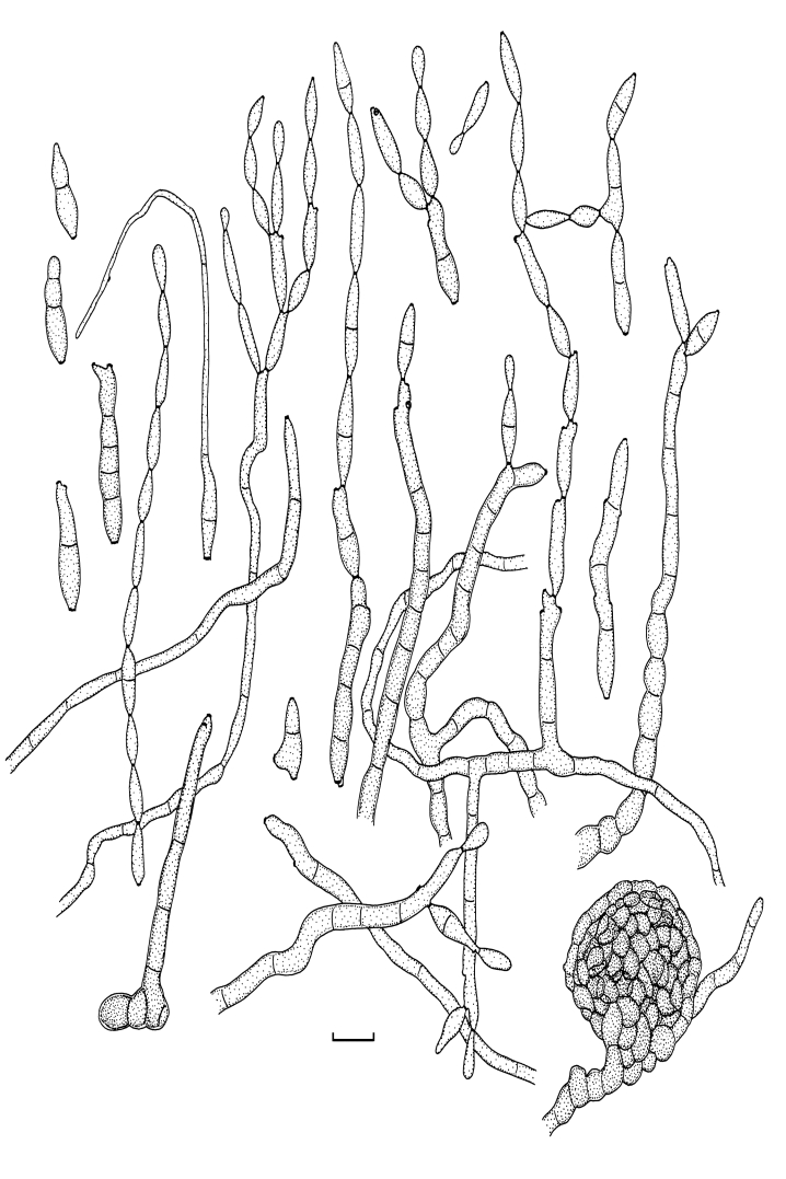
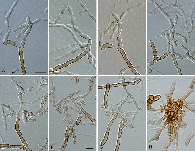
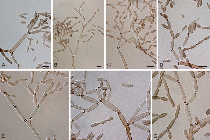
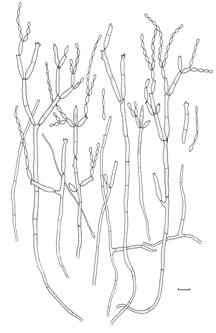
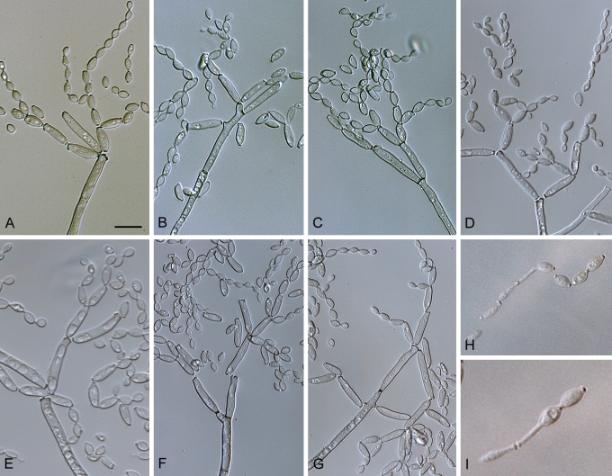
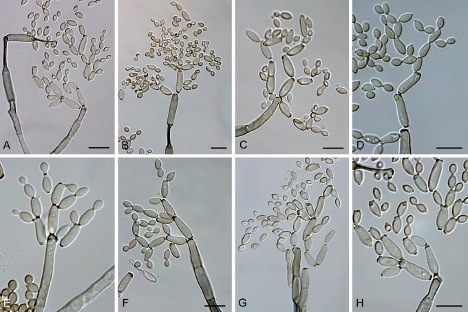
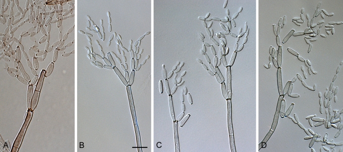
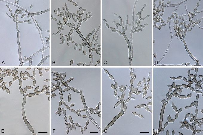
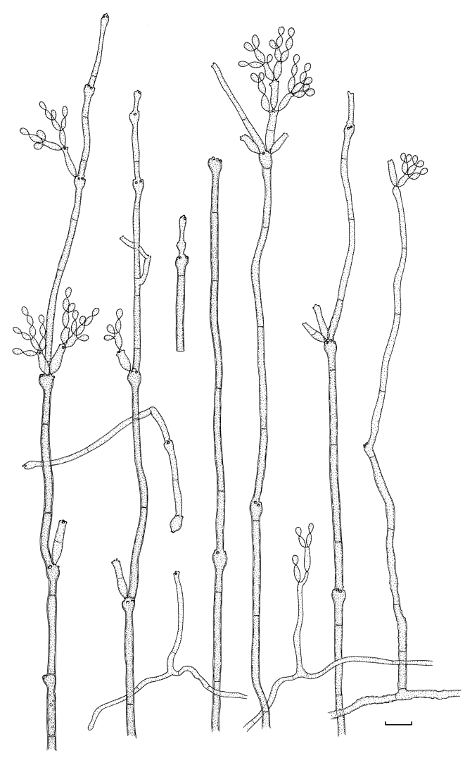
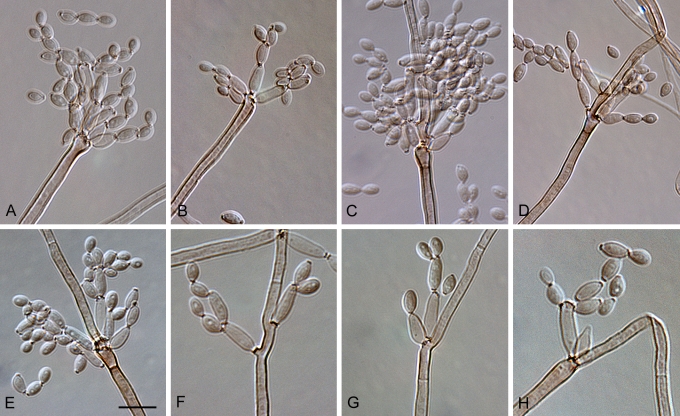
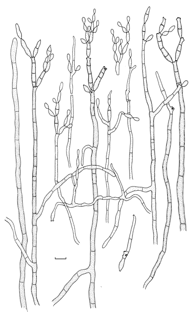
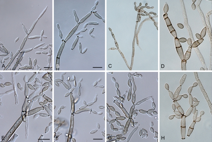
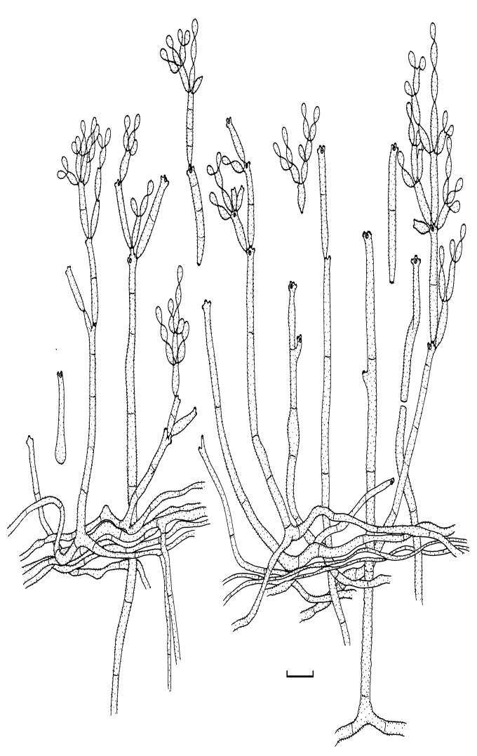
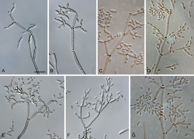
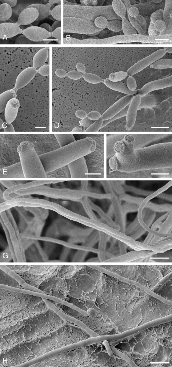
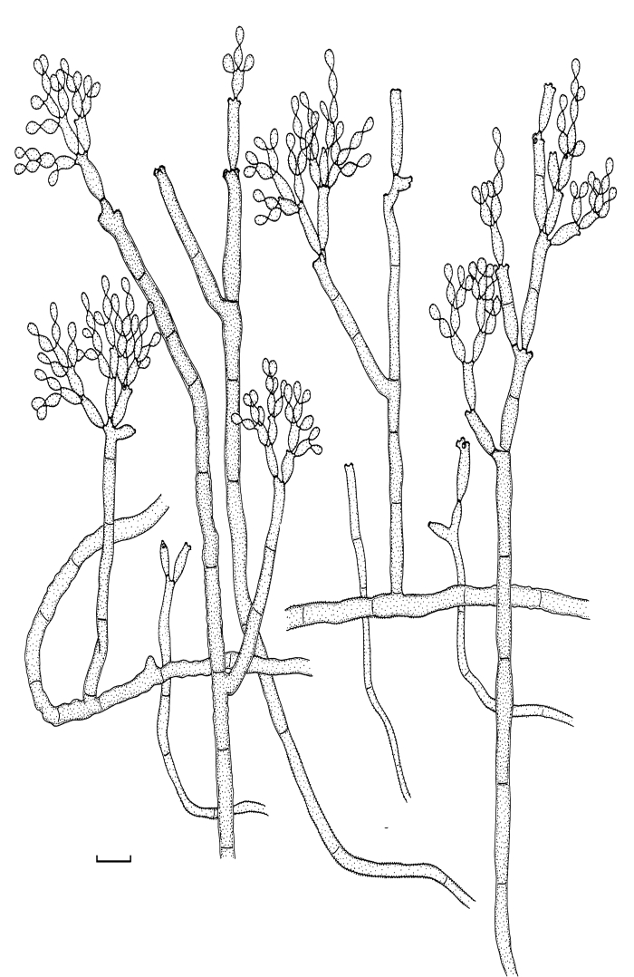
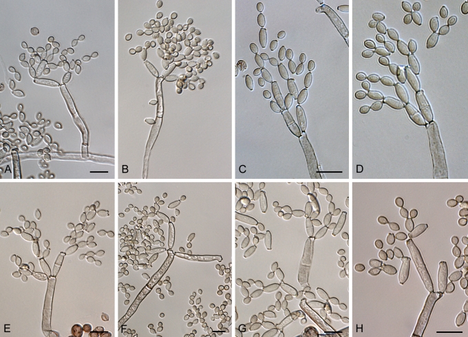
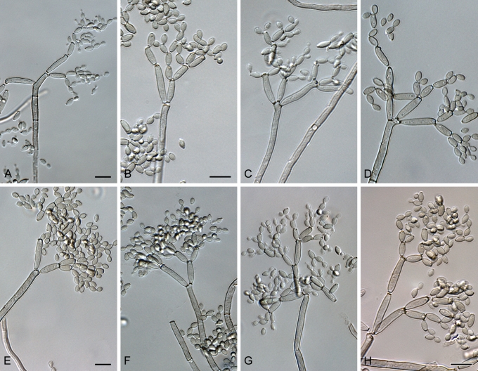
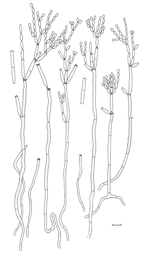
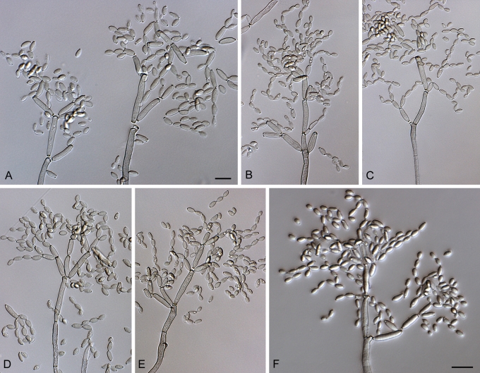
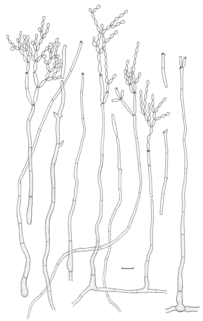
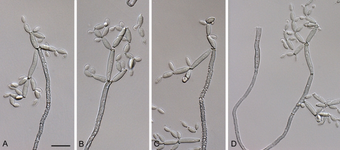
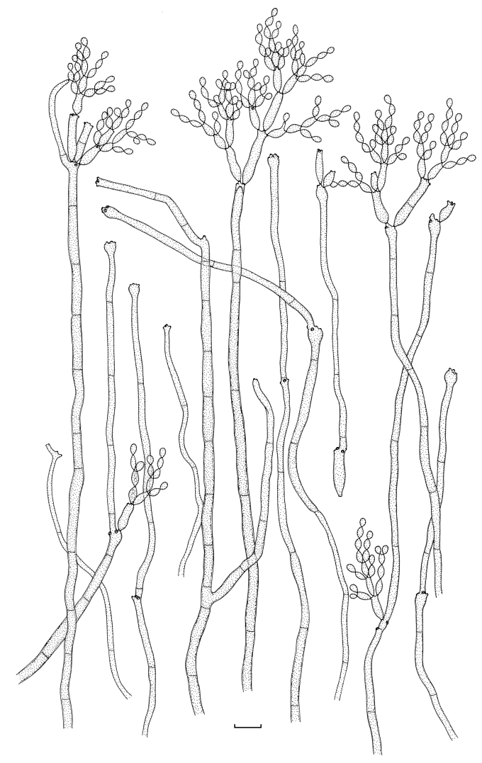
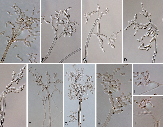
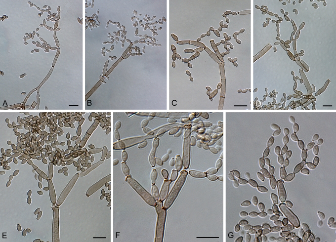
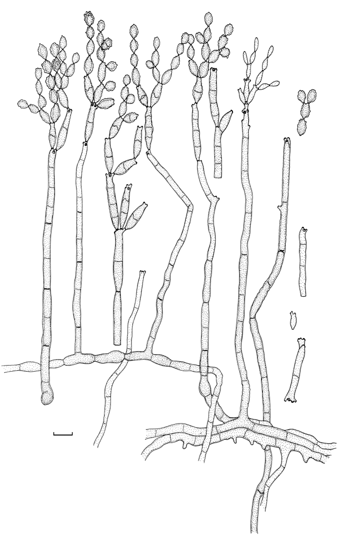
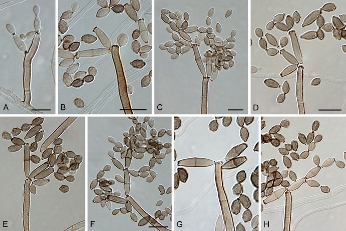
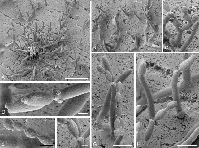
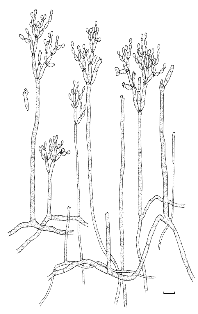
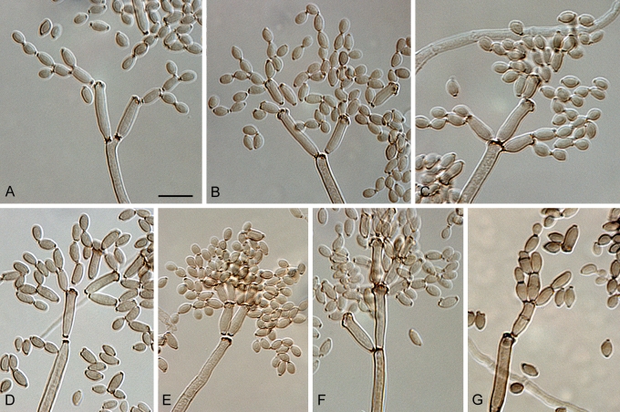
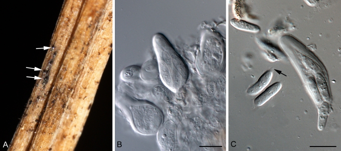
The genus Cladosporium is one of the largest genera of dematiaceous hyphomycetes, and is characterised by a coronate scar structure, conidia in acropetal chains and Davidiella teleomorphs. Based on morphology and DNA phylogeny, the species complexes of C. herbarum and C. sphaerospermum have been resolved, resulting in the elucidation of numerous new taxa. In the present study, more than 200 isolates belonging to the C. cladosporioides complex were examined and phylogenetically analysed on the basis of DNA sequences of the nuclear ribosomal RNA gene operon, including the internal transcribed spacer regions ITS1 and ITS2, the 5.8S nrDNA, as well as partial actin and translation elongation factor 1-α gene sequences. For the saprobic, widely distributed species Cladosporium cladosporioides, both a neotype and epitype are designated in order to specify a well established circumscription and concept of this species. Cladosporium tenuissimum and C. oxysporum, two saprobes abundant in the tropics, are epitypified and shown to be allied to, but distinct from C. cladosporioides. Twenty-two species are newly described on the basis of phylogenetic characters and cryptic morphological differences. The most important phenotypic characters for distinguishing species within the C. cladosporioides complex, which represents a monophyletic subclade within the genus, are shape, width, length, septation and surface ornamentation of conidia and conidiophores; length and branching patterns of conidial chains and hyphal shape, width and arrangement. Many of the treated species, e.g., C. acalyphae, C. angustisporum, C. australiense, C. basiinflatum, C. chalastosporoides, C. colocasiae, C. cucumerinum, C. exasperatum, C. exile, C. flabelliforme, C. gamsianum, and C. globisporum are currently known only from specific hosts, or have a restricted geographical distribution. A key to all species recognised within the C. cladosporioides complex is provided.

Keywords: Cladosporium oxysporum; Cladosporium tenuissimum; epitypification; new species; phylogeny; taxonomy.


Figures

Fig. 1.
Fig. 1.
(Parts a–c) Consensus phylogram of 26 002 trees resulting from a Bayesian analysis of 253 sequences in a combined ITS, ACT and TEF alignment. Bayesian posterior probabilities are colour-coded as indicated in the legend. Conidiophores are illustrated for all species treated in this study except for C. uredinicola and C. vignae which did not sporulate. The tree was rooted to sequences of Cercospora beticola strain CPC 11557 (GenBank accession numbers AY840527, AY840458, AY840494, respectively for ITS, ACT and TEF).
Fig. 1.
Fig. 1.
(Parts a–c) Consensus phylogram of 26 002 trees resulting from a Bayesian analysis of 253 sequences in a combined ITS, ACT and TEF alignment. Bayesian posterior probabilities are colour-coded as indicated in the legend. Conidiophores are illustrated for all species treated in this study except for C. uredinicola and C. vignae which did not sporulate. The tree was rooted to sequences of Cercospora beticola strain CPC 11557 (GenBank accession numbers AY840527, AY840458, AY840494, respectively for ITS, ACT and TEF).
Fig. 2.
Fig. 2.
Cladosporium acalyphae (CBS 125982). Macronematous conidiophores, mycelium, ramoconidia and conidial chains. Scale bar = 10 μm.
Fig. 3.
Fig. 3.
Cladosporium acalyphae (CBS 125982). A–G. Macronematous conidiophores and conidial chains. Scale bars = 10 μm.
Fig. 4.
Fig. 4.
Cladosporium acalyphae (CBS 125982). A. Secondary ramoconidia and conidia on a conidiophore. Note the smooth surface of the conidiophores. B–C. Secondary ramoconidia on smooth conidiophores and patterns of scars. D. Details of scars on a secondary ramoconidium. E. Secondary ramoconidia and scar. F. Conidia as seen with cryoSEM showing a reticulate surface ornamentation. G. SEM micrograph of “meristematic development” on the agar surface and two conidiophores formed. Scale bars = 2 (D, E), 5 (B, C, F), 10 (A, G) μm.
Fig. 5.
Fig. 5.
Cladosporium angustisporum (CBS 125983). Macro- and micronematous conidiophores, mycelium often forming ropes, ramoconidia and conidial chains. Scale bar = 10 μm.
Fig. 6.
Fig. 6.
Cladosporium angustisporum (CBS 125983). A–H. Macronematous conidiophores and conidial chains. Scale bars = 10 μm.
Fig. 7.
Fig. 7.
Cladosporium asperulatum (CBS 126340). Macronematous conidiophores, ramoconidia and conidial chains. Scale bar = 10 μm.
Fig. 8.
Fig. 8.
Cladosporium asperulatum (CBS 126340). A–D. Conidiophores and conidia. E–G. Secondary ramoconidia and conidia formed in branched chains. Scale bars = 10 μm.
Fig. 9.
Fig. 9.
Cladosporium asperulatum (CBS 126340). A–B. Conidiophores and very young conidia (A). C–D. Conidia and details of scars on a secondary ramoconidium. E. Conidia with sparse ornamentation. Note the round conidium-initial. F. Whorl of secondary ramoconidia formed at the tip of a conidiophore. G. Details of ornamentation showing loosely irregularly reticulate structures. H. Secondary ramoconidia. I. Overview of a conidiophore with scars on the tip of the conidiophore. J. Swollen cells at agar surface giving rise to conidiophores. Note the scar on the root structure. Scale bars = 1 (D), 2 (G), 5 (A, C, E–F, H–J), 10 (B) μm.
Fig. 10.
Fig. 10.
Cladosporium australiense (CBS 125984). Conidiophores, ramoconidia and conidial chains, mycelium sometimes forming ropes. Scale bar = 10 μm.
Fig. 11.
Fig. 11.
Cladosporium australiense (CBS 125984). A–B. Conidiophores and conidial chains. C. Conidiophore with a septate secondary ramoconidium still attached. D. Conidia. Scale bar = 10 μm.
Fig. 12.
Fig. 12.
Cladosporium basiinflatum (CBS 822.84). Mycelium, stromatic hyphal aggregations, conidiophores often with foot-like swollen base and conidial chains. Scale bar = 10 μm.
Fig. 13.
Fig. 13.
Cladosporium basiinflatum (CBS 822.84). A–D. Young conidiophores arising laterally from hyphae with conidial chains still attached. E–G. Older conidiophores formed in pairs arising from distinctly swollen hyphal cells or stromatic hyphal aggregations and numerous conidia. H. A single ramoconidium and numerous conidia. I. A conidial chain composed of intercalary and small terminal conidia. Scale bars = 10 μm.
Fig. 14.
Fig. 14.
Cladosporium chalastosporoides (CBS 125985). Conidiophores, conidia in long unbranched or dichotomously branched chains and microcyclic conidiogenesis. Scale bar = 10 μm.
Fig. 15.
Fig. 15.
Cladosporium chalastosporoides (CBS 125985). A–B. Conidiophores and conidia. C–D. Conidial chains. Scale bars = 10 μm.
Fig. 16.
Fig. 16.
Cladosporium chalastosporoides (CBS 125985). A–B. Examples of elongated conidia and chains. C. Branch formation on conidiophores. D. Branched conidiophore, ramoconidia and conidia. E. Elongated conidia. Note the broad areas of connection between the spores. F. Overview of a large cell mass that gives rise to conidiophores and spores. G. Detail of a cell mass showing meristematic growth. H. Differentiation of fungal cells into a cell mass. Scale bars = 5 (A–E), 10 (G–H), 50 (F) μm.
Fig. 17.
Fig. 17.
Cladosporium cladosporioides (CBS 112388). Macro- and micronematous conidiophores, ramoconidia and conidial chains. Scale bar = 10 μm.
Fig. 18.
Fig. 18.
Cladosporium cladosporioides (CBS 112388). A–F. Macronematous conidiophores and conidial chains. Scale bar = 10 μm.
Fig. 19.
Fig. 19.
Cladosporium cladosporioides (CBS 112388). A. Overview of a colony containing running differentiated conidiophore-forming hyphae and many aerial hyphae. B. Scars on a conidiophore. C. Top view of a conidiophore with scars and an aerial structure. D. Branching patterns of aerial hyphae intermingled with spore-forming structures. E. Aerial hyphae, conidiophores and spores. F. Conidiophores sprouting from agar with all types of dispersion structures. G. Conidial chains. H. Detail of conidial chains and ornamentation showing irregularly reticulate structures or embossed stripes probably caused by diminishing turgor and shriveling of tender young conidia. I. Three secondary ramoconidia. J. Secondary ramoconidia J. Secondary ramoconidia and conidia on agar with some irregularly reticulate ornamentation. Scale bars = 2 (B–C, H), 5 (F–G, I–J), 10 (D–E), 50 (A) μm.
Fig. 20.
Fig. 20.
Cladosporium colocasiae (A-D, G-H: CBS 386.64 and E-F, I: CBS 119542). A–D. Conidiophores and conidial chains. E–F. Intercalary conidiogenous cells with typical nodes and conidiogenous loci restricted to these swellings. G–H. Microcyclic conidiogenesis with conidia forming secondary conidiophores. I. Conidia. Scale bars = 10 μm.
Fig. 21.
Fig. 21.
Cladosporium cucumerinum (CBS 171.52). A–G. Conidiophores and conidial chains. Scale bars = 10 μm.
Fig. 22.
Fig. 22.
Cladosporium delicatulum (K 121551, holotype). Conidiophores and conidia in vivo. Scale bar = 10 μm. U. Braun del.
Fig. 23.
Fig. 23.
Cladosporium delicatulum (CBS H-20430, reference material). Conidiophores and conidia in vivo. Scale bar = 10 μm.
Fig. 24.
Fig. 24.
Cladosporium delicatulum (CBS 126344). Macro- and micronematous conidiophores and conidial chains in vitro. Scale bar = 10 μm.
Fig. 25.
Fig. 25.
Cladosporium delicatulum (CBS 126344). A–G. Macronematous conidiophores and conidial chains. Scale bar = 10 μm.
Fig. 26.
Fig. 26.
Cladosporium exasperatum (CBS 125986). Macro- and micronematous conidiophores, mycelium and conidial chains. Scale bar = 10 μm.
Fig. 27.
Fig. 27.
Cladosporium exasperatum (CBS 125986). A–C, E–H. Macronematous conidiophores and conidial chains. D. Ramoconidium seceding at the tip of a conidiophore. Scale bars = 10 μm.
Fig. 28.
Fig. 28.
Cladosporium exasperatum (CBS 125986). A. Ornamented conidia on aerial structures. Note the small scars with an ornamentation free zone. B. Secondary ramoconidium with a scar and aerial structures. C. Scars on a conidiophore. Note the smooth surface of the conidiophores. D. Ornamented secondary ramoconidium on a conidiophore with visible line of delineation. E. Septate aerial structure with conidiophores, conidia and scars as background. F. Two ornamented globose conidia adhered to an aerial structure showing an irregularly reticulate surface. G. Segmented differentiated hyphae on the agar surface giving rise to numerous conidiophores, conidiophore initials and aerial structures. H. Detail of differentiated substrate hyphae. I. Segmented differentiated hyphae in initials. Scale bars = 2 (A), 5 (B–F, H), 10 (G, I) μm.
Fig. 29.
Fig. 29.
Cladosporium exile (CBS 125987). Macro- and micronematous conidiophores, mycelium sometimes forming ropes and conidial chains. Scale bar = 10 μm.
Fig. 30.
Fig. 30.
Cladosporium exile (CBS 125987). A–G. Macronematous conidiophores and conidial chains. Scale bars = 10 μm.
Fig. 31.
Fig. 31.
Cladosporium exile (CBS 125987). A. Conidia and secondary ramoconidia with reticulate ornamentation, near long aerial hyphae or conidiophores. B. CryoSEM of different types of conidia on aerial structures. Note a remarkable pattern of blastoconidium formation (backwards) (arrow). C. Numerous hyphae and conidiophores in this overview of a colony of the fungus. D. Hyphae on the agar surface. E. Conidia and aerial structures. F. Detail of conidia and scars. Scale bars = 2 (F), 5 (A–B), 10 (E), 50 (C–D) μm.
Fig. 32.
Fig. 32.
Cladosporium flabelliforme (CBS 126345). Conidiophores and conidia in long often dichotomously branched chains. Scale bar = 10 μm.
Fig. 33.
Fig. 33.
Cladosporium flabelliforme (CBS 126345). A–H. Conidiophores and conidial chains. Scale bars = 10 μm.
Fig. 34.
Fig. 34.
Cladosporium funiculosum (CBS 122129). Macro- and micronematous conidiophores, mycelium sometimes formed in ropes and conidial chains. Scale bar = 10 μm.
Fig. 35.
Fig. 35.
Cladosporium funiculosum (CBS 122129). A–E, G. Macronematous conidiophores and conidial chains. F. Micronematous conidiophore with conidial chains. Scale bars = 10 μm.
Fig. 36.
Fig. 36.
Cladosporium gamsianum (CBS 125989). Conidiophores and conidial chains. Scale bar = 10 μm.
Fig. 37.
Fig. 37.
Cladosporium gamsianum (CBS 125989). A–G. Conidiophores and conidial chains. Scale bars = 10 μm.
Fig. 38.
Fig. 38.
Cladosporium globisporum (CBS 812.96). Macro- and micronematous conidiophores, ramoconidia and conidial chains. Scale bar = 10 μm.
Fig. 39.
Fig. 39.
Cladosporium globisporum (CBS 812.96). A–B. Delicately ornamented conidia showing a somewhat irregularly reticulate surface or slightly embossed stripes probably caused by diminishing turgor and shriveling of tender young conidia. C. Conidia, secondary ramoconidia and scars. D. Conidiophore with secondary ramoconidia. E. Globose ornamented small terminal conidia. F. Running hyphae on agar and conidia. Scale bars = 2 (B, E), 5 (A, C), 10 (D, F) μm.
Fig. 40.
Fig. 40.
Cladosporium globisporum (CBS 812.96). A–F. Macronematous conidiophores and conidial chains. Scale bar = 10 μm.
Fig. 41.
Fig. 41.
Cladosporium hillianum (CBS 125988). Mycelium, hyphal conglomeration, conidiophores and conidia. Scale bar = 10 μm.
Fig. 42.
Fig. 42.
Cladosporium hillianum (CBS 125988). A–G. Conidiophores and conidia. H. Hyphal conglomeration. Scale bars = 10 μm.
Fig. 43.
Fig. 43.
Cladosporium inversicolor (CBS 401.80). Conidiophores and conidial chains with intercalary conidia and small terminal conidia sometimes verruculose or irregularly rough-walled, rugose. Scale bar = 10 μm.
Fig. 44.
Fig. 44.
Cladosporium inversicolor (CBS 401.80). A–G. Conidiophores and conidial chains with intercalary conidia and small terminal conidia somewhat darker than ramoconidia and secondary ramoconidia. Scale bars = 10 μm.
Fig. 45.
Fig. 45.
Cladosporium iranicum (CBS 126346). Macro- and micronematous conidiophores, ramoconidia and conidial chains, intercalary conidia sometimes subrostrate. Scale bar = 10 μm.
Fig. 46.
Fig. 46.
Cladosporium iranicum (CBS 126346). A–D, F–G. Macronematous conidiophores and conidial chains. E. Conidia and conidiophore showing the often guttulate cell structure. H–I. Subrostrate intercalary conidia. Scale bar = 10 μm.
Fig. 47.
Fig. 47.
Cladosporium licheniphilum (CBS 125990). A–H. Macronematous conidiophores and conidial chains. Scale bars = 10 μm.
Fig. 48.
Fig. 48.
Cladosporium lycoperdinum (CBS 574.78C). A–D. Macronematous conidiophores and conidial chains. Scale bar = 10 μm.
Fig. 49.
Fig. 49.
Cladosporium myrtacearum (CBS 126350). A–H. Macronematous conidiophores and conidial chains. Scale bars = 10 μm.
Fig. 50.
Fig. 50.
Cladosporium oxysporum (CBS 125991). Macro- and micronematous conidiophores, conidia and microcyclic conidiogenesis with conidia forming secondary conidiophores. Scale bar = 10 μm.
Fig. 51.
Fig. 51.
Cladosporium oxysporum (CBS 125991). A–C. Tips of macronematous conidiophores, being typically nodulose in A and C. D–H. Intercalary conidiogenous cells and conidia. Scale bar = 10 μm.
Fig. 52.
Fig. 52.
Cladosporium paracladosporioides (CBS 171.54). Dimorphic mycelium, macro- and micronematous conidiophores, ramoconidia, conidia and microcyclic conidiogenesis. Scale bar = 10 μm.
Fig. 53.
Fig. 53.
Cladosporium paracladosporioides (CBS 171.54). A–C, E–F. Macronematous conidiophores and conidial chains. D, H. Conidial chains, septa of secondary ramoconidia distinctly darkened. G. Microcyclic conidiogenesis. Scale bars = 10 μm.
Fig. 54.
Fig. 54.
Cladosporium perangustum (CBS 125996). Macro- and micronematous conidiophores, mycelium often formed in dense ropes, ramoconidia and conidial chains. Scale bar = 10 μm.
Fig. 55.
Fig. 55.
Cladosporium perangustum (CBS 125996). A–G. Macronematous conidiophores and conidial chains. Scale bar = 10 μm.
Fig. 56.
Fig. 56.
Cladosporium perangustum (CBS 125996). A. Conidia with very gentle surface ornamentation showing irregularly reticulate structures. B. A coherent view on conidiophores, stipes, aerial hyphae and conidia. C. Secondary ramoconidia, conidia and scars. The conidia at the upper right show some cell wall structures. D. Conidiophore with secondary ramoconidia, intercalary and small terminal conidia. Note the disruptions of the cell walls between the conidia. E. Scars on very elongated secondary ramoconidia. F. Scar-pattern at the end of the conidiophores. Note the flattened separation domes. G. Ropes of aerial hyphae. H. Running segmented hyphae that may form conidiophores and not segmented aerial hyphae. Note the blastoconidium on one hypha. Scale bars = 2 (A, C, E–F), 5 (D, G), 10 (B, H) μm.
Fig. 57.
Fig. 57.
Cladosporium phyllactiniicola (CBS 126352). Macro- and micronematous conidiophores, mycelium and conidial chains. Scale bar = 10 μm.
Fig. 58.
Fig. 58.
Cladosporium phyllactiniicola (CBS 126352). A–H. Macronematous conidiophores and conidial chains. Scale bars = 10 μm.
Fig. 59.
Fig. 59.
Cladosporium phyllophilum (CBS 125992). A–H. Macronematous conidiophores and conidial chains. Scale bars = 10 μm.
Fig. 60.
Fig. 60.
Cladosporium pseudocladosporioides (CBS 125993). Macro- and micronematous conidiophores, ramoconidia and conidial chains. Scale bar = 10 μm.
Fig. 61.
Fig. 61.
Cladosporium pseudocladosporioides (CBS 125993). A–F. Macronematous conidiophores and conidial chains. Scale bars = 10 μm.
Fig. 62.
Fig. 62.
Cladosporium rectoides (CBS 125994). Conidiophores, ramoconidia and conidial chains. Scale bar = 10 μm.
Fig. 63.
Fig. 63.
Cladosporium rectoides (CBS 125994). A–G. Macronematous conidiophores and conidial chains; small terminal conidia sometimes with surface ornamentation indicated by the arrows in B. Scale bar = 10 μm.
Fig. 64.
Fig. 64.
Cladosporium scabrellum (CBS 126358). Macro- and micronematous conidiophores, ramoconidia and conidial chains. Scale bar = 10 μm.
Fig. 65.
Fig. 65.
Cladosporium scabrellum (CBS 126358). A–D (after 3 days). Macronematous conidiophores arising solitary or in small loose groups from hyphae or swollen hyphal cells and short conidial chains. E–H (after 7 days). Macronematous conidiophores and conidial chains. Scale bar = 10 μm.
Fig. 66.
Fig. 66.
Cladosporium scabrellum (CBS 126358). A. Globose conidia with nearly smooth surface. B. Overview of conidial chains. C. Elongated conidiophores. D. Secondary ramoconidium with large scars. E. Whorls of secondary ramoconidia and conidia. Note the presence of four scars on the top ramoconidium and the reticulate ornamentation of one of the conidia. F. Secondary ramoconidia of which one elongated. G. Conidiophore with secondary ramoconidia and large scar. Scale bars = 2 (A), 5 (D–G), 10 (B), 50(C) μm.
Fig. 67.
Fig. 67.
Cladosporium subuliforme (CBS 126500). Subulate conidiophores, ramoconidia and conidial chains. Scale bar = 10 μm.
Fig. 68.
Fig. 68.
Cladosporium subuliforme (CBS 126500). A–C. Tips of conidiophores with conidial chains. D. Subulate conidiophore with terminal and intercalary conidiogenous cell and conidia. Scale bar = 10 μm.
Fig. 69.
Fig. 69.
Cladosporium tenuissimum (CBS 125995). Macro- and micronematous conidiophores usually with a head-like swollen apex and sometimes additional intercalary nodules, conidial chains and microcyclic conidiogenesis. Scale bar = 10 μm.
Fig. 70.
Fig. 70.
Cladosporium tenuissimum (CBS 125995). A–H. Macronematous conidiophores and conidial chains. I–J. Micronematous conidiophores with conidia. Scale bars = 10 μm.
Fig. 71.
Fig. 71.
Cladosporium varians (CBS 126362). A–F. Macronematous conidiophores and conidial chains. G. Conidia. Scale bars = 10 μm.
Fig. 72.
Fig. 72.
Cladosporium verrucocladosporioides (CBS 126363). Macro- and micronematous conidiophores, mycelium sometimes formed in ropes, ramoconidia and conidial chains. Scale bar = 10 μm.
Fig. 73.
Fig. 73.
Cladosporium verrucocladosporioides (CBS 126363). A–H. Macronematous conidiophores and conidial chains. Scale bars = 10 μm.
Fig. 74.
Fig. 74.
Cladosporium verrucocladosporioides (CBS 126363). A. CryoSEM of a small colony illustrating the dense middle part with many young conidiophores and some disconnected masses of conidia. B. Stout erect conidiophores sprouting from rounded linearly oriented cells. C, G–H. Details of the fungal colony containing the structures on which conidiophores are formed and the different types of conidia. Note the ornamented conidia and the more or less smooth or only slightly ornamented conidiophores. D. Conidiophore, secondary ramoconidia and scars. E. Rounded conidia in a chain showing the reticulate surface ornamentation. F. Secondary ramoconidium and scars. Note the reduced ornamentation on this cell in comparison with the conidia. Scale bars = 2 (D, F), 5 (C, E), 10 (G–H), 20 (B), 50 (A) μm.
Fig. 75.
Fig. 75.
Cladosporium xylophilum (CBS 125997). Macro- and micronematous conidiophores, mycelium sometimes formed in ropes and conidial chains. Scale bar = 10 μm.
Fig. 76.
Fig. 76.
Cladosporium xylophilum (CBS 125997). A–G. Macronematous conidiophores and conidial chains. Scale bar = 10 μm.
Fig. 77.
Fig. 77.
Davidiella state (CBS H-19874) of Cladosporium cladosporioides-like strain CBS 109082. A. Ascomata on stems of Silene maritima. B–C. Asci and ascospores (arrow denotes mucoid layer). Scale bars = 10 μm.

References

    1. Anilkumar TB, Seshadri VS (1975). Cladosporium leaf spot of sunflower. Current Science 44(19): 722.
    1. Aptroot A (2006). Mycosphaerella and its anamorphs: 2. Conspectus of Mycosphaerella. CBS Biodiversity Series 5: 1–231.
    1. Arx JA von (1987). Plant pathogenic fungi. Beihefte zur Nova Hedwigia 87: 1–288.
    1. Arya C, Arya A (2003). New leaf spot diseases of social forestry trees – II. Journal of Mycology and Plant Pathology 33(2): 320–322.
    1. Arzanlou A, Groenewald JZ, Gams W, Braun U, Shin H-D, Crous PW (2007). Phylogenetic and morphotaxonomic revision of Ramichloridium and allied genera. Studies in Mycology 58: 57–93. - PMC - PubMed

LinkOut - more resources