. 2010 Sep 20;5(9):e12829.
doi: 10.1371/journal.pone.0012829.

NOF1 encodes an Arabidopsis protein involved in the control of rRNA expression

Affiliations

NOF1 encodes an Arabidopsis protein involved in the control of rRNA expression

Erwana Harscoët et al. PLoS One. .

Abstract

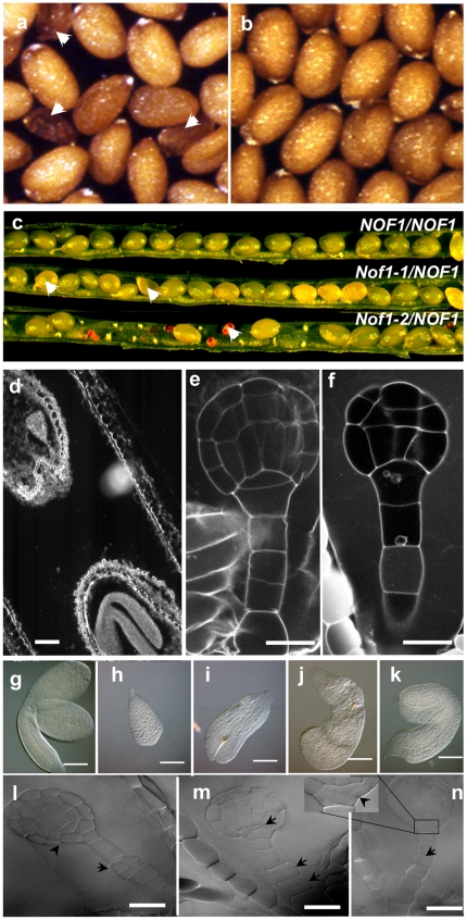
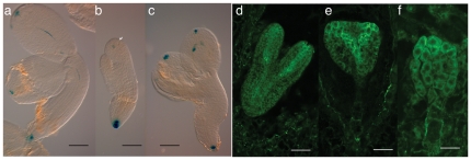
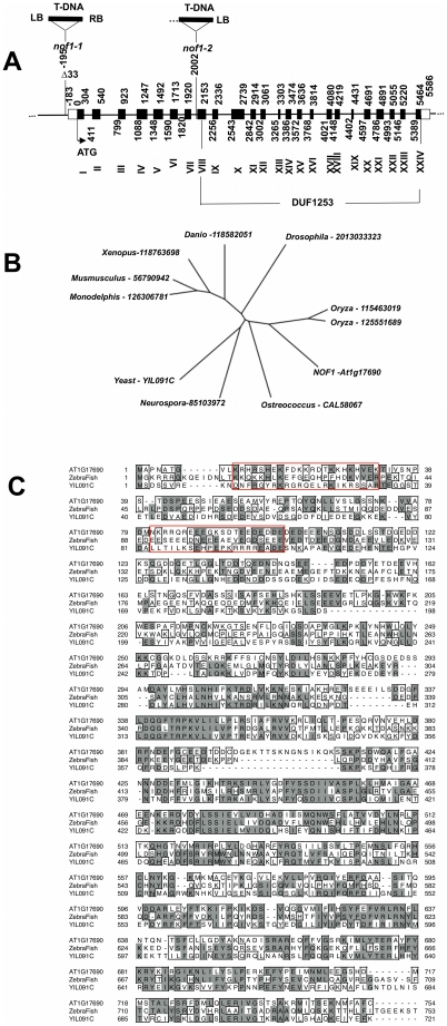
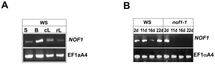
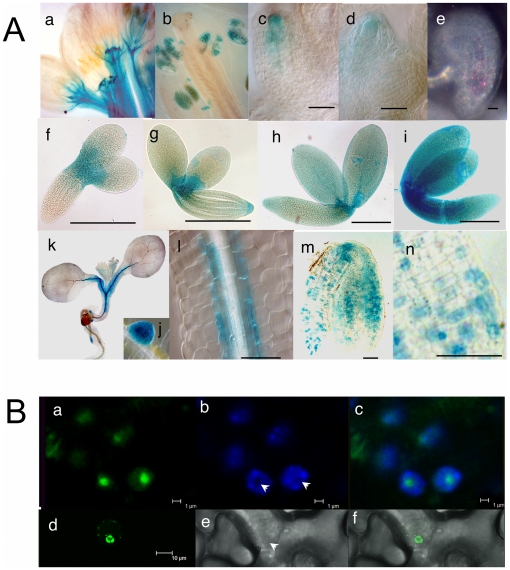
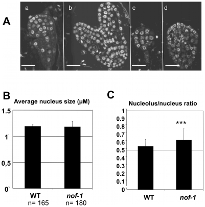
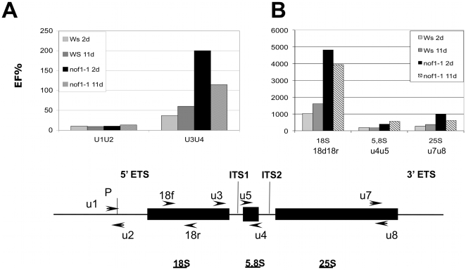
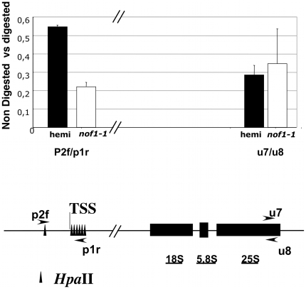
The control of ribosomal RNA biogenesis is essential for the regulation of protein synthesis in eukaryotic cells. Here, we report the characterization of NOF1 that encodes a putative nucleolar protein involved in the control of rRNA expression in Arabidopsis. The gene has been isolated by T-DNA tagging and its function verified by the characterization of a second allele and genetic complementation of the mutants. The nof1 mutants are affected in female gametogenesis and embryo development. This result is consistent with the detection of NOF1 mRNA in all tissues throughout plant life's cycle, and preferentially in differentiating cells. Interestingly, the closely related proteins from zebra fish and yeast are also necessary for cell division and differentiation. We showed that the nof1-1 mutant displays higher rRNA expression and hypomethylation of rRNA promoter. Taken together, the results presented here demonstrated that NOF1 is an Arabidopsis gene involved in the control of rRNA expression, and suggested that it encodes a putative nucleolar protein, the function of which may be conserved in eukaryotes.


Conflict of interest statement

Competing Interests: The authors have declared that no competing interests exist.

Figures

Figure 1
Figure 1. Phenotypic analyses of wild type and nof1-1 mutant seeds.
(a) Mature dry seeds from nof1-1/NOF1-1 hemizygous plants displaying a few dark brown mutant seeds (indicated with arrows) and (b) Wild-type (Ws) control seeds. (c) representative developping siliques of wild-type accession (Ws) that displays immature green seeds (top row); nof1-1/NOF1-1 (weak allele) genotype that display white (i.e. lethal) seeds (second row); and nof1-2/NOF1-2 genotype (third row) that display gaps (i.e. missing seed) and shrunken empty seed coat for the null allele nof1-2. (d) Late developmental stage in a single silique comparing a nof1-1 mutant embryo (top left) to a wild type embryo (bottom right). Laser scanning confocal image of Ws embryo (e) compared to nof1-1 globular embryo with abnormal cell divisions (f). Wild type embryo extracted from mature seed (g) compared to several nof1-1 embryos arrested at different stages of development (h–k). DIC images of nof1-1 embryos with abnormal cell divisions (arrows). Bars  = 600 µM (a, b, c), 10 µM (e, f, l, m, n), 100 µm (g–k).
Figure 2
Figure 2. Auxin signaling in nof1-1.
PRODR5: uidA expression in wild type (a) and mutants (b and c) embryos. Immunolocalization of PIN1 in wild type (d) and nof1-1 mutants (e and f) embryos. Bar  = 40 µm (a to c) and 100 µm (d to f).
Figure 3
Figure 3. Molecular characterization of NOF1.
A) Schematic representation of the structure of the NOF1 (At1g17690) locus in wild type and nof1-1 and nof1-2 mutants. The structure of the gene is deduced from the comparison between genomic and cDNA sequences. In nof 1-1, the T-DNA was inserted between positions −195 to −228 relative to the ATG (first codon), leading to a small deletion of 33 bp. In nof 1-2, a T-DNA is inserted after nucleotide 2002. Boxes are exons. The 5′UTR is predicted according to ESTs sequences. Location of the DUF1253 domain is indicated below the scheme. B) Unrooted neighbour-Joining tree representing the distance between NOF1 and the most closely related proteins from various organisms was obtained using the full amino-acid sequences, after clustal W alignment (http://align.genome.jp/). C) Deduced amino acid sequences of NOF1 and closely related proteins are presented (At1g17690: NOF1, Zebrafish (DEF): gi37046654, and yeast YIL091C). Identities between amino acid residues are shown with dark boxes and similarities with light boxes. The putative NoLS site is boxed.
Figure 4
Figure 4. Analyses of NOF1 mRNA accumulation in wild type and nof1-1.
RNA was extracted from various plant organs (A) and seeds (B) at different stages of development and used for reverse transcription. Primers specific for NOF1 and for EF1αA4 as control were used on the same set of first strand cDNA templates generated with dT primers. During silique development (B), WS or nof1-1 seeds were manually dissected based on seed phenotype to produce the NOF1 cDNA template at 2, 11, 16 and 22 days after fertilization. S: seeds, B: buds, cL: cauline leaves, rL: rosette leaves.
Figure 5
Figure 5. Cytological analyses of NOF1 expression and intracellular localization of the protein.
A The activity of the NOF1 promoter (pNOF:UidA) was investigated in various plant organs. The result of GUS activity was observed with Nomarski optics, except for (e) where dark field is used, on flowers (a–b) showing expression in buds and in pollen. Expression was found in the nucellus of developing ovules (c–d) and later in the embryo sac (e). During embryogenesis, GUS activity is found at the top of the hypocotyl and extends throughout the embryo during maturation (f to i). After 24 h of imbibition, the expression is found with a patchy pattern in the embryo (l to n) before disappearing. Five days after imbibition's start, the expression is found in root apex (k), lateral root initials (j) and around vascular bundles (l). Bars  = 10 µm (c to e), 100 µm (f to i) and 50 µm (l to n). B). Subcellular localization of NOF1:GFP in transgenic Arabidopsis lines expressing 35S:NOF1:GFP. GFP is detected in the nucleoli (a), DAPI staining (b) and merged image (c). Transient expression in tobacco leaves of 35S:NOF1:GFP showing nucleolar localization of NOF1:GFP (d), transmitted light (e), and merged pictures (f). Bar  = 1 µm (a, b, c) and 10 µm (d, e, f).
Figure 6
Figure 6. Nucleolus phenotypes.
A) Embryo development at the globular and heart stage of development, Ws (a and b respectively) and nof1-1 (c and d respectively) after DAPI staining and laser confocal imaging. Bar  = 5 µm. B) Average nucleus diameter was measured in Ws and nof1-1 embryos after DAPI staining and laser scanning confocal imaging. C) Average ratio of nucleolus vs nucleus diameters in Ws compared to nof1-1. A student test was performed to compare both populations of nucleoli, demonstrating a significant difference with p<0.0001 (t = −6.26).
Figure 7
Figure 7. Expression and processing of the 45S rRNA.
Accumulation of rRNA was monitored by qRT-PCR using specific primers for unprocessed (U1/U2, U3/U4) (A) and processed forms (i.e. 18S f/r, U5/U4 for 5.8S and U7/U8 for 25S) (B) of the 45S rRNA transcript. The cDNA templates were been obtained after manual seeds dissection from a hemizygous plants for the nof1-1 mutation or wild-type (Ws), at 2 or 11 days after fertilization. One representative experiment of three independent biological repeat is shown.
Figure 8
Figure 8. Quantification of DNA methylation at the 45S rDNA locus.
The level of methylation of the promoter region was estimated by qPCR. Genomic DNA was PCR amplified directly or after restriction with a methylation sensitive enzyme (HpaII) using specific primers (p2f and p2r). The amplification of the 25 s rDNA was used as internal control. Results are the means of 3 measurements (+/− standard deviation). Experiments have been made on two independent biological replicates showing similar results. TSS: transcription start site.

References

    1. Meinke DW. Molecular-Genetics of Plant Embryogenesis. Annual Review of Plant Physiol and Plant Mol Biol. 1995;46:369–394.
    1. Jurgens G, Mayer U, Busch M, Lukowitz W, Laux T. Pattern formation in the Arabidopsis embryo: a genetic perspective. Philos Trans R Soc Lond B Biol Sci. 1995;350:19–25. - PubMed
    1. Meinke DW, Meinke LK, Showalter TC, Schissel AM, Mueller LA, et al. A Sequence-Based Map of Arabidopsis Genes with Mutant Phenotypes. Plant Physiol. 2003;131:409–418. - PMC - PubMed
    1. Lepiniec L, Devic M, Berger F. Plant Functional Genomics. Binghamton: Food Products Press; 2005. Genetic and Molecular Control of Seed Development in Arabidopsis. pp. 511–564.
    1. Shi DQ, Liu J, Xiang YH, Ye D, Sundaresan V, et al. SLOW WALKER1, essential for gametogenesis in Arabidopsis, encodes a WD40 protein involved in 18S ribosomal RNA biogenesis. Plant Cell. 2005;17:2340–2354. - PMC - PubMed

Publication types

MeSH terms