A mouse strain where basal connective tissue growth factor gene expression can be switched from low to high
- PMID: 20877562
- PMCID: PMC2943916
- DOI: 10.1371/journal.pone.0012909
A mouse strain where basal connective tissue growth factor gene expression can be switched from low to high
Abstract
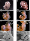
Connective tissue growth factor (CTGF) is a signaling molecule that primarily functions in extracellular matrix maintenance and repair. Increased Ctgf expression is associated with fibrosis in chronic organ injury. Studying the role of CTGF in fibrotic disease in vivo, however, has been hampered by perinatal lethality of the Ctgf null mice as well as the limited scope of previous mouse models of Ctgf overproduction. Here, we devised a new approach and engineered a single mutant mouse strain where the endogenous Ctgf-3' untranslated region (3'UTR) was replaced with a cassette containing two 3'UTR sequences arranged in tandem. The modified Ctgf allele uses a 3'UTR from the mouse FBJ osteosarcoma oncogene (c-Fos) and produces an unstable mRNA, resulting in 60% of normal Ctgf expression (Lo allele). Upon Cre-expression, excision of the c-Fos-3'UTR creates a transcript utilizing the more stable bovine growth hormone (bGH) 3'UTR, resulting in increased Ctgf expression (Hi allele). Using the Ctgf Lo and Hi mutants, and crosses to a Ctgf knockout or Cre-expressing mice, we have generated a series of strains with a 30-fold range of Ctgf expression. Mice with the lowest Ctgf expression, 30% of normal, appear healthy, while a global nine-fold overexpression of Ctgf causes abnormalities, including developmental delay and craniofacial defects, and embryonic death at E10-12. Overexpression of Ctgf by tamoxifen-inducible Cre in the postnatal life, on the other hand, is compatible with life. The Ctgf Lo-Hi mutant mice should prove useful in further understanding the function of CTGF in fibrotic diseases. Additionally, this method can be used for the production of mouse lines with quantitative variations in other genes, particularly with genes that are broadly expressed, have distinct functions in different tissues, or where altered gene expression is not compatible with normal development.
Conflict of interest statement
Figures


Similar articles
-
Selective expression of connective tissue growth factor in fibroblasts in vivo promotes systemic tissue fibrosis.Arthritis Rheum. 2010 May;62(5):1523-32. doi: 10.1002/art.27382. Arthritis Rheum. 2010. PMID: 20213804 Free PMC article.
-
Genetic Analysis of Connective Tissue Growth Factor as an Effector of Transforming Growth Factor β Signaling and Cardiac Remodeling.Mol Cell Biol. 2015 Jun;35(12):2154-64. doi: 10.1128/MCB.00199-15. Epub 2015 Apr 13. Mol Cell Biol. 2015. PMID: 25870108 Free PMC article.
-
Pivotal role of connective tissue growth factor in lung fibrosis: MAPK-dependent transcriptional activation of type I collagen.Arthritis Rheum. 2009 Jul;60(7):2142-55. doi: 10.1002/art.24620. Arthritis Rheum. 2009. PMID: 19565505
-
Connective tissue growth factor: context-dependent functions and mechanisms of regulation.Biofactors. 2009 Mar-Apr;35(2):200-8. doi: 10.1002/biof.30. Biofactors. 2009. PMID: 19449449 Review.
-
Transcriptional profiling of the scleroderma fibroblast reveals a potential role for connective tissue growth factor (CTGF) in pathological fibrosis.Keio J Med. 2004 Jun;53(2):74-7. doi: 10.2302/kjm.53.74. Keio J Med. 2004. PMID: 15247510 Review.
Cited by
-
Mammalian organ regeneration in spiny mice.J Muscle Res Cell Motil. 2023 Jun;44(2):39-52. doi: 10.1007/s10974-022-09631-3. Epub 2022 Sep 21. J Muscle Res Cell Motil. 2023. PMID: 36131170 Free PMC article. Review.
-
Selective deletion of connective tissue growth factor attenuates experimentally-induced pulmonary fibrosis and pulmonary arterial hypertension.Int J Biochem Cell Biol. 2021 May;134:105961. doi: 10.1016/j.biocel.2021.105961. Epub 2021 Mar 1. Int J Biochem Cell Biol. 2021. PMID: 33662577 Free PMC article.
-
Cysteine-rich protein 61 (CCN1) and connective tissue growth factor (CCN2) at the crosshairs of ocular neovascular and fibrovascular disease therapy.J Cell Commun Signal. 2013 Dec;7(4):253-63. doi: 10.1007/s12079-013-0206-6. Epub 2013 Jun 7. J Cell Commun Signal. 2013. PMID: 23740088 Free PMC article.
-
The CCN2/CTGF interactome: an approach to understanding the versatility of CCN2/CTGF molecular activities.J Cell Commun Signal. 2021 Dec;15(4):567-580. doi: 10.1007/s12079-021-00650-2. Epub 2021 Oct 6. J Cell Commun Signal. 2021. PMID: 34613590 Free PMC article. Review.
-
Eyeing the Cyr61/CTGF/NOV (CCN) group of genes in development and diseases: highlights of their structural likenesses and functional dissimilarities.Hum Genomics. 2015 Sep 23;9:24. doi: 10.1186/s40246-015-0046-y. Hum Genomics. 2015. PMID: 26395334 Free PMC article. Review.
References
-
- Thom T, Haase N, Rosamond W, Howard VJ, Rumsfeld J, et al. Heart disease and stroke statistics–2006 update: a report from the American Heart Association Statistics Committee and Stroke Statistics Subcommittee. Circulation. 2006;113:e85–151. - PubMed
-
- González A, López B, Díez J. Myocardial fibrosis in arterial hypertension. European Heart Journal Supplements. 2002;4:D18–D22.
-
- Sun Y, Weber KT. Infarct scar: a dynamic tissue. Cardiovasc Res. 2000;46:250–256. - PubMed
-
- Sun Y, Zhang JQ, Zhang J, Lamparter S. Cardiac remodeling by fibrous tissue after infarction in rats. J Lab Clin Med. 2000;135:316–323. - PubMed
-
- Sun Y, Zhang JQ, Zhang J, Ramires FJ. Angiotensin II, transforming growth factor-beta1 and repair in the infarcted heart. J Mol Cell Cardiol. 1998;30:1559–1569. - PubMed
Publication types
MeSH terms
Substances
Grants and funding
LinkOut - more resources
Full Text Sources
Molecular Biology Databases
Research Materials
Miscellaneous

