. 2010 Sep 22;5(9):e12871.
doi: 10.1371/journal.pone.0012871.

Human integrin α(3)β(1) regulates TLR2 recognition of lipopeptides from endosomal compartments

Affiliations

Human integrin α(3)β(1) regulates TLR2 recognition of lipopeptides from endosomal compartments

Meghan L Marre et al. PLoS One. .

Abstract

Background: Toll-like receptor (TLR)-2/TLR1 heterodimers recognize bacterial lipopeptides and initiate the production of inflammatory mediators. Adaptors and co-receptors that mediate this process, as well as the mechanisms by which these adaptors and co-receptors function, are still being discovered.

Methodology/principal findings: Using shRNA, blocking antibodies, and fluorescent microscopy, we show that U937 macrophage responses to the TLR2/1 ligand, Pam(3)CSK(4), are dependent upon an integrin, α(3)β(1). The mechanism for integrin α(3)β(1) involvement in TLR2/1 signaling is through its role in endocytosis of lipopeptides. Using inhibitors of endosomal acidification/maturation and physical tethering of the ligand, we show that the endocytosis of Pam(3)CSK(4) is necessary for the complete TLR2/1-mediated pro-inflammatory cytokine response. We also show that TLR2/1 signaling from the endosome results in the induction of different inflammatory mediators than TLR2/1 signaling from the plasma membrane.

Conclusion/significance: Here we identify integrin α(3)β(1) as a novel regulator for the recognition of bacterial lipopeptides. We demonstrate that induction of a specific subset of cytokines is dependent upon integrin α(3)β(1)-mediated endocytosis of the ligand. In addition, we address an ongoing controversy regarding endosomal recognition of bacterial lipopeptides by demonstrating that TLR2/1 signals from within endosomal compartments as well as the plasma membrane, and that downstream responses may differ depending upon receptor localization. We propose that the regulation of endosomal TLR2/1 signaling by integrin α(3)β(1) serves as a mechanism for modulating inflammatory responses.


Conflict of interest statement

Competing Interests: The authors have declared that no competing interests exist.

Figures

Figure 1
Figure 1. Integrin α3β1 mediates the U937 macrophage response to Pam3CSK4.
A) U937 macrophages stably transduced with integrin α3-specific shRNA (Itgα3 shRNA) or non-targeting shRNA (Ctrl. shRNA) were stimulated with 100 ng/ml Pam3CSK4 for 6 hours under serum-free conditions. Values represent mean secretion of IL-6 relative to control shRNA and S.E.M.of three independent experiments. Cells transduced with control shRNA secreted a mean of 350 pg/ml, and cells transduced with integrin α3-targeting shRNA secreted a mean of 61 pg/ml. * p = 0.014 B) U937 macrophages were treated with an integrin α3β1 blocking antibody (P1B5) or a control mouse ascites fluid (CMA) and stimulated with 100 ng/ml Pam3CSK4 for 6 hours under serum-free conditions. Values represent mean secretion of IL-6 relative to CMA-treated cells and S.E.M.of three independent experiments. CMA-treated cells secreted a mean of 1,068 pg/ml, and P1B5-treated cells secreted a mean of 607 pg/ml. * p = 0.014.
Figure 2
Figure 2. Exogenous serum proteins do not enhance the role of integrin α3β1.
A) U937 macrophages were stimulated with 100 ng/ml of Pam3CSK4 in the presence or absence of 1% FBS for 6 hours. Values represent mean secretion of IL-6 relative to cells stimulated under serum-free conditions and S.E.M. of three independent experiments. Cells stimulated under serum-free conditions secreted a mean of 1,048 pg/ml, and cells stimulated in the presence of 1% FBS secreted a mean of 975 pg/ml. B) U937 macrophages stably transduced with integrin α3-specific shRNA (Itgα3 shRNA) or non-targeting shRNA (Ctrl. shRNA) were stimulated with 100 ng/ml Pam3CSK4 in the presence or absence of 1% FBS for 6 hours. Values represent mean secretion of IL-6 relative to control cells and S.E.M. of three independent experiments. Under serum-free conditions, control cells secreted a mean of 1,048 pg/ml and cells transduced with integrin α3-targeting shRNA secreted a mean of 545 pg/ml. When stimulated in the presence of 1% FBS, control cells secreted a mean of 974 pg/ml and cells transduced with integrin α3-targeting shRNA secreted a mean of 556 pg/ml. * p = 0.037.
Figure 3
Figure 3. Integrin α3β1 mediates internalization, but not attachment, of Pam3CSK4.
A) U937 macrophages were stably transduced with integrin α3-targeting shRNA (Itgα3 shRNA) or non-targeting shRNA (Ctrl. shRNA), stimulated with 5 µg/ml Pam3CSK4-biotin for 60 minutes, and fixed and stained for immunofluorescent microscopy. Pam3CSK4-biotin was detected by α-biotin antibodies conjugated to Texas Red. Scale bars, 10 µm. Data are representative of three independent experiments. B) The association of Pam3CSK4-biotin to the macrophages was quantified by determining the association index (the number of cells associated with Pam3CSK4-biotin divided by total cells). Data represent the mean association index and S.E.M of three independent experiments. The mean association index for control cells was 54.6%, and the mean association index for cells transduced with integrin α3-targeting shRNA was 60.6%. C) U937 macrophages were stably transduced with integrin α3-targeting shRNA (Itgα3 shRNA) or non-targeting shRNA (Ctrl. shRNA) and stimulated with 5 µg/ml Pam3CSK4-biotin for 60 minutes. The cells were fixed and stained for immunofluorescent microscopy using α-biotin antibodies before (FITC) or after (Texas Red) permeabilization of the cells. Arrows represent internalized Pam3CSK4-biotin. Scale bars, 10 µm. Data are representative of three independent experiments. D) The endocytosis of Pam3CSK4-biotin was quantified by determining the endocytic index (the number of cells with internalized Pam3CSK4-biotin divided by number of cells with Pam3CSK4-biotin associated). Data represent the mean endocytic index and S.E.M. of three independent experiments. The mean endocytic index for control cells was 79.3%, and the mean endocytic index for cells transduced with integrin α3-targeting shRNA was 42.9%. * p = 0.037.
Figure 4
Figure 4. Pam3CSK4 co-localizes with TLR2 intracellularly.
U937 macrophages were stimulated with 5 µg/ml Pam3CSK4-rhodamine for 20 minutes. The cells were fixed and stained for immunofluorescent microscopy using α-TLR2 antibodies and secondary antibodies conjugated to Alexa Fluor 488. Images show one representative Z stack of 0.7 µm thickness. Scale bars, 8.61 µ µm.
Figure 5
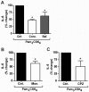
Figure 5. Pam3CSK4 induces signaling through TLR2/1 from endosomal compartments and is internalized through clathrin-mediated endocytosis.
A) U937 macrophages were treated with 100 ng/ml concanamycin A (Conc.), 500 µM bafilomycin A1 (Baf.), or control (Ctrl.), and stimulated with 100 ng/ml Pam3CSK4 for 6 hours under serum-free conditions. Values represent mean secretion of IL-6 relative to control cells and S.E.M. of three independent experiments. Control cells secreted a mean of 670 pg/ml, concanamycin A-treated cells secreted a mean of 320 pg/ml, and bafilomycin A1-treated cells secreted a mean of 430 pg/ml. * p = 0.037 B) U937 macrophages were treated with 1 µM monensin (Mon.) or control (Ctrl.), and stimulated with 100 ng/ml Pam3CSK4 for 6 hours under serum-free conditions. Values represent mean secretion of IL-6 relative to control cells and S.E.M. of three independent experiments. Control cells secreted a mean of 670 pg/ml, and monensin-treated cells secreted a mean of 420 pg/ml, * p = 0.037. C) U937 macrophages were treated with 5 µM chlorpromazine (CPZ) or control (Ctrl.) and stimulated with 100 ng/ml Pam3CSK4 for 6 hours under serum-free conditions. Values represent mean secretion of IL-6 relative to control cells and S.E.M. of three independent experiments. Control cells secreted a mean of 670 pg/ml and CPZ-treated cells secreted a mean of 340 pg/ml. * p = 0.037.
Figure 6
Figure 6. The IL-6 response is dependent upon internalization of Pam3CSK4.
A) Schematic of experiment comparing the inflammatory response in cells stimulated with either soluble Pam3CSK4-biotin or Pam3CSK4-biotin immobilized on streptavidin plates. U937 macrophages were stimulated with either soluble Pam3CSK4-biotin or Pam3CSK4-biotin immobilized on steptavidin plates for 6 hours under serum-free conditions. Values represent mean secretion of IL-6 relative to cells stimulated with soluble Pam3CSK4-biotin and S.E.M. of three independent experiments. Cells stimulated with soluble Pam3CSK4-biotin secreted a mean of 1,456 pg/ml, and cells stimulated with plate-bound Pam3CSK4-biotin secreted a mean of 620 pg/ml. * p = 0.037 B) Schematic of experiment comparing the inflammatory response in cells stimulated with Pam3CSK4-biotin in streptavidin wells either blocked or not with biotin-HRP prior to the addition of cells and Pam3CSK4-biotin simultaneously. U937 macrophages were stimulated with soluble Pam3CSK4-biotin in either unblocked streptavidin plates or streptavidin plates blocked with biotin-HRP for 6 hours under serum-free conditions. Values represent mean secretion of IL-6 relative to cells stimulated in blocked wells and S.E.M. of three independent experiments. Cells stimulated in blocked wells secreted a mean of 1,690 pg/ml and cells stimulated in unblocked wells secreted a mean of 895 pg/ml. * p = 0.037.
Figure 7
Figure 7. TLR2/1 transduces signals from the endosome for the induction of IFN-α1.
A) U937 macrophages were treated with 100 ng/ml of Pam3CSK4 under serum-free conditions for the indicated times. Expression of IFN-β was measured by qRT-PCR. Values represent mean induction of IFN-β expression relative to control cells and S.E.M. of three independent experiments. B) U937 macrophages were treated with 100 ng/ml concanamycin A (Conc.), 1 µM monensisn (Mon.), or control (Ctrl.), and stimulated with 100 ng/ml Pam3CSK4 under serum-free conditions for the indicated times. Expression of IFN-α1 was measured by qRT-PCR. Values represent mean induction of IFN-α1 expression relative to control cells and S.E.M. of three independent experiments. * p = 0.037.
Figure 8
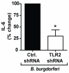
Figure 8. TLR2 mediates the inflammatory cytokine response to B. burgdorferi in U937 macrophages.
U937 macrophages were stably transduced with TLR2-specific shRNA (TLR2 shRNA) or non-targeting shRNA (Ctrl. shRNA) and stimulated with B. burgdorferi MOI 10 for 6 hours under serum-free conditions. Values represent mean secretion of IL-6 relative to control shRNA and S.E.M. of three independent experiments. Cells transduced with control shRNA secreted a mean of 553 pg/ml, and cells transduced with TLR2-targeting shRNA secreted a mean of 142 pg/ml. * p = 0.037.
Figure 9
Figure 9. Integrin α3β1 mediates the inflammatory response to B. burgdorferi in U937 macrophages.
A) U937 macrophages were stably transduced with integrin α3-specific shRNA (Itgα3 shRNA) or non-targeting shRNA (Ctrl. shRNA), and stimulated with B. burgdorferi MOI 10 for 6 hours under serum-free conditions. Values represent mean secretion of IL-6 relative to control shRNA and S.E.M. of three independent experiments. Cells transduced with control shRNA secreted a mean of 560 pg/ml, and cells transduced with integrin α3-targeting shRNA secreted a mean of 310 pg/ml. * p = 0.014 B) U937 macrophages were treated with an integrin α3β1 blocking antibody (P1B5) or control mouse ascites fluid (CMA) and stimulated with B. burgdorferi MOI 10 for 6 hours under serum-free conditions. Values represent mean secretion of IL-6 relative to CMA treated cells and S.E.M. of three independent experiments. CMA-treated cells secreted a mean of 1,500 pg/ml, and P1B5-treated cells secreted a mean of 560 pg/ml. * p = 0.014.
Figure 10
Figure 10. Integrin α3β1 mediates endocytosis of spirochetes.
A) U937 macrophages were stably transduced with integrin α3-specific shRNA (Itgα3 shRNA) or non-targeting shRNA (Ctrl. shRNA) and stimulated with B. burgdorferi MOI 10 for 60 minutes under serum-free conditions. Endocytosis of spirochetes was determined by immunofluorescent staining before (FITC-labeled) and after (Texas Red-labeled) permeabilization of the cells. Arrows indicate internalized spirochetes. Scale bars, 10 µm. Data are representative of three independent experiments. B) The association of B. burgdorferi with the macrophages was quantified by determining the association index (the number of cells associated with B. burgdorferi divided by the total number of cells). Data represent the mean association index and S.E.M. of three independent experiments. The mean association index for control cells was 51.1%, and the mean association index for cells transduced with integrin α3-targeting shRNA was 62.2%. C) The endocytosis of B. burgdorferi was quantified by determining the endocytic index (the number of cells with B. burgdorferi internalized divided by the number of cells with B. burgdorferi associated). Data represent the mean endocytic index and S.E.M. of three independent experiments. The mean endocytic index for control cells was 41.8%, and the mean endocytic index for cells transduced with integrin α3-targeting shRNA was 20.1%. * p = 0.037.
Figure 11
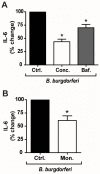
Figure 11. Induction of IL-6 by B. burgdorferi occurs downstream of endocytosis and endolysosomal processing of spirochetes.
A) U937 macrophages were treated with 100 ng/ml concanamycin A (Conc.), 500 µM bafilomycin A1 (Baf.), or control (Ctrl.) and stimulated with B. burgdorferi MOI 10 for 6 hours under serum-free conditions. Values represent mean secretion of IL-6 relative to control cells and S.E.M. of three independent experiments. Control cells secreted a mean of 955 pg/ml, concanamycin A-treated cells secreted a mean of 437 pg/ml, and bafilomycin A1-treated cells secreted a mean of 680 pg/ml. * p = 0.037 B) U937 macrophages were treated with 1 µM monensin (Mon.) or control (Ctrl.), and stimulated with B. burgdorferi MOI 10 for 6 hours under serum-free conditions. Values represent mean secretion of IL-6 relative to control cells and S.E.M. of three independent experiments. Control cells secreted a mean of 955 pg/ml and monensin-treated cells secreted a mean of 567 pg/ml. * p = 0.037.
Figure 12
Figure 12. Model.
Free bacterial lipopeptide is bound to host proteins which act as ligands for a “tethering receptor”, anchoring Pam3CSK4 to the cell and bringing it into proximity with TLR2 to initiate TLR2-mediated signaling at the cell surface. In addition to the induction of pro-inflammatory cytokines, the activation of cells via TLR2 may contribute to inside-out signaling, causing a shift in integrin α3β1 conformational equilibrium from an inactive (“closed”) to an active (“open”) conformation. Once active, integrin α3β1 serves as a tickling receptor, participating in the endocytosis of Pam3CSK4 through clathrin-mediated mechanisms. Upon internalization, Pam3CSK4 and TLR2 co-localize in the endosome. TLR2 signals from this endosomal compartment for the induction of a second subset pro-inflammatory cytokines such as IL-6 and type I interferons.

Similar articles

Cited by

References

    1. Takeuchi O, Akira S. Pattern recognition receptors and inflammation. Cell. 2010;140:805–820. - PubMed
    1. Ferwerda G, Meyer-Wentrup F, Kullberg BJ, Netea MG, Adema GJ. Dectin-1 synergizes with TLR2 and TLR4 for cytokine production in human primary monocytes and macrophages. Cell Microbiol. 2008;10:2058–2066. - PubMed
    1. Bowdish DM, Sakamoto K, Kim MJ, Kroos M, Mukhopadhyay S, et al. MARCO, TLR2, and CD14 are required for macrophage cytokine responses to mycobacterial trehalose dimycolate and Mycobacterium tuberculosis. PLoS Pathog. 2009;5:e1000474. - PMC - PubMed
    1. Schwandner R, Dziarski R, Wesche H, Rothe M, Kirschning CJ. Peptidoglycan- and lipoteichoic acid-induced cell activation is mediated by toll-like receptor 2. J Biol Chem. 1999;274:17406–17409. - PubMed
    1. Yoshimura A, Lien E, Ingalls RR, Tuomanen E, Dziarski R, et al. Cutting edge: recognition of Gram-positive bacterial cell wall components by the innate immune system occurs via Toll-like receptor 2. J Immunol. 1999;163:1–5. - PubMed

Publication types