

Light chain separated from the rest of the type a botulinum neurotoxin molecule is the most catalytically active form
- PMID: 20877571
- PMCID: PMC2943925
- DOI: 10.1371/journal.pone.0012872
Light chain separated from the rest of the type a botulinum neurotoxin molecule is the most catalytically active form
Abstract
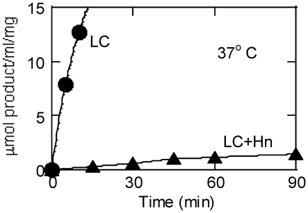
Botulinum neurotoxins (BoNT) are the most potent of all toxins. The 50 kDa N-terminal endopeptidase catalytic light chain (LC) of BoNT is located next to its central, putative translocation domain. After binding to the peripheral neurons, the central domain of BoNT helps the LC translocate into cytosol where its proteolytic action on SNARE (soluble NSF attachment protein receptor) proteins blocks exocytosis of acetyl choline leading to muscle paralysis and eventual death. The translocation domain also contains 105 Å -long stretch of ∼100 residues, known as "belt," that crosses over and wraps around the LC to shield the active site from solvent. It is not known if the LC gets dissociated from the rest of the molecule in the cytosol before catalysis. To investigate the structural identity of the protease, we prepared four variants of type A BoNT (BoNT/A) LC, and compared their catalytic parameters with those of BoNT/A whole toxin. The four variants were LC + translocation domain, a trypsin-nicked LC + translocation domain, LC + belt, and a free LC. Our results showed that K(m) for a 17-residue SNAP-25 (synaptosomal associated protein of 25 kDa) peptide for these constructs was not very different, but the turnover number (k(cat)) for the free LC was 6-100-fold higher than those of its four variants. Moreover, none of the four variants of the LC was prone to autocatalysis. Our results clearly demonstrated that in vitro, the LC minus the rest of the molecule is the most catalytically active form. The results may have implication as to the identity of the active, toxic moiety of BoNT/A in vivo.
Conflict of interest statement
Figures

References
-
- Simpson LL. Identification of the major steps in botulinum toxin action. Annu Rev Pharmacol Toxicol. 2004;44:167–193. - PubMed
-
- Montecucco C, Schiavo G. Structure and function of tetanus and botulinum neurotoxins. Q Rev Biophys. 1995;28:423–472. - PubMed
-
- Lacy DB, Tepp W, Cohen AC, DasGupta BR, Stevens RC. Crystal structure of botulinum neurotoxin type A and implications for toxicity. Nat Struct Biol. 1998;5:898–902. - PubMed
-
- Swaminathan S, Eswaramoorthy S. Structural analysis of the catalytic and binding sites of Clostridium botulinum neurotoxin B. Nat Struct Biol. 2000;7:693–699. - PubMed
-
- Ahmed SA, Olson MA, Ludivico ML, Gilsdorf J, Smith LA. Identification of Residues Surrounding the Active Site of Type A Botulinum Neurotoxin Important for Substrate Recognition and Catalytic Activity. Protein J. 2008;27:151–162. - PubMed
Publication types
MeSH terms
Substances
LinkOut - more resources
Full Text Sources
Medical
Miscellaneous
