

Schwannoma in head and neck: preoperative imaging study and intracapsular enucleation for functional nerve preservation
- PMID: 20879063
- PMCID: PMC2995979
- DOI: 10.3349/ymj.2010.51.6.938
Schwannoma in head and neck: preoperative imaging study and intracapsular enucleation for functional nerve preservation
Abstract
Purpose: In treating schwannoma patients, it is critical to determine the origin of the tumor to preserve nerve function. We evaluated the validity of preoperative imaging studies in distinguishing the neurological origin of the schwannomas of the head and neck, and the efficacy of intracapsular enucleation in preserving nerve function.
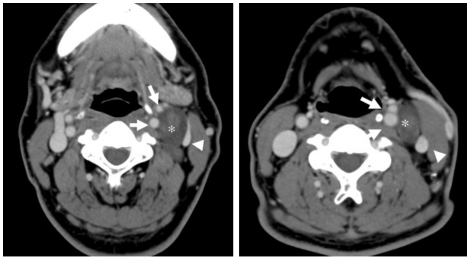
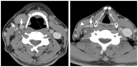
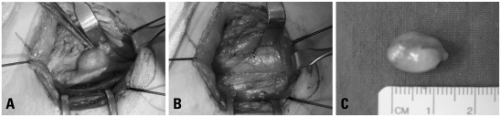
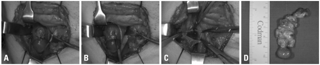
Materials and methods: In 7 cases of schwannomas in the head and neck region, we predicted whether the tumor originated from the vagus nerve or the cervical sympathetic chain through imaging studies including computed tomography (CT) and magnetic resonance imaging (MRI). All patients were performed intracapsular enucleation, and the function of the vagus nerve and the sympathetic nerve was evaluated preoperatively and postoperatively.
Results: Preoperative imaging studies showed 6 cases where the tumor was located between the carotid artery and the internal jugular vein, and 1 case where the tumor was located posteriorly, displacing the carotid artery and the internal jugular vein anteriorly. At the time of operation, we confirmed schwannoma originating from the vagus nerve on the first 6 cases, and schwannoma originating from the sympathetic nervous system on the last case. All patients went through successful intracapsular enucleation, and of the seven schwannoma cases, 6 patients maintained normal postoperative neurological function (85.7%).
Conclusion: Preoperative imaging studies offer valuable information regarding the location and origination of the tumor, and intracapsular enucleation helped us to preserve the nerve function.
Conflict of interest statement
The authors have no financial conflicts of interest.
Figures
References
-
- Ducatman BS, Scheithauer BW, Piepgras DG, Reiman HM, Ilstrup DM. Malignant peripheral nerve sheath tumors. A clinicopathologic study of 120 cases. Cancer. 1986;57:2006–2021. - PubMed
-
- Colreavy MP, Lacy PD, Hughes J, Bouchier-Hayes D, Brennan P, O'Dwyer AJ, et al. Head and neck schwannomas: a 10 year review. J Laryngol Otol. 2000;114:119–124. - PubMed
-
- Furukawa M, Furukawa MK, Katoh K, Tsukuda M. Differentiation between schwannoma of the vagus nerve and schwannoma of the cervical sympathetic chain by imaging diagnosis. Laryngoscope. 1996;106:1548–1552. - PubMed
-
- Fujino K, Shinohara K, Aoki M, Hashimoto K, Omori K. Intracapsular enucleation of vagus nerve-originated tumors for preservation of neural function. Otolaryngol Head Neck Surg. 2000;123:334–336. - PubMed
-
- Fornaro R, Frascio M, Stabilini C. Excision of a schwannoma of the head and neck: surgical technique. G Chir. 2006;27:428–432. - PubMed
MeSH terms
LinkOut - more resources
Full Text Sources
Medical
