

Sparse unbiased analysis of anatomical variance in longitudinal imaging
- PMID: 20879247
- PMCID: PMC3641640
- DOI: 10.1007/978-3-642-15705-9_40
Sparse unbiased analysis of anatomical variance in longitudinal imaging
Abstract
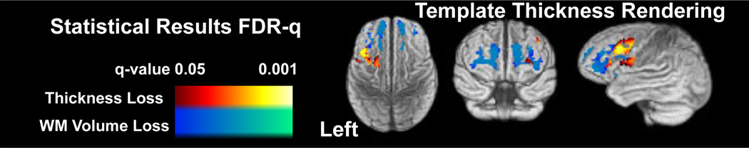
We present a new algorithm for reliable, unbiased, multivariate longitudinal analysis of cortical and white matter atrophy rates with penalized statistical methods. The pipeline uses a step-wise approach to transform and personalize template information first to a single-subject template (SST) and then to the individual's time series data. The first stream of information flows from group template to the SST; the second flows from the SST to the individual time-points and provides unbiased, prior-based segmentation and measurement of cortical thickness. MRI-bias correction, consistent longitudinal segmentation, cortical parcellation and cortical thickness estimation are all based on strong use of the subject-specific priors built from initial diffeomorphic mapping between the SST and optimal group template. We evaluate our approach with both test-retest data and with application to a driving biological problem. We use test-retest data to show that this approach produces (a) zero change when the retest data contains the same image content as the test data and (b) produces normally distributed, low variance estimates of thickness change centered at zero when test-retest data is collected near in time to test data. We also show that our approach--when combined with sparse canonical correlation analysis--reveals plausible, significant, annualized decline in cortical thickness and white matter volume when contrasting frontotemporal dementia and normal aging.
Figures


References
-
- Das SR, Avants BB, Grossman M, Gee JC. Registration based cortical thickness measurement. Neuroimage. 2009 Apr;45(3):867–879. http://dx.doi.org/10.1016/j.neuroimage.2008.12.016. - DOI - PMC - PubMed
-
- Fischl B, Dale AM. Measuring the thickness of the human cerebral cortex from magnetic resonance images. Proc. Natl. Acad. Sci. USA. 2000;97(20):11050–11055. http://dx.doi.org/10.1073/pnas.200033797. - DOI - PMC - PubMed
-
- Klein A, Andersson J, Ardekani BA, Ashburner J, Avants B, Chiang MC, Christensen GE, Collins DL, Gee J, Hellier P, Song JH, Jenkinson M, Lepage C, Rueckert D, Thompson P, Vercauteren T, Woods RP, Mann JJ, Parsey RV. Evaluation of 14 nonlinear deformation algorithms applied to human brain MRI registration. Neuroimage. 2009;46(3):786–802. http://dx.doi.org/10.1016/j.neuroimage.2008.12.037. - DOI - PMC - PubMed
-
- Klein A, Ghosh SS, Avants B, Yeo BTT, Fischl B, Ardekani B, Gee JC, Mann JJ, Parsey RV. Evaluation of volume-based and surface-based brain image registration methods. Neuroimage. 2010 Feb; http://dx.doi.org/10.1016/j.neuroimage.2010.01.091. - DOI - PMC - PubMed
MeSH terms
Grants and funding
LinkOut - more resources
Full Text Sources
Medical