

doi: 10.1007/978-3-642-15745-5_19.
A generative model for brain tumor segmentation in multi-modal images
Affiliations
- PMID: 20879310
- PMCID: PMC3050038
- DOI: 10.1007/978-3-642-15745-5_19
Item in Clipboard
A generative model for brain tumor segmentation in multi-modal images
Med Image Comput Comput Assist Interv.
2010.
Abstract
We introduce a generative probabilistic model for segmentation of tumors in multi-dimensional images. The model allows for different tumor boundaries in each channel, reflecting difference in tumor appearance across modalities. We augment a probabilistic atlas of healthy tissue priors with a latent atlas of the lesion and derive the estimation algorithm to extract tumor boundaries and the latent atlas from the image data. We present experiments on 25 glioma patient data sets, demonstrating significant improvement over the traditional multivariate tumor segmentation.
Figures
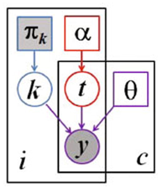
Graphical model for the proposed segmentation approach. Voxels are indexed with i, the channels are indexed with c. The known prior πk determines the label k of the normal, healthy tissue. The latent atlas α determines the channel-specific presence of tumor t. Normal state k, tumor state t, and intensity distribution parameters θ jointly determine the multi-modal image observations y. Observed (known) quantities are shaded. The tumor segmentation aims to estimate
, along with the segmentation of healthy tissue p(ki|y).
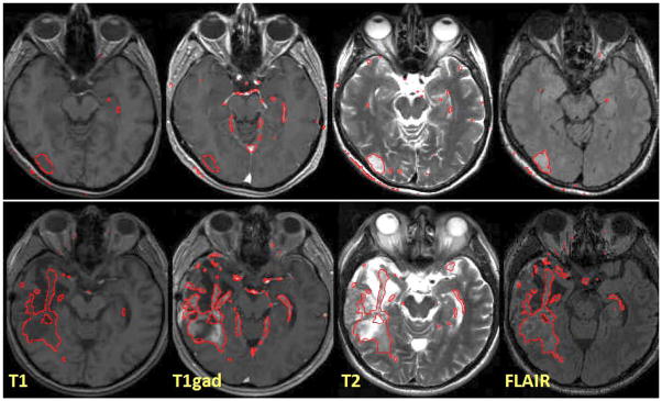
Examples of channel-specific segmentation results for four different modalities, in two patients. The outlines of regions with
are shown in red. The proposed method localizes the tumor reliably in post-therapeutic images (below), where surgery has led to significant deviations from normalcy for healthy tissues.
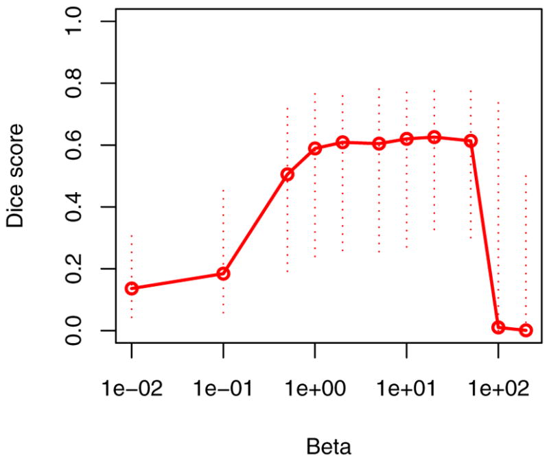
Sensitivity to the MRF parameter β. Indicated are the median (solid line) and the interquartile ranges of the average Dice scores of all 25 data set. While some regularization is beneficial, the segmentation performance is relatively insensitive to the choice of the only model parameter β.
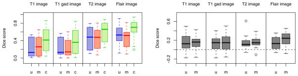
Benefits of the channel-specific segmentation. Boxplots show median, quartiles and outliers for the Dice scores of all 25 subjects, for all four modalities. Our channel-wise segmentation (c, green) improves over both multiple univariate (u, blue) and multivariate (m, red) segmentation, both in the absolute terms (left) and with respect to patient-specific differences (right). The right figure shows c–u and c–m.
Similar articles
-
Combining generative models for multifocal glioma segmentation and registration.Med Image Comput Comput Assist Interv. 2014;17(Pt 1):763-70. doi: 10.1007/978-3-319-10404-1_95. Med Image Comput Comput Assist Interv. 2014. PMID: 25333188 Free PMC article.
-
Low-rank to the rescue - atlas-based analyses in the presence of pathologies.Med Image Comput Comput Assist Interv. 2014;17(Pt 3):97-104. doi: 10.1007/978-3-319-10443-0_13. Med Image Comput Comput Assist Interv. 2014. PMID: 25320787 Free PMC article.
-
A unified framework for cross-modality multi-atlas segmentation of brain MRI.Med Image Anal. 2013 Dec;17(8):1181-91. doi: 10.1016/j.media.2013.08.001. Epub 2013 Aug 19. Med Image Anal. 2013. PMID: 24001931 Free PMC article.
-
Segmentation of image ensembles via latent atlases.Med Image Anal. 2010 Oct;14(5):654-65. doi: 10.1016/j.media.2010.05.004. Epub 2010 Jun 4. Med Image Anal. 2010. PMID: 20580305 Free PMC article.
-
A review of multivariate methods for multimodal fusion of brain imaging data.J Neurosci Methods. 2012 Feb 15;204(1):68-81. doi: 10.1016/j.jneumeth.2011.10.031. Epub 2011 Nov 11. J Neurosci Methods. 2012. PMID: 22108139 Free PMC article. Review.
Cited by
-
Diffusion-weighted imaging-based probabilistic segmentation of high- and low-proliferative areas in high-grade gliomas.Cancer Imaging. 2012 Apr 5;12(1):89-99. doi: 10.1102/1470-7330.2012.0010. Cancer Imaging. 2012. PMID: 22487677 Free PMC article.
-
GLISTR: glioma image segmentation and registration.IEEE Trans Med Imaging. 2012 Oct;31(10):1941-54. doi: 10.1109/TMI.2012.2210558. Epub 2012 Aug 13. IEEE Trans Med Imaging. 2012. PMID: 22907965 Free PMC article.
-
Concurrent tumor segmentation and registration with uncertainty-based sparse non-uniform graphs.Med Image Anal. 2014 May;18(4):647-59. doi: 10.1016/j.media.2014.02.006. Epub 2014 Feb 24. Med Image Anal. 2014. PMID: 24717540 Free PMC article.
-
Multifractal texture estimation for detection and segmentation of brain tumors.IEEE Trans Biomed Eng. 2013 Nov;60(11):3204-15. doi: 10.1109/TBME.2013.2271383. Epub 2013 Jun 27. IEEE Trans Biomed Eng. 2013. PMID: 23807424 Free PMC article.
-
Deformable templates guided discriminative models for robust 3D brain MRI segmentation.Neuroinformatics. 2013 Oct;11(4):447-68. doi: 10.1007/s12021-013-9190-5. Neuroinformatics. 2013. PMID: 23836390 Free PMC article.
References
-
- Van Leemput K, Maes F, Vandermeulen D, Suetens P. Automated model-based tissue classification of MR images of the brain. IEEE TMI. 1999;18:897–908. - PubMed
-
- Prastawa M, Bullitt E, Ho S, Gerig G. A brain tumor segmentation framework based on outlier detection. MedI A. 2004;8:275–283. - PubMed
-
- Bach Cuadra B, Pollo C, Bardera A, Cuisenaire O, Thiran JP. Atlas-based segmentation of pathological brain MR images using a model of lesion growth. IEEE TMI. 2004;23:1301–1314. - PubMed
Publication types
MeSH terms
Grants and funding
- P41-RR13218/RR/NCRR NIH HHS/United States
- R01-NS051826/NS/NINDS NIH HHS/United States
- P41-RR014075/RR/NCRR NIH HHS/United States
- U54 EB005149/EB/NIBIB NIH HHS/United States
- R01 NS052585/NS/NINDS NIH HHS/United States
- R01-EB006758/EB/NIBIB NIH HHS/United States
- R01-EB009051/EB/NIBIB NIH HHS/United States
- R01 EB006758/EB/NIBIB NIH HHS/United States
- U54-EB005149/EB/NIBIB NIH HHS/United States
- P41 RR014075/RR/NCRR NIH HHS/United States
- R01 NS051826/NS/NINDS NIH HHS/United States
- R01-NS052585/NS/NINDS NIH HHS/United States
- P41 RR013218/RR/NCRR NIH HHS/United States
LinkOut - more resources
Full Text Sources
Medical