

On super-resolution for fetal brain MRI
- PMID: 20879335
- PMCID: PMC3319126
- DOI: 10.1007/978-3-642-15745-5_44
On super-resolution for fetal brain MRI
Abstract
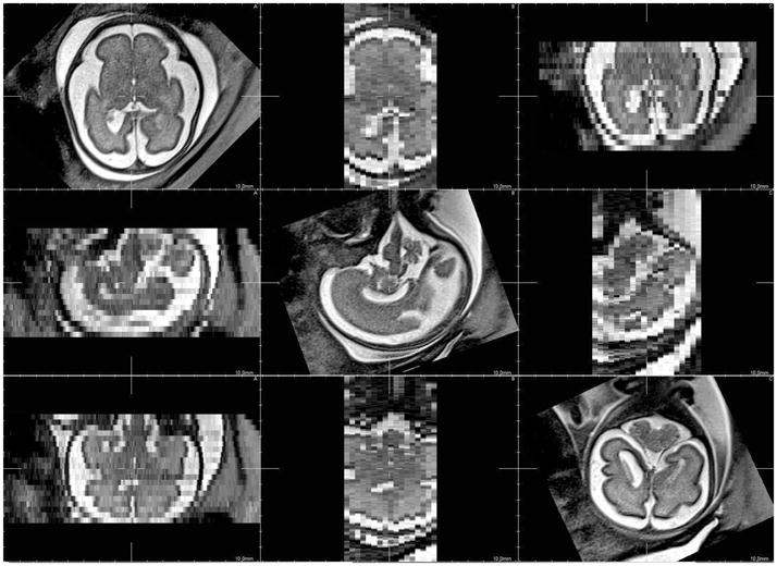
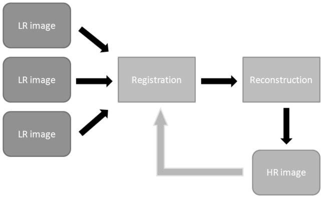
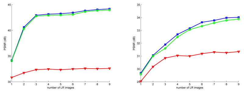
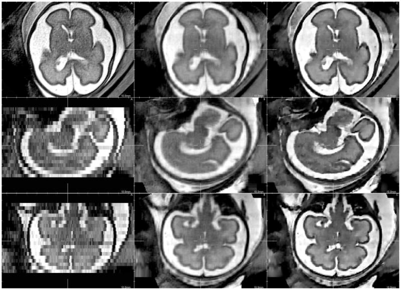
Super-resolution techniques provide a route to studying fine scale anatomical detail using multiple lower resolution acquisitions. In particular, techniques that do not depend on regular sampling can be used in medical imaging situations where imaging time and resolution are limited by subject motion. We investigate in this work the use of a super-resolution technique for anisotropic fetal brain MR data reconstruction without modifying the data acquisition protocol. The approach, which consists of iterative motion correction and high resolution image estimation, is compared with a previously used scattered data interpolation-based reconstruction method. To optimize acquisition time, an evaluation of the influence of the number of input images and image noise is also performed. Evaluation on simulated MR images and real data show significant improvements in performance provided by the super-resolution approach.
Figures
References
-
- Prayer D, Brugger PC, Prayer L. Fetal MRI: techniques and protocols. Pediatric Radiology. 2004 September;34(9):685–693. - PubMed
-
- Moore J, Drangova M, Wierzbicki M, Barron J, Peters T. A high resolution dynamic heart model based on averaged MRI data. Medical Image Computing and Computer-Assisted Intervention - MICCAI; 2003; 2003. pp. 549–555.
-
- Prager RW, Gee A, Berman L. Stradx: real-time acquisition and visualization of freehand three-dimensional ultrasound. Medical Image Analysis. 1999;3(2):129–140. - PubMed
-
- Rousseau F, Glenn OA, Iordanova B, Rodriguez-Carranza C, Vigneron DB, Barkovich JA, Studholme C. Registration-based approach for reconstruction of high-resolution in utero fetal MR brain images. Academic Radiology. 2006 September;13(9):1072–1081. - PubMed
-
- Jiang S, Xue H, Glover A, Rutherford M, Rueckert D, Hajnal JV. MRI of moving subjects using multislice snapshot images with volume reconstruction (SVR): application to fetal, neonatal, and adult brain studies. IEEE Transactions on Medical Imaging. 2007 July;26(7):967–980. - PubMed
Publication types
MeSH terms
Grants and funding
LinkOut - more resources
Full Text Sources
Medical