. 2011 Feb 1;350(1):13-23.
doi: 10.1016/j.ydbio.2010.09.016. Epub 2010 Sep 27.

APOBEC2, a selective inhibitor of TGFβ signaling, regulates left-right axis specification during early embryogenesis

Affiliations

APOBEC2, a selective inhibitor of TGFβ signaling, regulates left-right axis specification during early embryogenesis

Alin Vonica et al. Dev Biol. .

Abstract

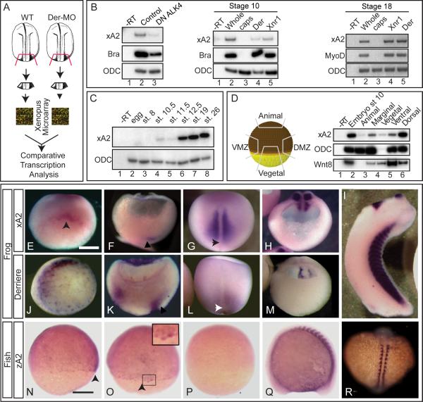
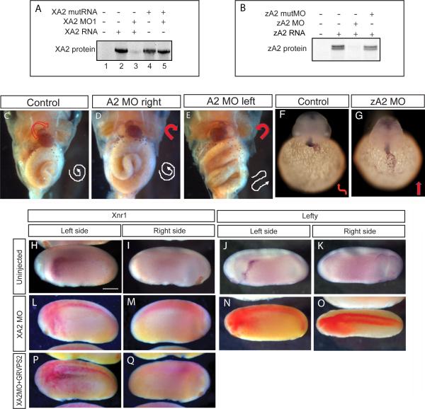
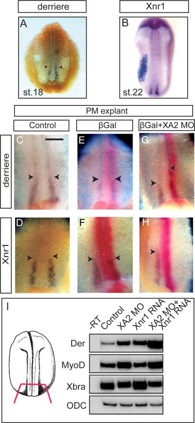
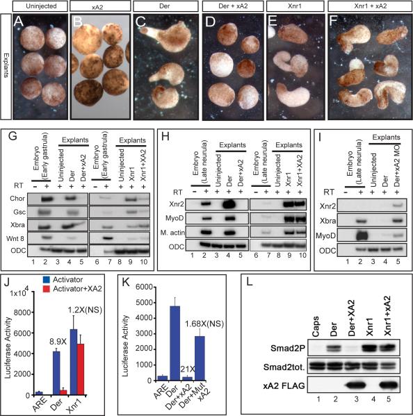
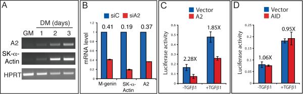
The specification of left-right asymmetry is an evolutionarily conserved developmental process in vertebrates. The interplay between two TGFβ ligands, Derrière/GDF1 and Xnr1/Nodal, together with inhibitors such as Lefty and Coco/Cerl2, have been shown to provide the signals that lead to the establishment of laterality. However, molecular events leading to and following these signals remain mostly unknown. We find that APOBEC2, a member of the cytidine deaminase family of DNA/RNA editing enzymes, is induced by TGFβ signaling, and that its activity is necessary to specify the left-right axis in Xenopus and zebrafish embryos. Surprisingly, we find that APOBEC2 selectively inhibits Derrière, but not Xnr1, signaling. The inhibitory effect is conserved, as APOBEC2 blocks TGFβ signaling, and promotes muscle differentiation, in a mammalian myoblastic cell line. This demonstrates for the first time that a putative RNA/DNA editing enzyme regulates TGFβ signaling and plays a major role in development.


Figures

Figure 1
Figure 1. APOBEC2 is a target of TGFβ signaling coexpressed with derrière in Xenopus embryos
A. Strategy for identification of genes regulated by derrière. Posterior dorsal fragments from wild-type and derrière -MO (Der-MO)-injected embryos were isolated at stage 18. B. TGFβ signaling and xA2 expression. Overexpression of a dominant negative type 1 receptor (DN ALK4) reduces expression of marginal xA2 in stage 10 whole embryos (left panel). Overexpressed Xnr1 (30 pg) and derrière (100 pg) RNA induce xA2 expression in stage 10 (central panel) and 18 (right side panel) animal caps. RT-PCR for xA2, MyoD, and Brachyury as markers of mesoderm induction, and ODC as loading control. C. Timing of xA2 expression. RT-PCR of embryos collected at the indicated developmental stages. D. Spatial expression of xA2 in stage 10 embryos. RT-PCR of embryonic explants (VMZ: ventral marginal zone; DMZ: dorsal marginal zone). E-M. Comparative expression of xA2 and derrière. In situ hybridization for xA2 (E-I), and derrière (JM) expression. (E, J) Stage 10 vegetal-dorsal views, arrowhead indicates the forming dorsal lip; (F, K) Stage 11 dorso-ventral sections (dorsal to the right). Arrowheads indicate recently involuted mesoderm; (G, L) dorsal views (anterior up). Arrowheads indicate the blastopore; (H, M) Stage 16 transversal sections, posterior fragments (dorsal is up). (I) Stage 32 lateral view. Overlap between xA2 and derrière occurs at stage 10 (dorsal marginal), stage 11 (involuted mesoderm), and stage 16 (paraxial mesoderm). N-R. Expression of zebrafish A2 (zA2). (N, O, P) 75% epiboly, in (N) lateral view (dorsal to the right) and (O) dorsal views. The arrowheads in N and O indicate the shield. (Q-R) 14 somite stage embryo. The inset in O (two fold magnification) shows shield cells with nuclear stain. (Q) Lateral view, anterior to the left, (R) dorsal view (anterior up). (P) Embryo stained with the sense probe as negative control. The bar in (E) indicates 0.3 mm, and in (N) 0.1 mm.
Figure 2
Figure 2. Effect of APOBEC2 protein depletion in Xenopus and zebrafish
A, B. Inhibition of in vitro translation by xA2 MO (A) and zA2 MO (B). C-E. Left side depletion of xA2 protein randomizes the left-right axis in Xenopus. Embryos injected on the left side with 10 ng xA2 MO were stained for light meromyosin at stage 46. (C) control embryo; (D) right side MO injection normal embryo; (E) left side MO injection, inverted heart and abnormally folded intestine. Arrows indicate the direction of the heart outflow tract and intestinal looping. F-G. Depletion of zA2 protein prevents heart looping in zebrafish. In situ hybridization with cmlc2 antisense probe for heart muscle on 36-40 hpf embryos. Arrows indicate ventricular looping. H-Q. xA2 depletion blocks the left side nodal signal. in situ hybridization for Xnr1 (H, I, L, M, P, Q), and Lefty (J, K, N, O) in purple, and injected LacZ RNA as tracer (L-Q) in red. Wild-type expression of Xnr1 (H) and Lefty (J) in the left lateral plate mesoderm was inhibited by injection of xA2 MO in the left paraxial mesoderm (L, N). Left side expression of Xnr1 was rescued by coinjection of GRVP16SMAD2Δ3 RNA (25 pg RNA, induced at stage 16; P). All views are lateral, except (O) (dorsal), anterior to the left. Embryos are stage 23 (Xnr1), and stage 24 (Lefty). The bar in (H) represents 0.3 mm.
Figure 3
Figure 3. xA2 depletion inhibits Xnr1 and increases derrière expression in posterior mesoderm
A-H. In situ hybridization for derrière (stage 18, A) and Xnr1 (stage 22, B), and double in situ hybridization for derrière (C, E, G), or Xnr1 (D, F, H), purple, and LacZ RNA coinjected as tracer (red). C-H are internal views of posterior dorsal fragments, anterior side up, of stage 18 embryos. The left side of each panel is the right side of each embryo. Xnr1 expression was inhibited by xA2 MO (n=36; H). Expression of derrière was expanded in anterior direction on the side injected with xA2 MO (n=33, arrowheads in G). The bar in C represents 0.1 mm. I. xA2 depletion synergizes with low levels of overexpressed Xnr1 RNA. RT-PCR of posterior poles injected bilaterally with xA2 MO (10 ng), Xnr1 RNA (1 pg), or both. The combination increased derrière and MyoD, but not Xbra expression.
Figure 4
Figure 4. APOBEC2 is a TGFβ inhibitor in Xenopus
A-F. xA2 prevents cap elongation induced by derrière but not Xnr1. RNAs were injected in the animal poles of 2 cell stage embryos (xA2 2ng, derrière 200pg, Xnr1 30pg). Caps were explanted at stage 10 and cultured to stage 18. G, H. Expression of xA2 RNA represses derrière-, but not Xnr1-dependent gene induction. RT-PCR of early gastrula (G) and late neurula (H) stage animal caps injected with derrière RNA (200 pg), or Xnr1 RNA (20 pg), with or without xA2 RNA (2 ng). I. xA2 depletion increased mesoderm induction by derrière RNA. RT-PCR for mesodermal genes (Xnr2, Xbra and MyoD), with ODC as loading control, in stage 18 animal caps injected with derrière RNA (100 pg) and xA2 MO (10 ng). J. xA2 specifically inhibits Derrière-induced transcription. Transcription assays for the Activin/nodal reporter ARE. Transcriptional inhibition by xA2 was statistically significant with derrière (200 pg RNA), but not with Xnr1 (20 pg RNA). K. An intact putative deaminase domain is required for the inhibitory activity of xA2. Wild-type, but not the triple mutant xA2 (Mut xA2, 2 ng RNA), inhibited transcription induced by derrière (200 pg RNA). The difference in transcriptional activity induced by Derrière alone vs. Derrière+xA2 wild-type was statistically significant (P<0.001), while that vs. the mutant xA2 was not. L. xA2 blocks the C-terminal phosphorylation of endogenous Smad2/3 induced by derrière. Western blot for total Smad2/3 and C-terminally phosphorylated Smad2/3 in stage 10.5 animal caps. No effect was seen on total Smad2/3 protein levels.
Figure 5
Figure 5. mAPOBEC2 inhibits TGFβ signaling in C2C12 myoblasts
A. RT-PCR analysis of mA2 and the muscle differentiation marker Skeletal-α-Actin mRNA levels during C2C12 differentiation. B. Real-time qPCR analysis of the mRNA levels of A2 and the indicated muscle markers in C2C12 cells transfected either with control (siC) or anti-A2 (siA2) siRNAs and differentiated for 2 days in DM. Numbers above the histogram bars indicate the relative decrease in mRNA levels. C, D. A2, but not AID, inhibits TGFβ signaling in C2C12 cells. Cells were transfected with the p3TP-lux reporter in combination with an empty vector (Vector) or an A2 (C) or AID (D) expressing vector. After transfection, cells were cultured with or without 5ng/ml recombinant TGF-β1 for 24 hours before harvesting for luciferase assay. Numbers above the histogram bars indicate the fold reduction in luciferase activity.

Similar articles

Cited by

References

    1. Amthor H, Macharia R, Navarrete R, Schuelke M, Brown SC, Otto A, Voit T, Muntoni F, Vrbova G, Partridge T, Zammit P, Bunger L, Patel K. Lack of myostatin results in excessive muscle growth but impaired force generation. Proc Natl Acad Sci U S A. 2007;104:1835–40. - PMC - PubMed
    1. Anant S, Mukhopadhyay D, Sankaranand V, Kennedy S, Henderson JO, Davidson NO. ARCD-1, an apobec-1-related cytidine deaminase, exerts a dominant negative effect on C to U RNA editing. Am J Physiol Cell Physiol. 2001;281:C1904–16. - PubMed
    1. Artaza JN, Bhasin S, Mallidis C, Taylor W, Ma K, Gonzalez-Cadavid NF. Endogenous expression and localization of myostatin and its relation to myosin heavy chain distribution in C2C12 skeletal muscle cells. J Cell Physiol. 2002;190:170–9. - PubMed
    1. Bell E, Munoz-Sanjuan I, Altmann CR, Vonica A, Brivanlou AH. Cell fate specification and competence by Coco, a maternal BMP, TGFbeta and Wnt inhibitor. Development. 2003;130:1381–9. - PubMed
    1. Bisgrove BW, Yost HJ. Classification of left-right patterning defects in zebrafish, mice, and humans. American Journal of Medical Genetics. 2001;101:315–23. - PubMed

Publication types

MeSH terms