. 2010 Oct 12;107(41):17785-90.
doi: 10.1073/pnas.1004361107. Epub 2010 Sep 28.

Disrupted-in-schizophrenia 1 (DISC1) plays essential roles in mitochondria in collaboration with Mitofilin

Affiliations

Disrupted-in-schizophrenia 1 (DISC1) plays essential roles in mitochondria in collaboration with Mitofilin

Young-Un Park et al. Proc Natl Acad Sci U S A. .

Abstract

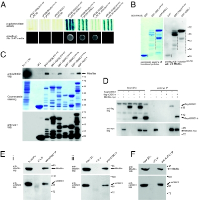
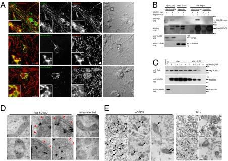
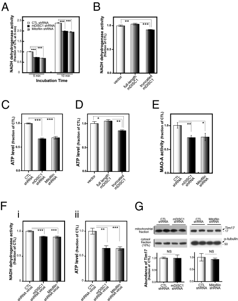
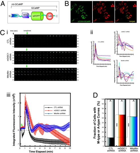
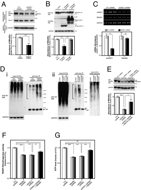
Disrupted-in-schizophrenia 1 (DISC1) has emerged as a schizophrenia-susceptibility gene affecting various neuronal functions. In this study, we characterized Mitofilin, a mitochondrial inner membrane protein, as a mediator of the mitochondrial function of DISC1. A fraction of DISC1 was localized to the inside of mitochondria and directly interacts with Mitofilin. A reduction in DISC1 function induced mitochondrial dysfunction, evidenced by decreased mitochondrial NADH dehydrogenase activities, reduced cellular ATP contents, and perturbed mitochondrial Ca(2+) dynamics. In addition, deficiencies in DISC1 and Mitofilin induced a reduction in mitochondrial monoamine oxidase-A activity. The mitochondrial dysfunctions evoked by the deficiency of DISC1 were partially phenocopied by an overexpression of truncated DISC1 that is associated with schizophrenia in human. DISC1 deficiencies induced the ubiquitination of Mitofilin, suggesting that DISC1 is critical for the stability of Mitofilin. Finally, the mitochondrial dysfunction induced by DISC1 deficiency was partially reversed by coexpression of Mitofilin, confirming a functional link between DISC1 and Mitofilin for the normal mitochondrial function. According to these results, we propose that DISC1 plays essential roles for mitochondrial function in collaboration with a mitochondrial interacting partner, Mitofilin.


Conflict of interest statement

The authors declare no conflict of interest.

Figures

Fig. 1.
Fig. 1.
Interactions among DISC1, NDEL1, and Mitofilin. (A) Interactions of DISC1, NDEL1, and Mitofilin in the yeast two-hybrid assay. (Upper) Interaction-dependent β-galactosidase expression. (Lower) Growth on HIS selective media containing 20 mM 3-amino-1,2,4-triazol (3-AT). hDISC1, human DISC1. (B) Interaction of Mitofilin with DISC1 and NDEL1 in vitro analyzed by a blot overlay assay. The PVDF membrane with the purified hDISC1 and NDEL1 protein bands transferred from an SDS/PAGE was first stained with Coomassie Blue R250 (Left) and subjected to GST–Mitofilin blot overlay followed by anti-Mitofilin Western blot analyses (Right). Dashed lines are the distribution of purified recombinant proteins with degradation products. WB, Western blotting. (C) Associations of Mitofilin with DISC1 and NDEL1 determined by GST pull-down assays. Lysates were prepared from SH-SY5Y cells that were differentiated for 36 h. (D) Coimmunoprecipitation of Mitofilin with full-length and truncated DISC1. HEK293 cells were transfected with flag-hDISC1 or flag-hDISC1-tr (amino acid residues 1–598), and Mitofilin-myc constructs as indicated. IP, immunoprecipitation. (E) Coimmunoprecipitation of endogenous DISC1 and Mitofilin from the differentiated CAD cells (i) and cultured mouse primary neurons (ii). Lysates prepared from CAD cells differentiated in the serum-free media for 36 h or mouse primary neurons cultured for 5 d in vitro (DIV5) were used. Anti-mDISC1 IP, IP with goat anti-mDISC1 antibody (N-16; Santa Cruz); CTL IP, control IP with goat anti-PhyA (Phytochrome A, Arabidopsis thaliana) antibody. (F) Coimmunoprecipitation of DISC1 and Mitofilin in the mouse brain lysate. Anti-mDISC1 IP, IP with goat anti-mDISC1 antibody (N-16; Santa Cruz); CTL IP, control IP with goat anti-human DISC1 antibody (K-20; Santa Cruz).
Fig. 2.
Fig. 2.
Localization of the DISC1–Mitofilin complex in mitochondria. (A) Colocalization of Mitofilin and DISC1 in mitochondria. Mouse primary neurons in culture were transfected with Mitofilin–EGFP plasmid on the DIV3 (green; Middle and Bottom), treated with mitotracker (red; Top and Middle) on the DIV4 as indicated, and applied to immunocytochemistry using goat anti-mDISC1 antibody (N-16; Santa Cruz; Top and Bottom), and Mitofilin–EGFP was detected by fluorescence from EGFP (Middle and Bottom). (Scale bar: 10 μm.) (B) Coimmunoprecipitation of Mitofilin with DISC1 from the mitochondrial fraction. The mitochondrial and cytosolic fractions from HEK293 cells transfected as indicated were subjected to immunoprecipitation followed by Western blotting. The same membrane was reprobed for Tom20, a mitochondrial marker, and α-tubulin. (C) Increased trypsin sensitivity of mitochondrial DISC1 by Triton X-100 treatment. The mitochondrial fraction was prepared from flag-hDISC1–transfected HEK293 cells en masse and divided into aliquots for the treatments of Triton X-100 (final 2%) and trypsin followed by Western blotting. (D) Localization of overexpressed DISC1 to the inside of mitochondria. HEK293 cells transfected with the flag-hDISC1 construct (Left) along with untransfected control cells (Right) were subjected to anti-flag IGEM. Mitochondrial outer membrane is indicated by red arrowheads. (Scale bar: 100 nm.) (E) Localization of endogenous DISC1 to the inside of mitochondria. Mouse brain sections were subjected to goat anti-mDISC1 IGEM (Left). The clusters of 10-nm immuno-gold particles for endogenous mDISC1 in mitochondria are indicated by arrows. CTL, IGEM with goat anti-PhyA antibody (Right).
Fig. 3.
Fig. 3.
Compromised mitochondrial function by deficiencies in DISC1 and Mitofilin. (A) Down-regulation of mitochondrial NADH dehydrogenase activity by mDISC1 or Mitofilin knockdown. CAD cells were transfected with shRNA constructs and differentiated for an additional 5 d. Media were replaced with the assay solution, and the time-dependent conversion of soluble formazan from a tetrazolium salt was measured at OD495. Error bars are mean ± SEM. ***P < 0.001, two-tailed t test (n = 6). (B) Down-regulation of mitochondrial NADH dehydrogenase activity by an overexpression of truncated mDISC1. CAD cells were transfected with plasmids as indicated and differentiated for 72 h before NADH-dehydrogenase activity assay. Incubation time was 10 min. Error bars are mean ± SEM. **P < 0.01; ***P < 0.001, two-tailed t test (n = 6). (C) Reduction of ATP contents in the mDISC1 or Mitofilin knockdown cells. CAD cells were transfected with shRNA constructs and differentiated for an additional 5 d. ATP contents in the cell lysates were measured. Error bars are mean ± SEM. ***P < 0.001, two-tailed t test (n = 6). (D) Reduction of ATP contents by an overexpression of truncated DISC1. CAD cells were transfected with full-length mDISC1 or truncated mDISC1, and after 72 h of differentiation in serum-free media, the ATP contents in lysates were measured. Error bars are mean ± SEM. *P < 0.05; **P < 0.01, two-tailed t test (n = 6). (E) Decreased monoamine oxidase-A (MAO-A) activities in the mDISC1 or Mitofilin knockdown cells. SN4741 cells were transfected with shRNA constructs and differentiated for an additional 5 d. Lysates were subjected to an MAO-A activity assay. Error bars are mean ± SEM. *P < 0.05; **P < 0.01, two-tailed t test (n = 6). (F) Reduction of NADH dehydrogenase activity and ATP contents by DISC1 and Mitofilin knockdown in cultured primary neurons. Cultured mouse cortical neurons (DIV2) were infected with lentiviruses containing mDISC1 or Mitofilin shRNA and cultured for an additional 6 d before the NADH dehydrogenase activity assays (i) and ATP measurement (ii). Error bars are mean ± SEM. **P < 0.01; ***P < 0.001, two-tailed t test (n = 6). (G) Unaltered mitochondrial contents in mDISC1 and Mitofilin knockdown cells. CAD cells were transfected with mDISC1 and Mitofilin shRNAs and differentiated for 5 d. The Tim17 levels in mitochondrial and α-tubulin in corresponding cytosol fractions were analyzed. Ratios of Tim17/α-tubulin were subjected to statistical analysis. Error bars are mean ± SEM. NS, not significant, two-tailed t test (n = 4).
Fig. 4.
Fig. 4.
Abnormal mitochondrial Ca2+ buffering by deficiencies in DISC1 and Mitofilin. (A) A schematic diagram of mt-GCaMP, a mitochondrial Ca2+ sensor. (B) Restricted expression of mt-GCaMP in mitochondria. Mitotracker (red) and anti-GFP antibody (green) in differentiated CAD cells. (C) Time-lapse mitochondrial Ca2+ imaging. CAD cells were cotransfected with mt-GCaMP constructs in combination with shRNA constructs (mt-GCaMP:shRNA plasmid = 1:5, molar ratio), and changes in fluorescence in response to ionomycin treatment were measured by time-lapse imaging 5 d after transfection. (i) Examples of time-lapse images for 495 s. Numbers, the time elapsed in seconds; green arrows, time point of ionomycin addition. (ii) Individual mitochondrial Ca2+ in response to ionomycin; six representative measurements of each are shown. Fluorescence intensity was normalized by fluorescence at the resting condition. (iii) Mean values of the curves shown in ii. Error bars are mean ± SEM. (D) The increased fraction of the cells showing abnormal fluctuation of Ca2+ (M-type curves) in mDISC1 and Mitofilin shRNA-transfected cells. ***P < 0.001, Fisher's exact test (n = 16 for CTL shRNA, n = 23 for mDISC1 shRNA, n = 10 for Mitofilin shRNA).
Fig. 5.
Fig. 5.
Down-regulation of Mitofilin by aberrant DISC1 function. (A) Down-regulation of Mitofilin protein levels by knockdown of DISC1. CAD cells were transfected with control or mDISC1 shRNA constructs and differentiated for 5 d. Mitochondrial fraction was isolated, and endogenous Mitofilin protein levels were analyzed by anti-Mitofilin Western blotting. Error bars are mean ± SEM. **P < 0.01, two-tailed t test (n = 6). (B) Down-regulation of Mitofilin protein levels by overexpression of truncated DISC1 protein. CAD cells were transfected with constructs of flag-mDISC1, truncated flag-mDISC1 (amino acid residues 1–598), and Mitofilin-myc and were differentiated for 72 h. Whole-cell lysates were analyzed. Ratios of Mitofilin/GAPDH were subjected to statistical analyses. Error bars are mean ± SEM. **P < 0.01, two-tailed t test (n = 4). (C) Unaffected Mitofilin mRNA level in DISC1 knockdown cells. CAD cells were transfected with shRNA constructs and differentiated for 5 d. mDISC1 and Mitofilin mRNA levels were measured by RT-PCR. Error bars are mean ± SEM. **P < 0.01, two-tailed t test (n = 4). (D) Increased ubiquitination of Mitofilin by DISC1 knockdown or overexpression of truncated DISC1. (i) CAD cells were cotransfected with shRNA, HA-Ub, and Mitofilin-myc constructs, and after 68 h of differentiation, anti-Myc immunoprecipitates from the cell lysates were analyzed by anti-Ub Western blotting. (ii) CAD cells were transfected with truncated mDISC1 or full-length mDISC1 along with Mitofilin-myc constructs, differentiated, and analyzed as in a. A lower exposure blot of input is also shown. Brackets indicate ubiquitinated Mitofilin. (E) Inhibition of down-regulation of Mitofilin by a proteasome inhibitor. CAD cells were transfected with control or mDISC1 shRNA constructs and differentiated for 5 d. MG132 (Sigma, 25 μM) was treated for 8 h ahead of the preparation of lysates. Error bars are mean ± SEM. *P < 0.05; ***P < 0.001, two-tailed t test (n = 4). (F and G) Partial rescue of the DISC1 knockdown-mediated reduction of NADH dehydrogenase activity and ATP contents by coexpression of Mitofilin. CAD cells were transfected with the constructs as indicated and differentiated for 5 d, and NADH dehydrogenase activity (F) and ATP contents (G) were measured. Error bars are mean ± SEM. **P < 0.01; ***P < 0.001, NS, not significant, two-tailed t test (n = 6).

References

    1. American Psychiatric Association—Task Force on DSM-IV . Diagnostic and Statistical Manual of Mental Disorders: DSM-IV-TR. 4th Ed. Washington, DC: American Psychiatric Association; 2000. p. xxxvii.
    1. Cardno AG, Gottesman II. Twin studies of schizophrenia: From bow-and-arrow concordances to star wars Mx and functional genomics. Am J Med Genet. 2000;97:12–17. - PubMed
    1. Harrison PJ, Weinberger DR. Schizophrenia genes, gene expression, and neuropathology: On the matter of their convergence. Mol Psychiatry. 2005;10:40–68. - PubMed
    1. Mackie S, Millar JK, Porteous DJ. Role of DISC1 in neural development and schizophrenia. Curr Opin Neurobiol. 2007;17:95–102. - PubMed
    1. Kamiya A, et al. A schizophrenia-associated mutation of DISC1 perturbs cerebral cortex development. Nat Cell Biol. 2005;7:1167–1178. - PubMed

Publication types

MeSH terms