. 2010 Dec 3;285(49):38502-10.
doi: 10.1074/jbc.M110.131201. Epub 2010 Sep 29.

The solution structure of a plant calmodulin and the CaM-binding domain of the vacuolar calcium-ATPase BCA1 reveals a new binding and activation mechanism

Affiliations

The solution structure of a plant calmodulin and the CaM-binding domain of the vacuolar calcium-ATPase BCA1 reveals a new binding and activation mechanism

Hiroaki Ishida et al. J Biol Chem. .

Abstract

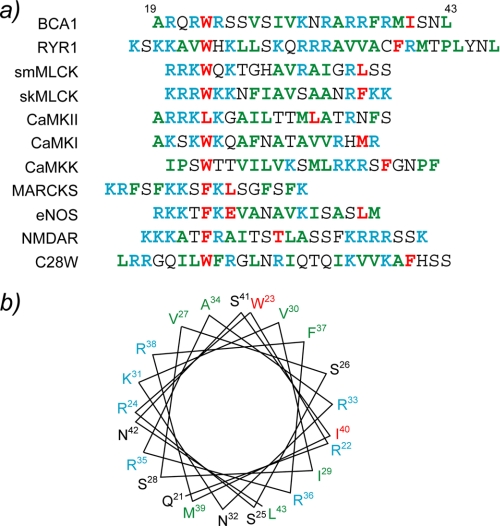
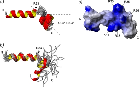
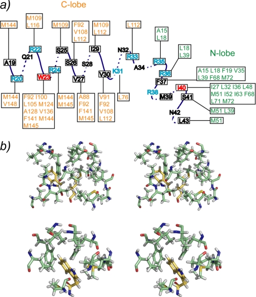
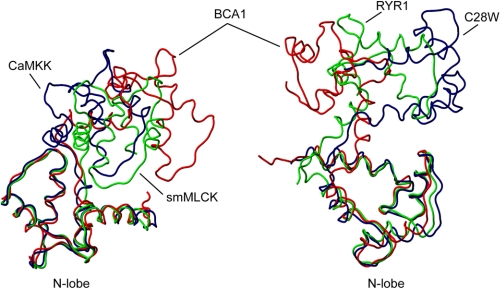
The type IIb class of plant Ca(2+)-ATPases contains a unique N-terminal extension that encompasses a calmodulin (CaM) binding domain and an auto-inhibitory domain. Binding of Ca(2+)-CaM to this region can release auto-inhibition and activates the calcium pump. Using multidimensional NMR spectroscopy, we have determined the solution structure of the complex of a plant CaM isoform with the CaM-binding domain of the well characterized Ca(2+)-ATPase BCA1 from cauliflower. The complex has a rather elongated structure in which the two lobes of CaM do not contact each other. The anchor residues Trp-23 and Ile-40 form a 1-8-18 interaction motif. Binding of Ca(2+)-CaM gives rise to the induction of two helical parts in this unique target peptide. The two helical portions are connected by a highly positively charged bend region, which represents a relatively fixed angle and positions the two lobes of CaM in an orientation that has not been seen before in any complex structure of calmodulin. The behavior of the complex was further characterized by heteronuclear NMR dynamics measurements of the isotope-labeled protein and peptide. These data suggest a unique calcium-driven activation mechanism for BCA1 and other plant Ca(2+)-ATPases that may also explain the action of calcium-CaM on some other target enzymes. Moreover, CaM activation of plant Ca(2+)-ATPases seems to occur in an organelle-specific manner.


Figures

FIGURE 1.
FIGURE 1.
a, amino acid sequence alignment of CaMBDs from various CaM target proteins. The anchoring residues are highlighted in red. The basic and acidic residues are shown in cyan and magenta, respectively, whereas hydrophobic residues are displayed in green. b, a helical wheel representation of the CaMBD of BCA1. The residues are colored in the same manner as in a. NOS, endothelial nitric oxide synthase.
FIGURE 2.
FIGURE 2.
The 25 lowest energy structures of SCaM4·BCA1 are superimposed. The N- and C-terminal lobe of SCaM4 are shown in light green and purple, respectively, whereas the BCA1 peptide is shown in yellow. Only the folded regions of the N-terminal unit (SCaM4 residues 6–74 and BCA1 peptide residues 38–43) and the C-terminal unit (SCaM4 residues 81–145 and BCA1 residues 20–33) are superimposed in the right and left panel, respectively. b, ribbon structures of the lowest energy structure of SCaM4·BCA1 at different angles. The regions are colored in the same manner as in a. The side chains of the anchor residues of the BCA1 peptide, Trp-23 and Ile-40, are shown. The side chain of Phe-37, which also has several hydrophobic contacts to SCaM4, is indicated as well.
FIGURE 3.
FIGURE 3.
a, the bound BCA1 peptide structures from the 25 lowest energy structures of SCaM4·BCA1. Only the first α-helical region (residues 20–33) is superimposed. The averaged angle between the first and the second α-helices for all the structures is 48.4° ± 5.3°. b, 20 BCA1 peptide structures determined in 30% trifluoroethanol are superimposed using the first α-helical region (residues 20–22). These structures were taken from Yamniuk and Vogel (20). c, the surface electrostatic properties of the BCA1 peptide. The residues that form a basic cluster in the middle of BCA1 peptide are labeled. C, C-terminal; N, N-terminal.
FIGURE 4.
FIGURE 4.
Schematic showing the observed intermolecular NOEs between SCaM4 and the BCA1 peptide. The two anchor residues are depicted in red, whereas the basic residues are shown in cyan. b, stereo views of the local interactions between SCaM4 and BCA1 residues, Phe-37 and Ile-40 (top panel), and Trp-23 (bottom panel).
FIGURE 5.
FIGURE 5.
The structure of SCaM4 in SCaM4·BCA1 (red) is compared with CaM in CaM·CaMKK (navy; Protein Data Bank code 1IQ5), CaM·smMLCK (green; Protein Data Bank code 1CDL) in the left panel and CaM·C28W (navy; Protein Data Bank code 2KNE) and CaM·RYR1 (green; Protein Data Bank code 2BCX) in the right panel. Only the well folded regions of the N-terminal lobe (residues 6–74) were overlaid in both panels.
FIGURE 6.
FIGURE 6.
The principle axis of the diffusion tensor is calculated separately for the N- and C-terminal lobe for SCaM4·BCA1 and displayed on the five lowest energy structures. Please note that the N- and C-terminal lobe were superimposed in the left and right panel, respectively.
FIGURE 7.
FIGURE 7.
15N-relaxation data for BCA1 peptide bound to SCaM4. The measured {1H}-15N NOE, T2, T, and T/T2 are plotted as a function of the residue number. A diagram showing the positions of the two helices and the bend in the BCA1 peptide structure is also displayed on top of the panels. The relaxation data show that the bending region of BCA1 peptide is not flexible in the complex.
FIGURE 8.
FIGURE 8.
The surface structure of SCaM4 is displayed together with a ribbon structure of SCaM4·BCA1. The side chains of the two Ser residues that have been reported to become phosphorylated by protein kinase C are shown (53). The acidic side chains of SCaM4 and the basic side chains of the BCA1 peptide that can form electrostatic interactions are also highlighted.
FIGURE 9.
FIGURE 9.
The interaction between the N-terminal lobe of SCaM4 and the BCA1 peptide are compared with those of CaM·smMLCK (a) and CaM·NMDA receptor (b). The N-terminal lobe of SCaM4 is colored green in both panels. c, the sequence alignment of the CaMBD of BCA1 with those of other type IIB Ca2+-ATPases. The conserved residues among all of the CaMBDs are boxed. d, the CaMBDs are classified into three groups according to the sequence alignment in b.
FIGURE 10.
FIGURE 10.
Model for a possible target activation mechanism from SCaM4·BCA1. a, only the N-terminal cytosolic domain, which contains the CaMBD and AID, and the second cytosolic domain, which contains the catalytic domain, are shown. b, at a low level of Ca2+, SCaM4 would be either free from the CaMBD or bound to the CaMBD only through its C-terminal lobe. c, at a high level of Ca2+, the N-terminal lobe binds Ca2+ and interacts with the CaMBD of BCA1 and induces a bend causing the release of the AID from the active site region leading to the full activation of BCA1.

References

    1. Reddy A. S. (2001) Plant Sci. 160, 381–404 - PubMed
    1. Lecourieux D., Ranjeva R., Pugin A. (2006) New Phytol. 171, 249–269 - PubMed
    1. Snedden W. A., Fromm H. (2001) New Phytol. 151, 35–66 - PubMed
    1. DeFalco T. A., Bender K. W., Snedden W. A. (2010) Biochem. J. 425, 27–40 - PubMed
    1. Zielinski R. E. (1998) Annu. Rev. Plant Physiol. Plant Mol. Biol. 49, 697–725 - PubMed

Publication types

MeSH terms

Associated data

LinkOut - more resources